. 2020 Aug 18;11(4):e01153-20.
doi: 10.1128/mBio.01153-20.

The sps Genes Encode an Original Legionaminic Acid Pathway Required for Crust Assembly in Bacillus subtilis

Affiliations

The sps Genes Encode an Original Legionaminic Acid Pathway Required for Crust Assembly in Bacillus subtilis

Thomas Dubois et al. mBio. .

Abstract

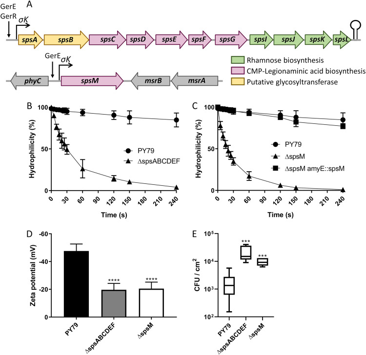
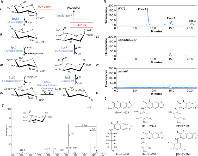
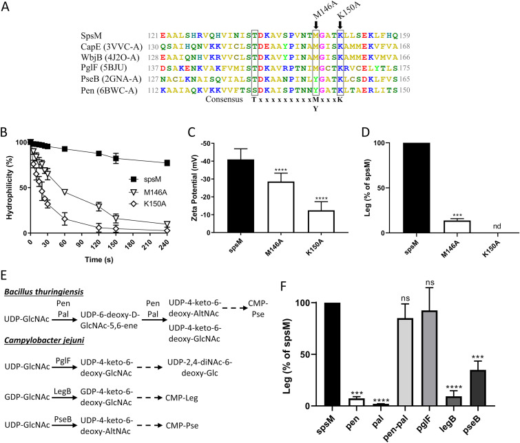
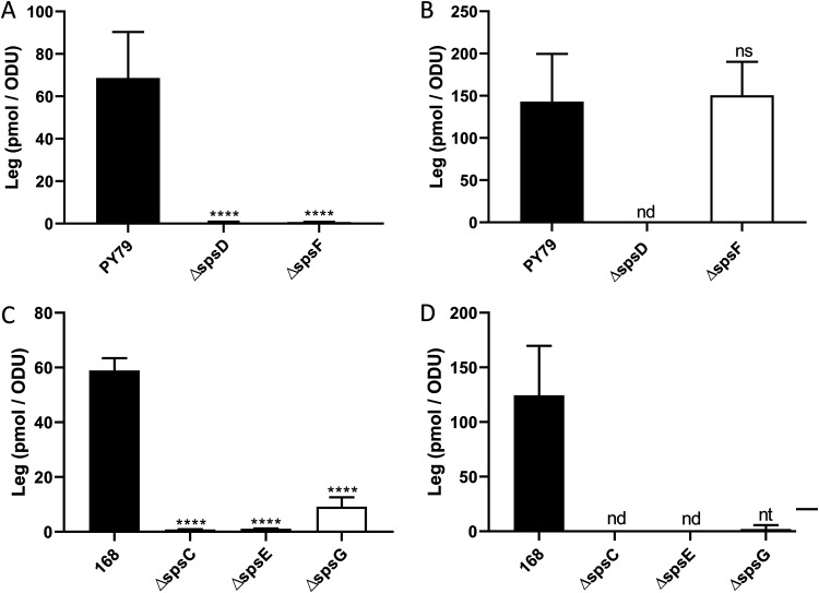
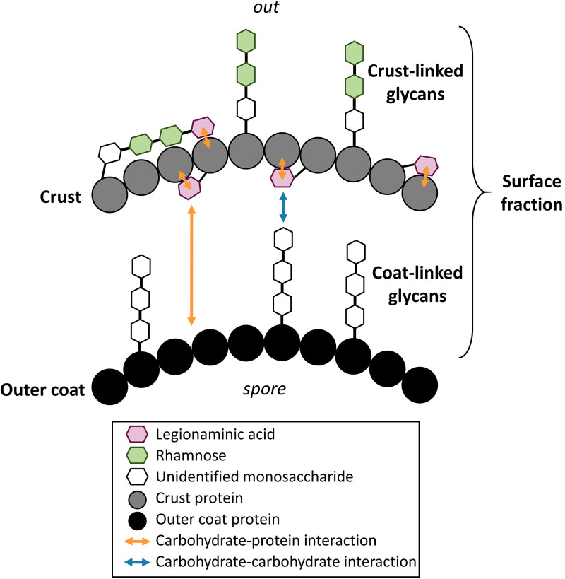
The crust is the outermost spore layer of most Bacillus strains devoid of an exosporium. This outermost layer, composed of both proteins and carbohydrates, plays a major role in the adhesion and spreading of spores into the environment. Recent studies have identified several crust proteins and have provided insights about their organization at the spore surface. However, although carbohydrates are known to participate in adhesion, little is known about their composition, structure, and localization. In this study, we showed that the spore surface of Bacillus subtilis is covered with legionaminic acid (Leg), a nine-carbon backbone nonulosonic acid known to decorate the flagellin of the human pathogens Helicobacter pylori and Campylobacter jejuni We demonstrated that the spsC, spsD, spsE, spsG, and spsM genes of Bacillus subtilis are required for Leg biosynthesis during sporulation, while the spsF gene is required for Leg transfer from the mother cell to the surface of the forespore. We also characterized the activity of SpsM and highlighted an original Leg biosynthesis pathway in B. subtilis Finally, we demonstrated that Leg is required for the assembly of the crust around the spores, and we showed that in the absence of Leg, spores were more adherent to stainless steel probably because of their reduced hydrophilicity and charge.IMPORTANCEBacillus species are a major economic and food safety concern of the food industry because of their food spoilage-causing capability and persistence. Their persistence is mainly due to their ability to form highly resistant spores adhering to the surfaces of industrial equipment. Spores of the Bacillus subtilis group are surrounded by the crust, a superficial layer which plays a key role in their adhesion properties. However, knowledge of the composition and structure of this layer remains incomplete. Here, for the first time, we identified a nonulosonic acid (Leg) at the surfaces of bacterial spores (B. subtilis). We uncovered a novel Leg biosynthesis pathway, and we demonstrated that Leg is required for proper crust assembly. This work contributes to the description of the structure and composition of Bacillus spores which has been under way for decades, and it provides keys to understanding the importance of carbohydrates in Bacillus adhesion and persistence in the food industry.

Keywords: Bacillus subtilis; bacterial adhesion; crust; legionaminic acid; nonulosonic acid; spores.


Figures

FIG 1
FIG 1
The sps genes participate in the surface and adhesion properties of B. subtilis spores. (A) Schematic representation of the spsABCDEFGIJKL and spsM genes in B. subtilis PY79. Transcriptional activators and promoters are shown with arrows and broken arrows, respectively. Potential stem-loop structure is indicated with a lollipop. The annotation of the sps genes is presented in Table S1 in the supplemental material. (B) Surface hydrophilicity of spores of the PY79 and PY79 ΔspsABCDEFspsABCDEF) strains evaluated by MATH assays. (C) Surface hydrophilicity of spores of the PY79, PY79 ΔspsMspsM), and PY79 ΔspsM amyE::spsMspsM amyE::spsM) strains. (D) Surface charge of spores evaluated by zetametry assays. (E) Adhesion of spores to stainless steel coupons. The results are expressed in CFU per square centimeter of stainless steel. The data are averages from at least three independent experiments performed with spores prepared independently. The error bars represent the standard deviations (SDs) of the means. ***, P ≤ 0.001; ****, P ≤ 0.0001 for Δ versus PY79 by Mann-Whitney test.
FIG 2
FIG 2
The sps genes are required for crust assembly. TEM images of spore sections after ruthenium red staining. The experiments were performed with the spores of the PY79, PY79 ΔspsABCDEFspsABCDEF), and PY79 ΔspsMspsM) strains. White arrows, coat layers; black arrows, crust; blue arrows, cap-like structures.
FIG 3
FIG 3
The sps genes encode a pathway required for legionaminic acid addition on the spore surface of B. subtilis. (A) Predicted Leg pathway in B. subtilis: I, UDP-N-acetyl-α-d-glucosamine (UDP-GlcNAc); II, UDP-2-acetamido-2,6-dideoxy-α-d-xylo-hexose-4-ulose (UDP-4-keto-6-deoxy-GlcNAc); III, UDP-4-amino-4,6-dideoxy-N-acetyl-α-d-glucosamine (UDP-4-amino-6-deoxy-GlcNAc); IV, UDP-2,4-diacetamido-2,4,6-trideoxy-α-d-glucose (UDP-2,4-diNAc-6-deoxy-Glc); V, 2,4-diacetamido-2,4,6-trideoxy-d-mannose (2,4-diNAc-6-deoxy-Man); VI, 5,7-diacetamido-3,5,7,9-tetradeoxy-d-glycero-β-d-galacto-nonulosonic acid (legionaminic acid or Leg); VII, CMP-5,7-diacetamido-3,5,7,9-tetradeoxy-d-glycero-β-d-galacto-nonulosonic acid (CMP-legionaminic acid or CMP-Leg). (B) Chromatograms of the RP-HPLC-FL experiments performed on the surface fractions of spores of the PY79, PY79 ΔspsABCDEFspsABCDEF), and PY79 ΔspsMspsM) strains. The RP-HPLC-FL experiments were carried out on at least three independent surface fractions for each strain. One representative chromatogram is presented. (C) Identification by LC/ESI-MS3 of DMB-Leg5Ac7Ac. Representative MS3 spectra of DMB-Leg5Ac7Ac, [M+H]+ at m/z 451. The ion [M+H-18]+ at m/z 433 is indicated by the black diamond. (D) Structure of DMB ([M+H]+ = 229), DMB-Leg ([M+H]+ = 541), and of the major structure fragments ([M+H]+ = 433, [M+H]+ = 415.1, [M+H]+ = 374, [M+H]+ = 356).
FIG 4
FIG 4
SpsM is a C-4/C-6 dehydratase using UDP-GlcNAc as a substrate. (A) Alignment of the protein sequence of SpsM with the protein sequences of CapE (Staphylococcus aureus), WbjB (Acinetobacter baumannii), PglF (Campylobacter jejuni), PseB (Helicobacter pylori), and Pen (Bacillus thuringiensis). The PDB number corresponding to each sequence is indicated in parentheses. The three conserved residues of the catalytic triad are black boxed. The arrows indicate residues modified by directed mutagenesis. The experiments presented in panels B, C, and D were carried out with PY79 ΔspsM amyE::spsM (spsM), PY79 ΔspsM amyE::spsM M146A (M146A), and PY79 ΔspsM amyE::spsM K150A (K150A) strains. (B) Surface hydrophilicity of spores evaluated by MATH assays. (C) Surface charge of spores evaluated by zetametry assays. ****, P ≤ 0.0001 for M146A or K150A versus spsM by Mann-Whitney test. (D) Relative amounts of Leg in the surface fractions of spores measured by RP-HPLC-FL. The results are relative to the amount of Leg of the PY79 ΔspsM amyE::spsM strain. nd, not detectable; ***, P ≤ 0.001 for M146A or K150A versus spsM by Welch’s t test. (E) Enzymatic reactions catalyzed by Pen-Pal, PglF, LegB, and PseB in B. thuringiensis and C. jejuni. The solid arrows represent the enzymatic reactions catalyzed by the enzymes. The dotted arrows represent the remaining parts of the pathways and point to the final products. (F) Relative amounts of Leg in the mother cells of the PY79 ΔspsM amyE::spsM (spsM), PY79 ΔspsM amyE::pen (pen), PY79 ΔspsM amyE::pal (pal), PY79 ΔspsM amyE::pen-pal (pen-pal), PY79 ΔspsM amyE::pglF (pglF), PY79 ΔspsM amyE::legB (legB), and PY79 ΔspsM amyE::pseB (pseB) strains measured at t10 by RP-HPLC-FL. The results are relative to the amount of Leg in the PY79 ΔspsM amyE::spsM strain. ns, not significant; ***, P ≤ 0.001; ****, P ≤ 0.0001 for pen, pal, pen-pal, pglF, legB, or pseB versus spsM by Welch’s t test. The error bars represent the SDs of the means.
FIG 5
FIG 5
The spsC, spsD, spsE, and spsG genes are required for Leg biosynthesis, while spsF is required for Leg transfer to the forespore surface. The amounts of Leg were measured in the surface fractions of spores (A and C) and the mother cells of sporulating B. subtilis cells at t10 (B and D) by RP-HPLC-FL. The experiments were performed with PY79, PY79 ΔspsDspsD), and PY79 ΔspsFspsF) strains (A and B) or 168, 168 ΔspsCspsC), 168 ΔspsEspsE), and 168 ΔspsGspsG) strains (C and D). The results were standardized by the OD600 of the spore preparations (A and C) or the OD600 of the cultures at t10 (B and D). ns, not significant; nd, not detected; nt, not statistically tested because the data set did not pass the Shapiro-Wilk normality test (two values of three were equal to zero). The error bars represent the SDs of the means. ****, P ≤ 0.0001 for Δ versus PY79 or 168 by Welch’s t test.
FIG 6
FIG 6
Legionaminic acid is linked to the crust. The amounts of Leg were measured in the surface fractions of spores by RP-HPLC-FL. The experiments were performed with PY79, PY79 ΔspsAspsA), and PY79 ΔspsBspsB) strains (A) or PY79, PY79 cotE::erm (cotE::erm), PY79 cotX::erm (cotX::erm), and PY79 cotZ::erm (cotZ::erm) strains (B). The results were standardized by the OD600 of the spore preparations. ns, not significant.; ****, P ≤ 0.0001 for Δ versus PY79 by Welch’s t test. The error bars represent the SDs of the means.
FIG 7
FIG 7
Hypothetical model of the B. subtilis spore surface organization. In this model, one or several crust proteins are glycosylated by Leg or by a crust-linked glycan containing Leg. Leg could participate in the assembly and/or the stabilization of the interactions between the crust proteins possibly through a carbohydrate-protein interaction. The crust proteins and the crust-linked glycan give the spores their hydrophilicity and negative charge that define the adhesion properties of B. subtilis spores. Leg might also participate in the anchoring of the crust proteins to the outer coat by interacting through a carbohydrate-protein interaction with the outer coat proteins or by a carbohydrate-carbohydrate interaction with a coat-linked glycan. The interactions presented in this model are speculative. They are extrapolated from current knowledge about NulOs in bacteria (see Discussion). In the absence of data about the structure of the coat-linked and crust-linked glycans, the number of monosaccharides that make up the glycans and the position of the bonds between monosaccharides are just indicative of a possible structure.

References

    1. Gopal N, Hill C, Ross PR, Beresford TP, Fenelon MA, Cotter PD. 2015. The prevalence and control of Bacillus and related spore-forming bacteria in the dairy industry. Front Microbiol 6:1418. doi:10.3389/fmicb.2015.01418. - DOI - PMC - PubMed
    1. Warth AD, Strominger JL. 1969. Structure of the peptidoglycan of bacterial spores: occurrence of the lactam of muramic acid. Proc Natl Acad Sci U S A 64:528–535. doi:10.1073/pnas.64.2.528. - DOI - PMC - PubMed
    1. Warth AD, Strominger JL. 1972. Structure of the peptidoglycan from spores of Bacillus subtilis. Biochemistry 11:1389–1396. doi:10.1021/bi00758a010. - DOI - PubMed
    1. Driks A, Eichenberger P. 2016. The spore coat. Microbiol Spectr 4:TBS-0023-2016. doi:10.1128/microbiolspec.TBS-0023-2016. - DOI - PubMed
    1. Driks A, Roels S, Beall B, Moran CP, Losick R. 1994. Subcellular localization of proteins involved in the assembly of the spore coat of Bacillus subtilis. Genes Dev 8:234–244. doi:10.1101/gad.8.2.234. - DOI - PubMed

Publication types

MeSH terms

Substances

LinkOut - more resources