. 2020 Aug 18;11(4):e01453-20.
doi: 10.1128/mBio.01453-20.

A Plant Virus Ensures Viral Stability in the Hemolymph of Vector Insects through Suppressing Prophenoloxidase Activation

Affiliations

A Plant Virus Ensures Viral Stability in the Hemolymph of Vector Insects through Suppressing Prophenoloxidase Activation

Xiaofang Chen et al. mBio. .

Abstract

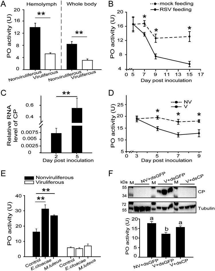
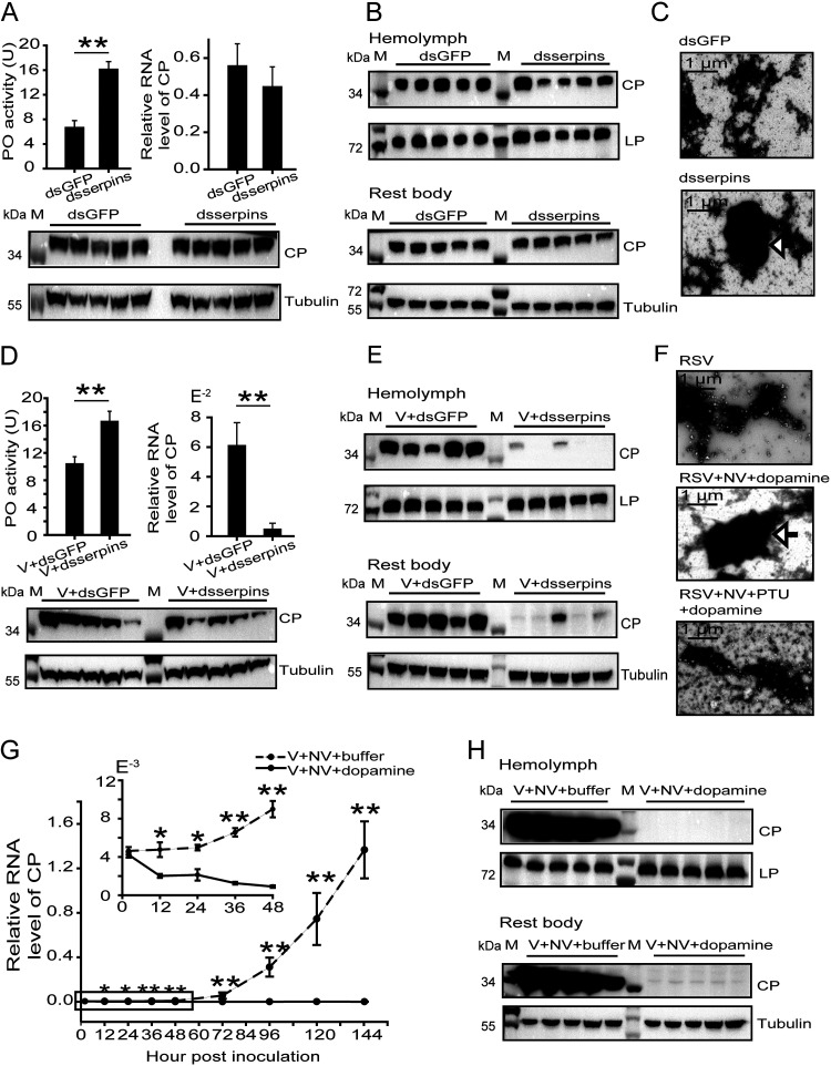
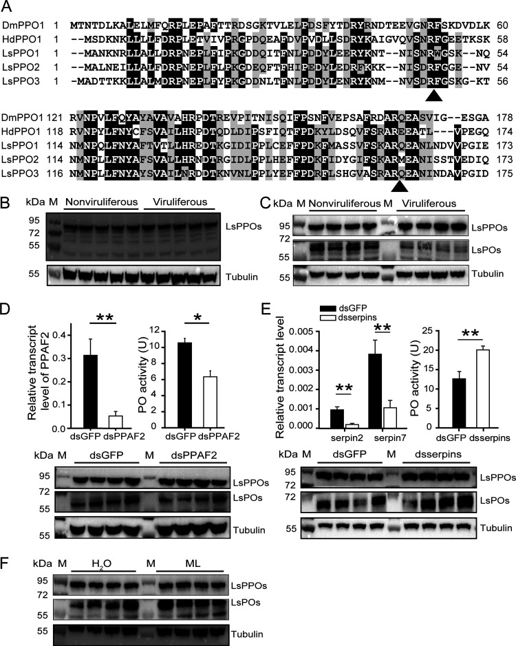
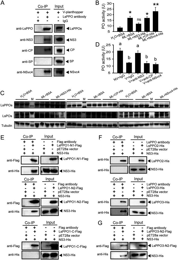
Most plant viruses require vector insects for transmission. Viral stability in the hemolymph of vector insects is a prerequisite for successful transmission of persistent plant viruses. However, knowledge of whether the proteolytic activation of prophenoloxidase (PPO) affects the stability of persistent plant viruses remains elusive. Here, we explored the interplay between rice stripe virus (RSV) and the PPO cascade of the vector small brown planthopper. Phenoloxidase (PO) activity was suppressed by RSV by approximately 60%. When the PPO cascade was activated, we found distinct melanization around RSV particles and serious damage to viral stability in the hemolymph. Viral suppression of PO activity was derived from obstruction of proteolytic cleavage of PPOs by binding of the viral nonstructural protein NS3. These results indicate that RSV attenuates the PPO response to ensure viral stability in the hemolymph of vector insects. Our research provides enlightening cues for controlling the transmission of vector-borne viruses.IMPORTANCE Large ratios of vector-borne plant viruses circulate in the hemolymph of their vector insects before entering the salivary glands to be transmitted to plants. The stability of virions in the hemolymph is vital in this process. Activation of the proteolytic prophenoloxidase (PPO) to produce active phenoloxidase (PO) is one of the major innate immune pathways in insect hemolymph. How a plant virus copes with the PPO immune reaction in its vector insect remains unclear. Here, we report that the PPO affects the stability of rice stripe virus (RSV), a notorious rice virus, in the hemolymph of a vector insect, the small brown planthopper. RSV suppresses PPO activation using viral nonstructural protein. Once the level of PO activity is elevated, RSV is melanized and eliminated from the hemolymph. Our work gives valuable clues for developing novel strategies for controlling the transmission of vector-borne plant viruses.

Keywords: NS3; hemolymph; plant virus; prophenoloxidase; rice stripe virus; small brown planthopper; vector insect.


Figures

FIG 1
FIG 1
Phylogenetic trees of PPO activation cascade members of the small brown planthopper and other insect species. The neighbor-joining method (pairwise deletion and p-distance model) was used. Bootstrap analysis (1,000 replicates) was applied to evaluate the internal support of the tree topology. Bootstrap values higher than 60% were present at the nodes. (A) HPs. (B) PPAPs/PPAFs. (C) PPOs. (D) Serpins. The cascade members of the small brown planthopper are highlighted in italics and bold letters. The GenBank accession numbers of all of the proteins are listed in Table S1. Aa, Aedes aegypti; Ag, Anopheles gambiae; Bm, Bombyx mori; Dm, Drosophila melanogaster; Hd, Holotrichia diomphalia; Ha, Helicoverpa armigera; Hc, Hyphantria cunea; Ls, Laodelphax striatellus; Ms, Manduca sexta; Of, Ostrinia furnacalis; Tm, Tenebrio molitor.
FIG 2
FIG 2
Differential responses of PPO activation pathway genes to RSV in the fat body and hemocytes of planthoppers measured by quantitative real-time PCR. The relative transcript level of each gene to that of EF2 is reported as the mean ± SE. Fat bodies from 20 to 30 individuals and hemocytes from 100 to 150 individuals in a replicate and eight replicates for each organ were used. FB, fat body. H, hemocytes. *, P < 0.05; **, P < 0.01.
FIG 3
FIG 3
PO activity was inhibited by RSV in planthoppers. (A) PO activity in the hemolymph and whole body of viruliferous and nonviruliferous planthoppers. Totals of 10 and 12 replicates were used for hemolymph and whole-body samples, respectively. (B) PO activity was measured in the whole body of planthoppers fed on RSV-infected rice seedlings (RSV feeding), and the virus was allowed to incubate in insects for different times. The control group was nonviruliferous insects fed on rice seedlings without RSV infection (mock feeding). Six to nine replicates were used for each group. (C) Relative RNA levels of CP in the planthoppers injected with RSV crude preparations. Six to 10 replicates were tested for each group. (D) PO activity in the whole body of planthoppers injected with RSV crude preparations (V) or crude preparations from nonviruliferous insects (NV). Five to eight replicates were used for each group. (E) PO activity in nonviruliferous or viruliferous planthoppers infected with E. cloacae or M. luteus at 24 h. The control group was injected with water. Five or six replicates were used for each group. *, P < 0.05; **, P < 0.01. (F) Variations in CP and PO activity in planthoppers injected with the mixture of V and the double-stranded RNA of CP (dsCP). The control group was injected with a mixture of the double-stranded RNA of GFP (dsGFP) and V or NV. Five to eight replicates were used for each group. A homemade anti-CP polyclonal antibody was applied to quantify CP. An anti-human β-tubulin monoclonal antibody was used to measure tubulin as an internal control. Different letters indicate statistically significant differences in PO activity. M, marker.
FIG 4
FIG 4
Adverse effects of POs on RSV stability in the hemolymph of planthoppers. (A) Variations in PO activity and CP RNA and protein levels in the whole body of viruliferous planthoppers after injection of the mixture of double-stranded RNAs of serpin2 and serpin7 (dsserpins) compared to those after injection of double-stranded RNA of GFP (dsGFP). The PO activity was measured with seven replicates. The RNA levels of CP were quantified with eight replicates. A homemade anti-CP polyclonal antibody was applied to quantify CP, and an anti-human β-tubulin monoclonal antibody was used to measure tubulin as an internal control via Western blotting. (B) Western blot to show CP in the hemolymph and rest body of viruliferous planthoppers after injection of dsserpins or dsGFP. A homemade anti-lipoprotein polyclonal antibody was used to quantify lipoprotein (LP) as an internal control. (C) Transmission electron microscopy images to show melanization around RSV particles isolated from the hemolymph of viruliferous planthoppers after injection of dsserpins or dsGFP. The arrow indicates melanized viruses. (D) Variations in PO activity and CP RNA and protein levels in the whole body of planthoppers 4 days after injection of a mixture of RSV crude preparations (V) and dsserpins or dsGFP. PO activity was assayed with eight replicates. The RNA levels of CP were quantified with six or seven replicates. (E) Western blot to show CP in the hemolymph and rest body of planthoppers injected with a mixture of V and dsserpins or dsGFP. (F) Transmission electron microscopy images to show melanization around purified RSV particles that were incubated with crude preparations from nonviruliferous planthoppers (NV) and dopamine in the presence or absence of phenylthiourea (PTU). The arrow indicates melanized viruses. (G) qRT-PCR showing the relative RNA levels of CP in planthoppers injected with a mixture of V, NV, and dopamine at different time points with 6 to 12 replicates. Dopamine was replaced by Tris-HCl buffer in the control group. The boxed time points are magnified in the upper-left panel. (H) Western blot to show CP in the hemolymph and rest body of planthoppers injected with a mixture of V, NV, and dopamine or Tris-HCl buffer at 144 h postinoculation. M, marker. *, P < 0.05; **, P < 0.01.
FIG 5
FIG 5
Reduction in PO production by RSV in planthoppers. (A) Sequence alignment of the PPO N terminus of several insects. Possible proteolytic cleavage sites are marked with arrows. Ls, L. striatellus. Dm, D. melanogaster. Hd, H. diomphalia. (B) Western blot assay showing the protein levels of LsPPOs and LsPOs in nonviruliferous and viruliferous planthoppers using an anti-LsPPO antibody. An anti-human β-tubulin monoclonal antibody was used to measure tubulin as an internal control. (C) Western blot assay showing the protein levels of LsPPOs and LsPOs by separate blottings. The PVDF membrane was excised at the site of the 72-kDa marker, and then the two pieces of membrane were separately incubated with the anti-LsPPO antibody. (D) Variations in PO activity and PO protein level in nonviruliferous planthoppers after injection of double-stranded RNAs of PPAF2 (dsPPAF2) compared to those after injection of double-stranded RNA of GFP (dsGFP). PO activity was assayed with six replicates. The relative transcript level of PPAF2 compared to that of EF2 was measured by qRT-PCR with six replicates and reported as the mean ± SE. (E) Variations in PO activity and PO protein level in nonviruliferous planthoppers after injection of a mixture of double-stranded RNAs of serpin2 and serpin7 (dsserpins) compared to those after injection of dsGFP. PO activity was assayed with five replicates. The relative transcript level of serpin2 and serpin7 compared to that of EF2 was measured by qRT-PCR with seven or eight replicates and reported as the mean ± SE. (F) Western blot assay showing the protein levels of LsPOs in nonviruliferous planthoppers that were inoculated with M. luteus (ML) or water. M, marker. *, P < 0.05; **, P < 0.01.
FIG 6
FIG 6
NS3 was responsible for the reduction in PO production. (A) Coimmunoprecipitation (Co-IP) assay for the interaction between LsPPOs and RSV proteins in vivo using an anti-LsPPO polyclonal antibody. Rabbit IgG was used as a negative control. Homemade monoclonal anti-CP, anti-NS3, and anti-SP antibodies and polyclonal anti-NSvc4 and anti-LsPPO antibodies were used in Western blot analysis. V-planthopper, the total proteins from viruliferous adult planthoppers. (B) Comparisons of PO activity in nonviruliferous planthoppers injected with a mixture of M. luteus (ML) and recombinantly expressed NS3-His, CP-His, NSvc4-His, or BSA compared to that of the control group insects injected with BSA and water. Seven to 10 replicates were used. *, P < 0.05; **, P < 0.01. ns, no significant difference. (C) Western blot assay showing the variation in LsPOs in the nonviruliferous planthoppers that were injected with a mixture of ML and NS3-His, CP-His, or NSvc4-His. The control group was injected with BSA and water or ML and BSA. An anti-LsPPO polyclonal antibody and anti-human β-tubulin monoclonal antibody were used. M, marker. (D) Comparisons of PO activity among planthoppers injected with a mixture of crude preparations from nonviruliferous planthoppers (NV) and IgG, RSV crude preparations (V) and IgG, V and anti-NS3 polyclonal antibody, V and anti-CP polyclonal antibody, or V and anti-NSvc4 polyclonal antibody. Five to seven replicates were used. Different letters indicate statistically significant differences in PO activity. (E) Co-IP assay for the interaction between a recombinantly expressed fragment of LsPPO1 (LsPPO1-N1-Flag, LsPPO1-N2-Flag, or LsPPO1-C-Flag) and NS3-His using an anti-Flag monoclonal antibody. The total proteins from E. coli expressing empty pET28a vector were used as a negative control. An anti-Flag or anti-His monoclonal antibody was used in Western blot analysis. (F) Co-IP assay for the interaction of recombinantly expressed LsPPO2-His or LsPPO3-His with NS3-His using an anti-LsPPO polyclonal antibody. (G) Co-IP assay for the interaction between LsPPO3-N2-Flag and NS3-His using an anti-Flag monoclonal antibody.

Similar articles

Cited by

References

    1. Bragard C, Caciagli P, Lemaire O, Lopez-Moya JJ, MacFarlane S, Peters D, Susi P, Torrance L. 2013. Status and prospects of plant virus control through interference with vector transmission. Annu Rev Phytopathol 51:177–201. doi:10.1146/annurev-phyto-082712-102346. - DOI - PubMed
    1. Hogenhout SA, Ammar el D, Whitfield AE, Redinbaugh MG. 2008. Insect vector interactions with persistently transmitted viruses. Annu Rev Phytopathol 46:327–359. doi:10.1146/annurev.phyto.022508.092135. - DOI - PubMed
    1. Liu W, Gray S, Huo Y, Li L, Wei T, Wang X. 2015. Proteomic analysis of interaction between a plant virus and its vector insect reveals new functions of hemipteran cuticular protein. Mol Cell Proteomics 14:2229–2242. doi:10.1074/mcp.M114.046763. - DOI - PMC - PubMed
    1. Kingsolver MB, Huang Z, Hardy RW. 2013. Insect antiviral innate immunity: pathways, effectors, and connections. J Mol Biol 425:4921–4936. doi:10.1016/j.jmb.2013.10.006. - DOI - PMC - PubMed
    1. Kumar A, Srivastava P, Sirisena P, Dubey SK, Kumar R, Shrinet J, Sunil S. 2018. Mosquito innate immunity. Insects 9:95. doi:10.3390/insects9030095. - DOI - PMC - PubMed

Publication types

LinkOut - more resources