Review
. 2020 Aug 18;29(157):200021.
doi: 10.1183/16000617.0021-2020. Print 2020 Sep 30.

Lung complications of Sjogren syndrome

Affiliations
Review

Lung complications of Sjogren syndrome

Fabrizio Luppi et al. Eur Respir Rev. .

Abstract

Primary Sjogren syndrome (pSS) is a systemic autoimmune disease characterised by lymphocytic infiltration of exocrine glands and by a number of systemic manifestations, including those regarding the lung. Pulmonary involvement in pSS includes interstitial lung disease (ILD) and airway disease, together with lymphoproliferative disorders.Patients with pSS-ILD report impaired health-related quality of life and a higher risk of death, suggesting the importance of early diagnosis and treatment of this type of pulmonary involvement. In contrast, airway disease usually has little effect on respiratory function and is rarely the cause of death in these patients.More rare disorders can be also identified, such as pleural effusion, cysts or bullae.Up to date, available data do not allow us to establish an evidence-based treatment strategy in pSS-ILD. No data are available regarding which patients should be treated, the timing to start therapy and better therapeutic options. The lack of knowledge about the natural history and prognosis of pSS-ILD is the main limitation to the development of clinical trials or shared recommendations on this topic. However, a recent trial showed the efficacy of the antifibrotic drug nintedanib in slowing progression of various ILDs, including those in pSS patients.


Conflict of interest statement

Conflict of interest: F. Luppi reports personal fees from Roche and Boheringer-Ingelheim, outside the submitted work. Conflict of interest: M. Sebastiani has nothing to disclose. Conflict of interest: N. Sverzellati has nothing to disclose. Conflict of interest: A. Cavazza has nothing to disclose. Conflict of interest: C. Salvarani has nothing to disclose. Conflict of interest: A. Manfredi has nothing to disclose.

Figures
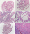
FIGURE 1
FIGURE 1
Some examples of surgical lung biopsies showing different interstitial lung disease in patients with Sjogren syndrome. a) Mixed cellular/fibrosing nonspecific interstitial pneumonia (NSIP), consisting of a combination of lymphoplasmacytic infiltrate and uniform interstitial fibrosis. b,c) A fibrosing process with usual interstitial pneumonia pattern, consisting of patchy scars (b) and fibroblastic foci (c). The lymphoplasmacytic infiltrate present in (c) is a clue, suggesting an underlying autoimmune disease. d) A lymphoplasmacytic interstitial infiltrate consistent with lymphocytic interstitial pneumonia, associated with small cysts of probable bronchiolar/alveolar duct origin. In this case, the limit with cellular NSIP is blurred; when the infiltrate is denser, the differential diagnosis with lymphoma (particularly mucosa-associated lymphoid tissue (MALT) lymphoma) can be difficult and requires immunohistochemical and sometimes molecular evaluation. e) Transbronchial biopsy showing organising pneumonia, consisting of intra-alveolar plugs of loose connective tissue. Some fibrin is frequently present in organising pneumonia; when fibrin is prominent, the term acute fibrinous and organising pneumonia can be applied. f) Nodular amyloidosis, consisting of deposition of acellular jaline material. The peripheral giant cell reaction is a clue pointing to the correct diagnosis. In nodular amyloidosis, any associated lymphoid infiltrate should be regarded with suspicion and should induce the pathologist to order the appropriate immunohistochemical stains to evaluate the possibility of a concomitant indolent B-cell lymphoma, particularly of the MALT type: in this case, the lymphoid infiltrate was polyclonal.
FIGURE 2
FIGURE 2
A 63-year-old woman with Sjogren syndrome. Coronal computed tomography image shows numerous cysts admixed with fine reticular abnormalities in the lower lobes. In the upper lobes, a few centrilobular branching opacities (circle) consistent with bronchiolitis (likely follicular) can be appreciated. This patient also suffered from pulmonary hypertension, which is responsible for pulmonary artery enlargement (arrow).
FIGURE 3
FIGURE 3
A 51-year-old woman with Sjogren syndrome. Axial computed tomography shows peribronchovascular reticular and ground-glass opacities in the lower lobes. This pattern is consistent with nonspecific interstitial pneumonia.
FIGURE 4
FIGURE 4
Histological findings of airway pathology in Sjogren syndrome. a) Bronchial biopsy showing a dense periglandular lymphoid infiltrate, similar to that seen in minor salivary gland biopsies in Sjogren syndrome. b) Surgical lung biopsy showing cellular bronchiolitis, consisting of a centrilobular cellular lymphoplasmacytic infiltrate, associated with bronchioloectasia. In the upper part of the picture a small granuloma is present (shown at higher magnification in c). Small non-necrotising granulomas are more frequent in Sjogren syndrome than in other connective tissue disease, and the histologic distinction with other granulomatous diseases (particularly infections and sarcoidosis) can be difficult. Occasionally Sjogren syndrome and sarcoidosis coexist in the same patient [86]. d) Surgical lung biopsy showing follicular bronchiolitis, a variant of cellular bronchiolitis in which the lymphoid infiltrate is organised in follicles with germinal centres. In Sjogren syndrome, cellular/follicular bronchiolitis can be the only lesion present in the biopsy, or can coexist with other lesions (cellular nonspecific interstitial pneumonia in this case). e) Surgical lung biopsy showing constrictive bronchiolitis, consisting of granulation tissue obliterating the bronchiolar lumen. Constrictive bronchiolitis is quite rare in Sjogren syndrome, cellular/follicular bronchiolitis can be the only lesion present in the biopsy, or can coexist, and clinically corresponds to the most severe forms of airway disease.

References

    1. Fox RI. Sjögren's syndrome. Lancet 2005; 366: 321–331. doi:10.1016/S0140-6736(05)66990-5 - DOI - PubMed
    1. Qin B, Wang J, Yang Z, et al. . Epidemiology of primary Sjogren's syndrome: a systematic review and meta-analysis. Ann Rheum Dis 2015; 74: 1983–1989. doi:10.1136/annrheumdis-2014-205375 - DOI - PubMed
    1. Patel R, Shahane A. The epidemiology of Sjögren's syndrome. Clin Epidemiol 2014; 6: 247–255. - PMC - PubMed
    1. Natalini J, Johr C, Kreider M. Pulmonary involvement in Sjögren syndrome. Clin Chest Med 2019; 40: 531–544. doi:10.1016/j.ccm.2019.05.002 - DOI - PubMed
    1. Ramos-Casals M, Brito-Zerón P, Seror R, et al. . Characterization of systemic disease in primary Sjögren's syndrome: EULAR-SS task force recommendations for articular, cutaneous, pulmonary and renal involvements. Rheumatology (Oxford) 2015; 54: 2230–2238. - PMC - PubMed