. 2020 Oct 16;295(42):14522-14535.
doi: 10.1074/jbc.RA120.014684. Epub 2020 Aug 19.

Alternative N-terminal regions of Drosophila myosin heavy chain II regulate communication of the purine binding loop with the essential light chain

Affiliations

Alternative N-terminal regions of Drosophila myosin heavy chain II regulate communication of the purine binding loop with the essential light chain

Marieke J Bloemink et al. J Biol Chem. .

Abstract

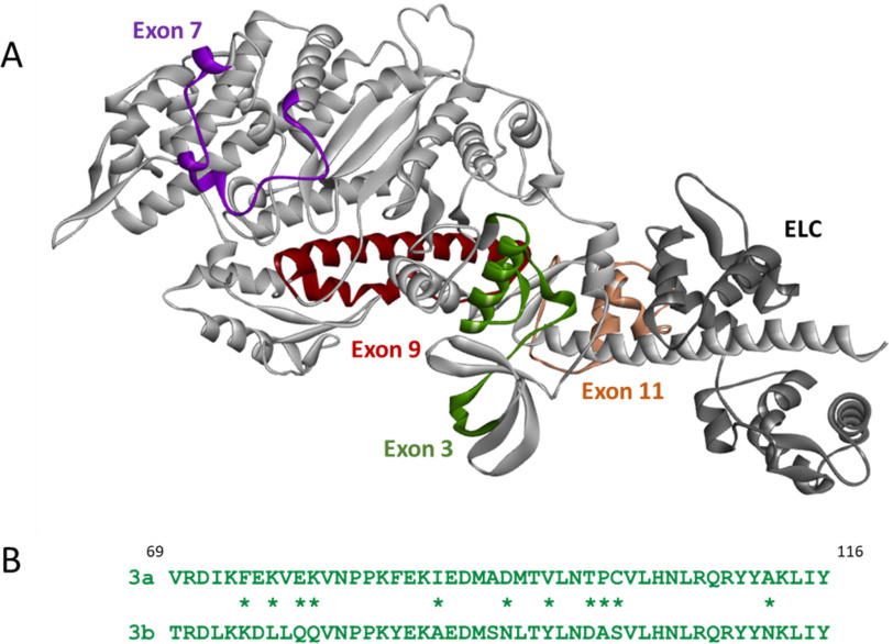
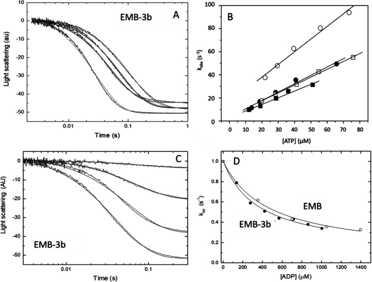
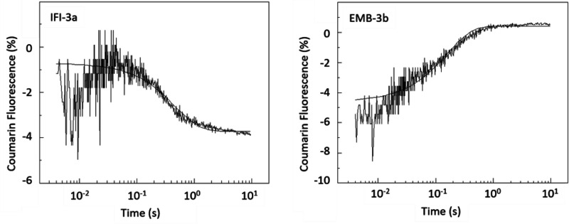
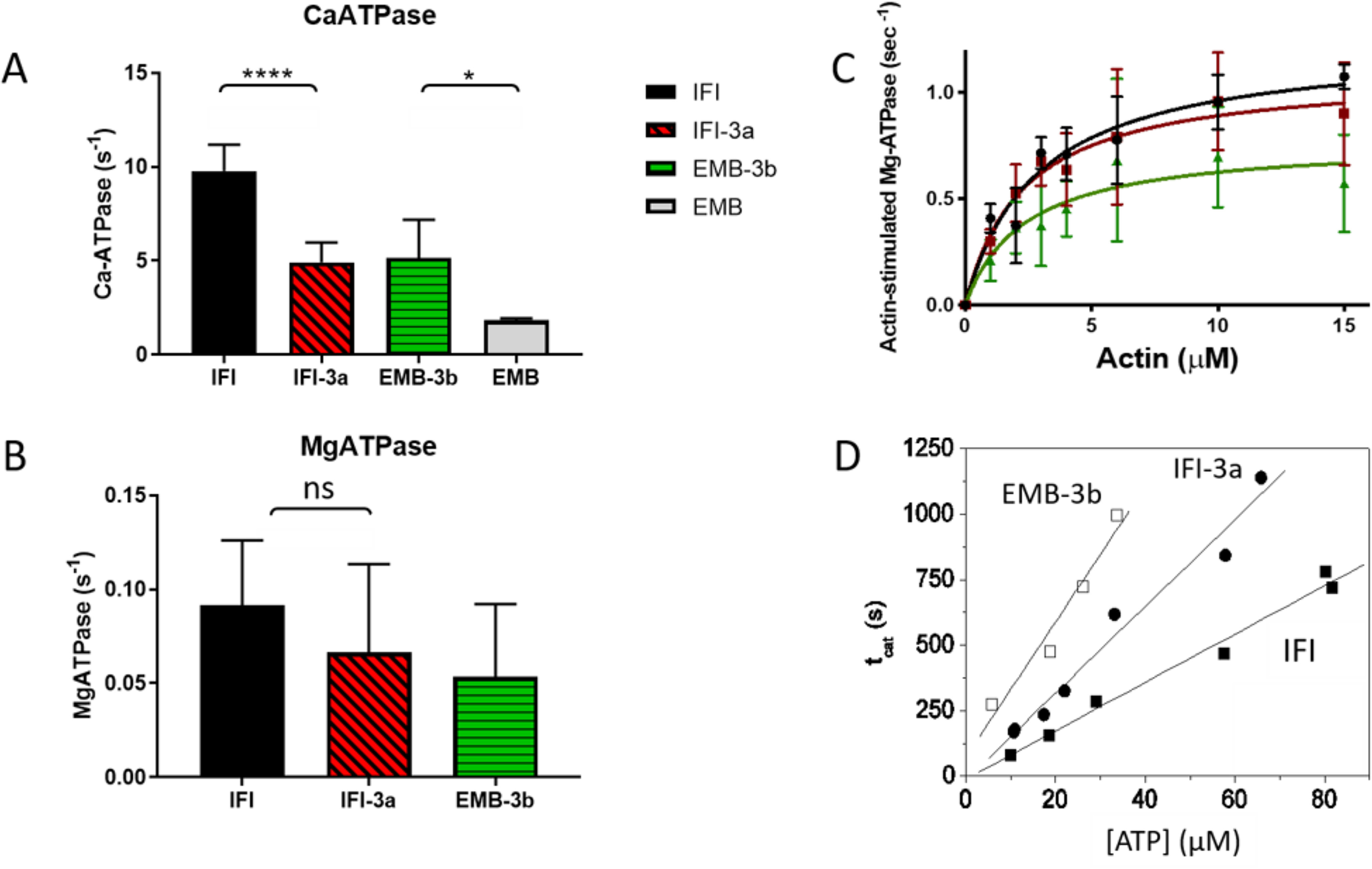
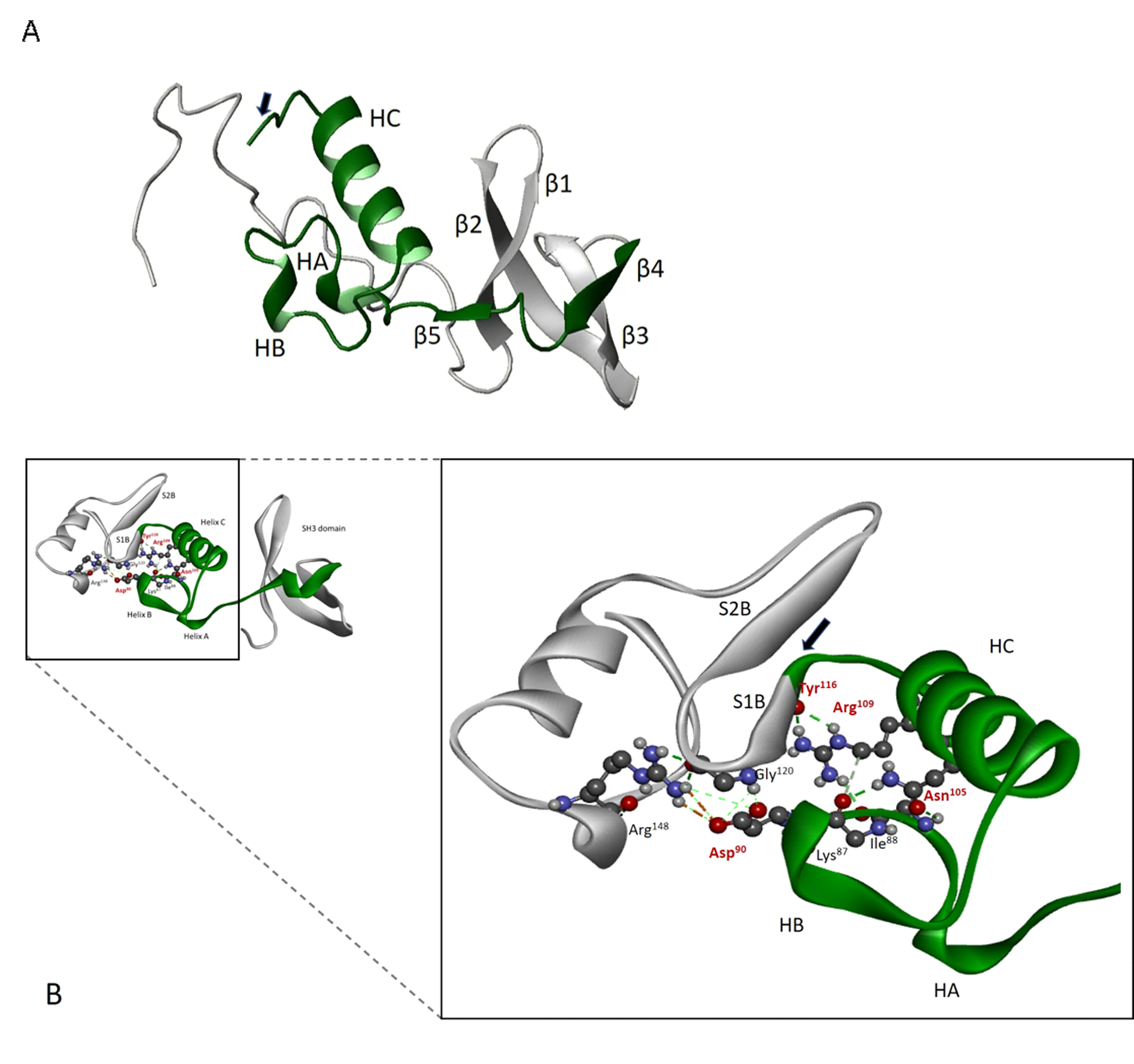
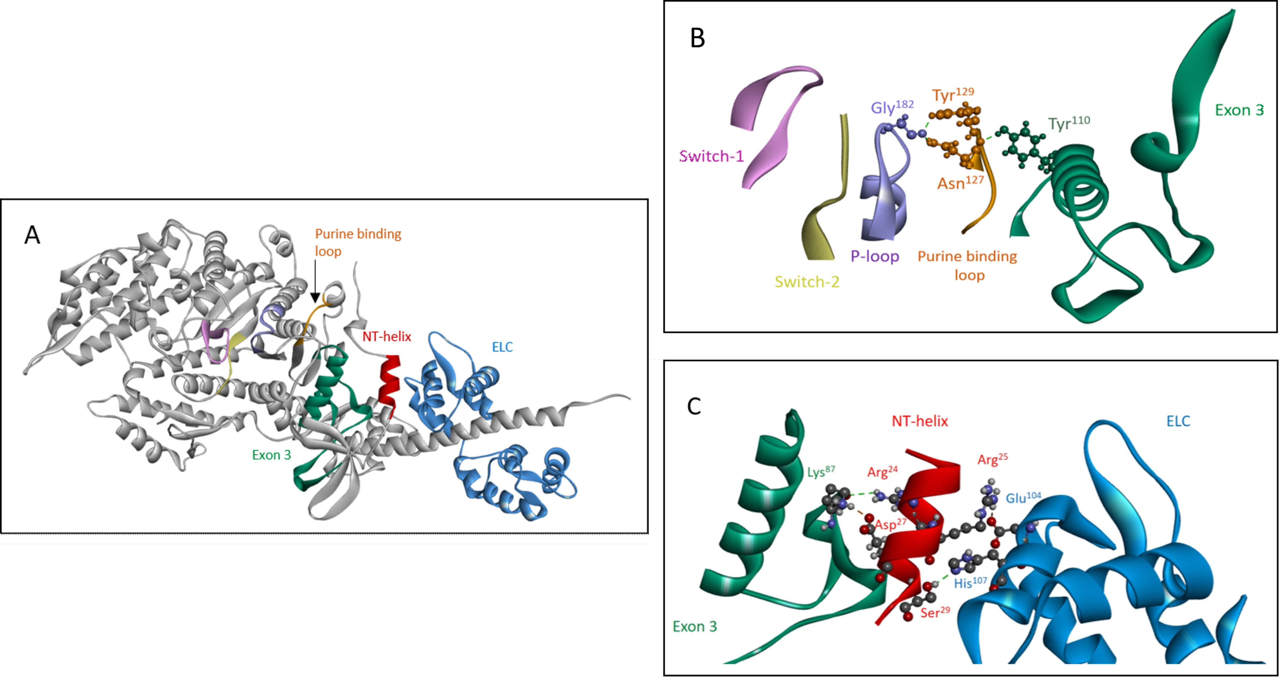
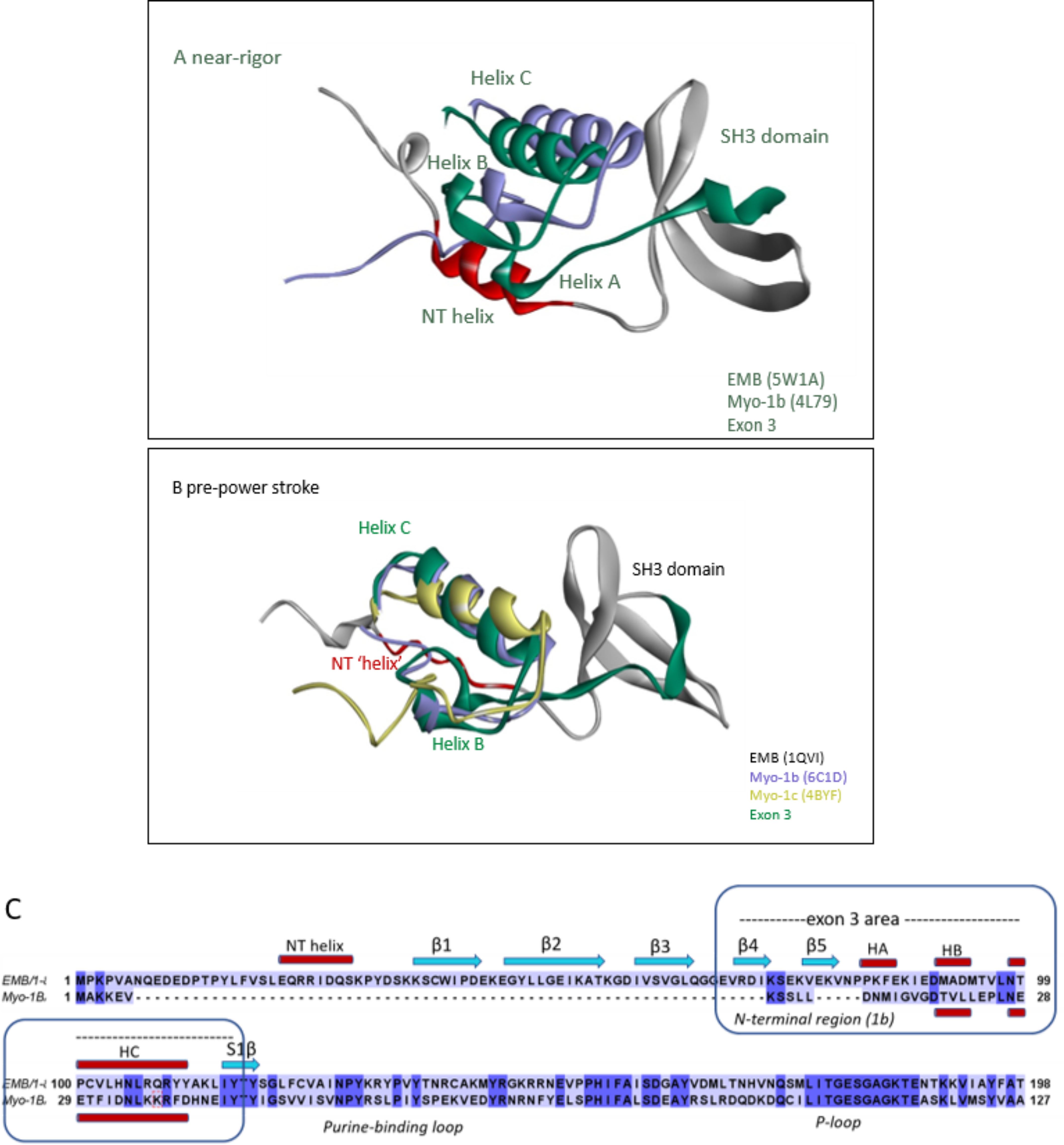
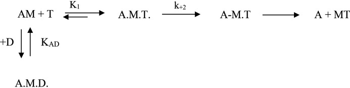
We investigated the biochemical and biophysical properties of one of the four alternative exon-encoded regions within the Drosophila myosin catalytic domain. This region is encoded by alternative exons 3a and 3b and includes part of the N-terminal β-barrel. Chimeric myosin constructs (IFI-3a and EMB-3b) were generated by exchanging the exon 3-encoded areas between native slow embryonic body wall (EMB) and fast indirect flight muscle myosin isoforms (IFI). We found that this exchange alters the kinetic properties of the myosin S1 head. The ADP release rate (k-D ) in the absence of actin is completely reversed for each chimera compared with the native isoforms. Steady-state data also suggest a reciprocal shift, with basal and actin-activated ATPase activity of IFI-3a showing reduced values compared with wild-type (WT) IFI, whereas for EMB-3b these values are increased compared with wild-type (WT) EMB. In the presence of actin, ADP affinity (KAD ) is unchanged for IFI-3a, compared with IFI, but ADP affinity for EMB-3b is increased, compared with EMB, and shifted toward IFI values. ATP-induced dissociation of acto-S1 (K1k+2 ) is reduced for both exon 3 chimeras. Homology modeling, combined with a recently reported crystal structure for Drosophila EMB, indicates that the exon 3-encoded region in the myosin head is part of the communication pathway between the nucleotide binding pocket (purine binding loop) and the essential light chain, emphasizing an important role for this variable N-terminal domain in regulating actomyosin crossbridge kinetics, in particular with respect to the force-sensing properties of myosin isoforms.

Keywords: actin; fluorescence; force-sensing; homology modeling; kinetics; muscle; myosin; protein structure-function; sequence alignment.


Conflict of interest statement

Conflict of interest—The authors declare that they have no conflicts of interest with the contents of this article.

Figures

Figure 1.
Figure 1.
A, structure of the EMB isoform in the rigor-like conformation, with the location of the four variable regions in the myosin head indicated: exon 3 region (green), exon 7 region (purple), exon 9 region (dark red), and exon 11 region (light brown). The ELC is shown in dark gray (the coordinates of the EMB crystal structure (PDB ID: 5W1A) were used to generate this image). B, alternative sequences encoded by the exon 3a and 3b regions, with nonconservative differences denoted by asterisks: 3a is found in the slow EMB isoform and 3b is present in the fast IFI isoform.
Figure 2.
Figure 2.
ATP-induced dissociation (K1k+2) and ADP affinity (KAD) of the two myosin S1 exon 3 chimeras. A, example of light-scattering traces for acto-S1 dissociation with EMB-3b S1. B, the second-order rate constant (K1k+2) for the ATP-induced dissociation of S1 from actin is determined from a linear fit to the plot of the kobs versus [ATP] (see “Experimental Procedures”). The linear fits yielded values of 0.71 ± 0.12 μm−1 s−1 for EMB-3b (filled circles) as compared with 0.91 ± 0.13 μm−1 s−1 for EMB (open circles). For IFI-3a the linear fits yielded mean values of 0.66 ± 0.08 μm−1 s−1 (filled squares) as compared with 0.75 ± 0.08 μm−1 s−1 for IFI S1 (open squares). C, light-scattering traces for acto-S1 dissociation with EMB-3b S1 at various ADP concentrations. D, comparison of the affinity of ADP for acto-S1 (KAD) for EMB and EMB-3b, with the relative kobs (krel) values shown. Hyperbolic fits resulted in KAD values of 587 ± 48 μm for EMB (open circles) and 496 ± 79 μm for EMB-3b S1 (filled circles).
Figure 3.
Figure 3.
Rate of ADP release (k-D) from Drosophila S1 isoforms. The rate constant for cmADP dissociation (k-D) from S1 in the absence of actin was determined using flash photolysis. After release of ATP (15 μm) from caged-ATP (100 μm), a fluorescent ADP analog (eda-deac ADP) bound to S1 was displaced by ATP. The change in fluorescence upon release of eda-deac ADP from S1 was used to determine k-D. Exchange of either the exon 3a or 3b domain resulted in a complete reversal of the ADP release rate (see also Table 1). The dissociation rate for EMB-3b (k-D, 7.0 s−1) is not significantly different to IFI (k-D, 7.5 s−1) whereas that of IFI-3a (k-D, 2.1 s−1) is similar to EMB (k-D, 1.8 s−1).
Figure 4.
Figure 4.
Steady-state ATPase activity of IFI, EMB, and exon 3 chimeric S1 isoforms. AD, basal Ca-ATPase activity (A), basal Mg-ATPase (B), actin-activated Mg-ATPase activity (Vmax) (C), and the turnover number (kcat) for acto-S1 (D) were determined as described under “Experimental Procedures.” Notations above histograms indicate the level of statistically significant differences (*, p < 0.05; ****, p < 0.0001; ns, not statistically significant). Significant differences were assumed for p < 0.05.
Figure 5.
Figure 5.
Location and structure of exon 3 domain (green) within the myosin heavy chain N terminus. A, secondary structure elements encoded by exon 3 (residues 69–116) include β4 (69–73), β5 (77–79), helix A (HA, 83–85), helix B (HB, 91–93), helix C (HC, 98–111), and the first β-strand of the seven-stranded β-sheet S1β (starts at 115, indicated with an arrow). The full SH3 element is also depicted (β1, Lys36–Glu43; β2, Glu45–Lys56; β3, Ile59–Gln65). B, conserved interactions within exon 3 regions involve residues Asp90, Asn105, Arg109, and Tyr116. Asn105 forms hydrogen bonds with backbone oxygens of three residues close to Asp90 (Lys87, Ile/Ala88, and Met91) whereas Arg109 forms H bonds with Tyr116.
Figure 6.
Figure 6.
The exon 3 area is part of the communication pathway between the essential light chain and the purine binding loop. A, overview of elements in the communication pathway between the ELC (blue) and purine binding loop (127–135, orange), shown for rigor-like EMB myosin S1 (PDB ID: 5W1A). In addition to the exon 3 area (69–116, green), the small N-terminal helix (21–30, red) is also involved in signal transfer from the ELC toward the nucleotide binding pocket. B, detailed view of purine binding loop interactions with the exon 3 area and the P-loop (PDB ID: 5W1A). Fig. 6B is reused and extended in the supplementary section to demonstrate that interaction of the exon 3 region with the purine binding loop depends on conformational state of the myosin head (Figs. S2 and S3). C, close-up of N-terminal helix interactions with the ELC (Glu104 and His107) and the exon 3 region (Lys87) (PDB ID: 5W1A).
Figure 7.
Figure 7.
Exon 3 interaction with helix NT is distinct between IFI and EMB and depends on conformational state. Comparison of helix NT interaction with exon 3 residues and the ELC throughout the crossbridge cycle for IFI and EMB myosin isoforms, with exon 3 area (residues 69–116) in green, the NT–helix (residues 21–30) in red and the ELC in blue. A, near rigor state: EMB crystal structure (PDB ID: 5W1A) and corresponding IFI homology model show similar interaction of helix NT with exon 3 region and ELC. B, pre-power stroke state: interaction of helix NT (Arg24) with the exon 3 area is present for IFI (Asn112), whereas for EMB (Ala112) this interaction is absent (PDB ID: 1QVI). C, post-power stroke state: IFI maintains contacts between the sidechains of variable exon 3 residue Asn112 and helix NT residue Arg24. Additional contacts are found for exon 3 residues Pro83 and Lys84 (backbone oxygen) with helix NT residue Arg25 (side chain). No exon 3–helix NT contacts were found for EMB (PDB ID: 1KK8).
Figure 8.
Figure 8.
Overlay of N-terminal regions of EMB and Myo-1b or Myo-1c. A and B, EMB (gray) with the exon 3 area (green) and the N-terminal helix (NT) shown in red, Myo-1b (purple), and Myo-1c (yellow). Note the similar orientation of exon 3 secondary structure elements (helix B and C) of EMB with respect to the NTR of Myo-1b and/or Myo-1c in both near-rigor and pre-power stroke state. A, overlay of crystal structures of Myo-1b (PDB ID: 4L79) with EMB (rigor-like, PDB ID: 5W1A). B, overlay of Myo-1b (PDB ID: 6C1D) and Myo-1c (PDB ID: 4BYF) with EMB homology model (pre-power stroke, PDB ID: 1QVI template). C, sequence alignment of N-terminal regions of EMB and Myo-1b showing conserved secondary structure elements (HB and HC) for the exon-3 encoded region (EMB) and the NTR (Myo-1b).
Scheme 1.
Scheme 1.
The interaction of S1 with actin, ATP, and ADP. M, A, T, and D symbolize myosin S1, actin, ATP, and ADP, respectively.

References

    1. George E. L., Ober M. B., and Emerson C. P. (1989) Functional domains of the Drosophila melanogaster muscle myosin heavy-chain gene are encoded by alternatively spliced exons. Mol. Cell Biol. 9, 2957–2974 10.1128/mcb.9.7.2957 - DOI - PMC - PubMed
    1. Bernstein S. I., and Milligan R. A. (1997) Fine tuning a molecular motor: The location of alternative domains in the Drosophila myosin head. J. Mol. Biol. 271, 1–6 10.1006/jmbi.1997.1160 - DOI - PubMed
    1. Wells L., Edwards K. A., and Bernstein S. I. (1996) Myosin heavy chain isoforms regulate muscle function but not myofibril assembly. EMBO J. 15, 4454–4459 10.1002/j.1460-2075.1996.tb00822.x - DOI - PMC - PubMed
    1. Swank D. D. M., Knowles A. F. A., Suggs J. A. J., Sarsoza F., Lee A., Maughan D. W., and Bernstein S. I. (2002) The myosin converter domain modulates muscle performance. Nat. Cell Biol. 4, 312–316 10.1038/ncb776 - DOI - PubMed
    1. Miller B. M., Nyitrai M., Bernstein S. I., and Geeves M. A. (2003) Kinetic analysis of Drosophila muscle myosin isoforms suggests a novel mode of mechanochemical coupling. J. Biol. Chem. 278, 50293–50300 10.1074/jbc.M308318200 - DOI - PubMed

Publication types

MeSH terms

LinkOut - more resources