. 2020 Oct 14;94(21):e01051-20.
doi: 10.1128/JVI.01051-20. Print 2020 Oct 14.

Molecular Characterization of Influenza C Viruses from Outbreaks in Hong Kong SAR, China

Affiliations

Molecular Characterization of Influenza C Viruses from Outbreaks in Hong Kong SAR, China

Rodney S Daniels et al. J Virol. .

Abstract

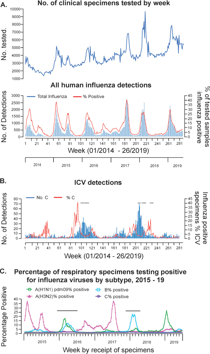
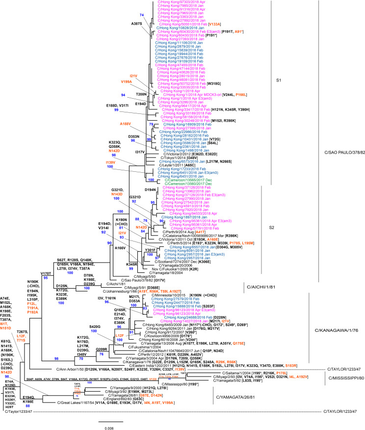
In 2014, the Centre for Health Protection in Hong Kong introduced screening for influenza C virus (ICV) as part of its routine surveillance for infectious agents in specimens collected from patients presenting with symptoms of respiratory viral infection, including influenza-like illness (ILI). A retrospective analysis of ICV detections up to week 26 of 2019 revealed persistent low-level circulation, with two outbreaks having occurred in the winters of 2015 to 2016 and 2017 to 2018. These outbreaks occurred at the same time as, and were dwarfed by, seasonal epidemics of influenza types A and B. Gene sequencing studies on stored ICV-positive clinical specimens from the two outbreaks have shown that the hemagglutinin-esterase (HE) genes of the viruses fall into two of the six recognized genetic lineages (represented by C/Kanagawa/1/76 and C/São Paulo/378/82), with there being significant genetic drift compared to earlier circulating viruses within both lineages. The location of a number of encoded amino acid substitutions in hemagglutinin-esterase fusion (HEF) glycoproteins suggests that antigenic drift may also have occurred. Observations of ICV outbreaks in other countries, with some of the infections being associated with severe disease, indicates that ICV infection has the potential to have significant clinical and health care impacts in humans.IMPORTANCE Influenza C virus infection of humans is common, and reinfection can occur throughout life. While symptoms are generally mild, severe disease cases have been reported, but knowledge of the virus is limited, as little systematic surveillance for influenza C virus is conducted and the virus cannot be studied by classical virologic methods because it cannot be readily isolated in laboratories. A combination of systematic surveillance in Hong Kong SAR, China, and new gene sequencing methods has been used in this study to assess influenza C virus evolution and provides evidence for a 2-year cycle of disease outbreaks. The results of studies like that reported here are key to developing an understanding of the impact of influenza C virus infection in humans and how virus evolution might be associated with epidemics.

Keywords: genome sequencing; influenza C virus; outbreak surveillance; virus evolution.


Figures

FIG 1
FIG 1
ICV surveillance in Hong Kong 2014 to 2019. Results of weekly influenza surveillance in Hong Kong covering the period from week 01 of 2014 to week 26 of 2019 are shown for (A) all human influenza virus detections in terms of numbers tested, numbers detected, and the percentage of tested specimens showing positivity and (B) ICV detections by number and percentage of the total number of influenza detections. Clinical specimens from the weeks indicated (*) were analyzed by gene sequencing. (C) For the period from week 01 of 2015 to week 26 of 2019 the proportions of influenza types/subtypes detected by week are shown with the ICV outbreak periods indicated above the virus detection profiles. All data were taken from Flu Express reports published by the Centre for Health Protection, Hong Kong, and panel C is reproduced from reference .
FIG 2
FIG 2
Phylogenetic analysis of ICV HE genes. The phylogeny was generated as described in Materials and Methods, and reference sequences from different clades were downloaded from the EpiFlu Database of GISAID. Viruses from the 2015 to 2016 and 2017 to 2018 outbreaks in Hong Kong are shown in blue and pink, respectively, and other viruses sequenced at WHO Collaborating Centre, London, are shown in green. Group-defining amino acid substitutions are shown on nodes, together with bootstrap values of 70 and above, and virus specific substitutions are shown after virus names, with HEF2 substitutions being in orange and those affecting N-linked glycosylation indicated (±CHO). Bar indicates the proportion of nucleotide changes between sequences. We gratefully acknowledge the authors and originating and submitting laboratories of the sequences from the EpiFlu Database of GISAID that were downloaded for use in the preparation of this study (all submitters of data may be contacted directly via the GISAID website [http://gisaid.org/] and the relevant sequence accession numbers are given in Table S5 in the supplemental material).
FIG 3
FIG 3
HEF structures indicating the amino acid substitutions that define the genetic/antigenic lineages. The structure of an HEF monomer derived from C/Johannesburg/1/66 (51) was downloaded from the Protein Data Bank (PDB identifier 1FLC) and annotated using PyMOL. Amino acid substitutions (compared to HEF of C/Taylor/1233/47) that define later genetic/antigenic lineages are shown as follows: (A) Mississippi, (B) Yamagata, (C) Kanagawa, (D) Aichi, (E) Sao Paulo (C/Tokyo/1/2014, S1 sublineage), and (F) Sao Paulo (C/Fukuoka/1/2005, S2 sublineage). Each cartoon shows the receptor-binding domain (HEF1) in green, the esterase domain (HEF1) in red and the fusion domain (HEF2) in blue. Residues involved directly in receptor binding are shown as bright green sticks, and those involved directly in esterase activity as bright red sticks. Amino acid substitutions are shown as spheres, color-coded as follows: pink, substitutions conserved across all six groups; yellow, lineage-defining substitutions; bright green, substitution (K172G) at a residue involved directly in receptor binding (which is found in Mississippi, Aichi, and Kanagawa lineages only). Amino acid substitutions are color-coded as follows: brown, HEF2; black, HEF1 with those in designated antigenic sites indicated in green (site A2), red (site A1), and purple (site Y1), with substitutions at HEF1 positions 193 and 198 forming parts of sites A1 and A3, respectively (40). Amino acid substitutions falling in esterase domain 1 (*), esterase domain 2 (&), and the receptor-binding domain (•) are indicated (51), as are those under positive selection (P) (39).

References

    1. Taylor RM. 1949. Studies on survival of influenza virus between epidemics and antigenic variants of the virus. Am J Public Health Nations Health 39:171–178. doi:10.2105/ajph.39.2.171. - DOI - PMC - PubMed
    1. Jennings R. 1968. Respiratory viruses in Jamaica: a virologic and serologic study. 3. Hemagglutination-inhibiting antibodies to type B and C influenza viruses in the sera of Jamaicans. Am J Epidemiol 87:440–446. doi:10.1093/oxfordjournals.aje.a120834. - DOI - PubMed
    1. Dykes AC, Cherry JD, Nolan CE. 1980. A clinical, epidemiologic, serologic, and virologic study of influenza C virus infection. Arch Intern Med 140:1295–1298. doi:10.1001/archinte.1980.00330210043021. - DOI - PubMed
    1. O’Callaghan RJ, Gohd RS, Labat DD. 1980. Human antibody to influenza C virus: its age-related distribution and distinction from receptor analogs. Infect Immun 30:500–505. - PMC - PubMed
    1. Homma M, Ohyama S, Katagiri S. 1982. Age distribution of the antibody to type C influenza virus. Microbiol Immunol 26:639–642. doi:10.1111/mim.1982.26.7.639. - DOI - PubMed

Publication types

MeSH terms