. 2021 Sep 1;106(9):2405-2416.
doi: 10.3324/haematol.2019.236455.

A three-gene signature based on MYC, BCL-2 and NFKBIA improves risk stratification in diffuse large B-cell lymphoma

Affiliations

A three-gene signature based on MYC, BCL-2 and NFKBIA improves risk stratification in diffuse large B-cell lymphoma

Enrico Derenzini et al. Haematologica. .

Abstract

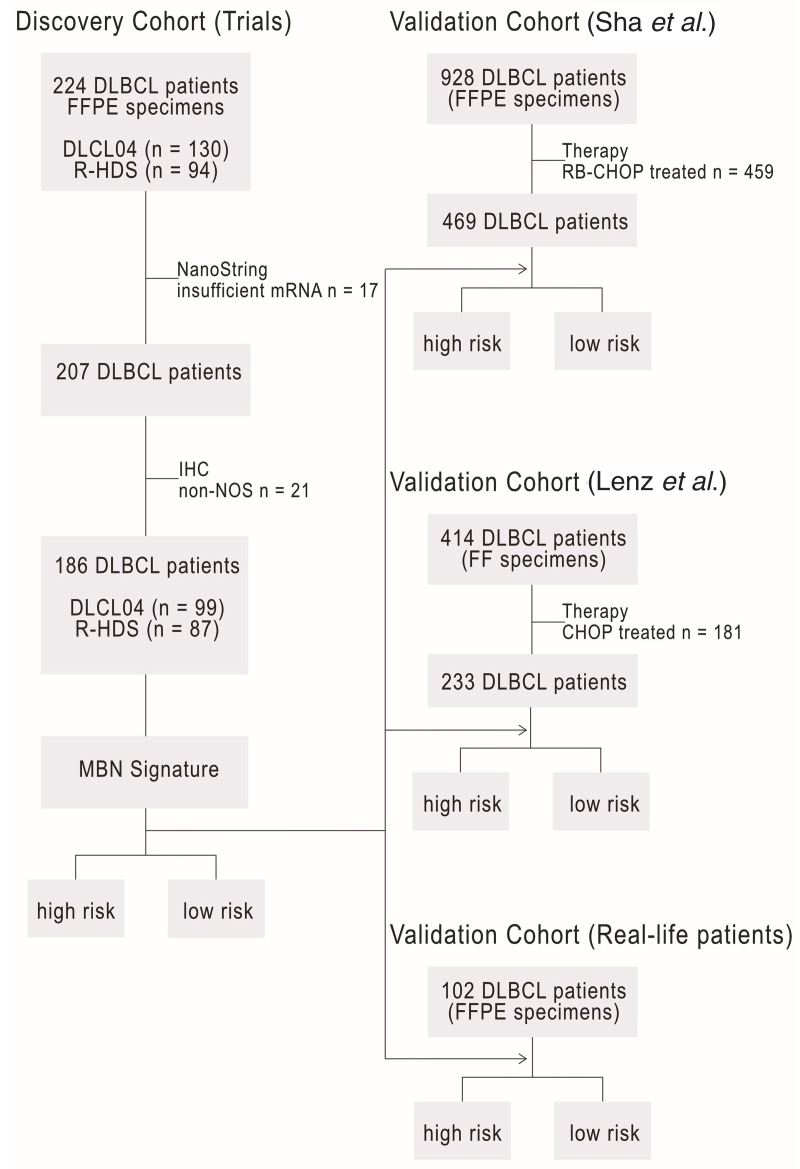
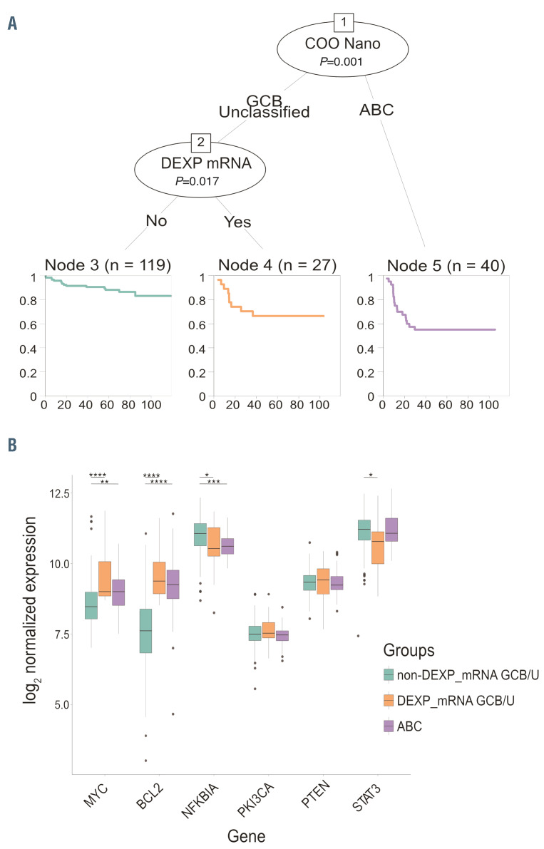
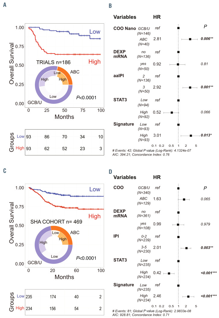
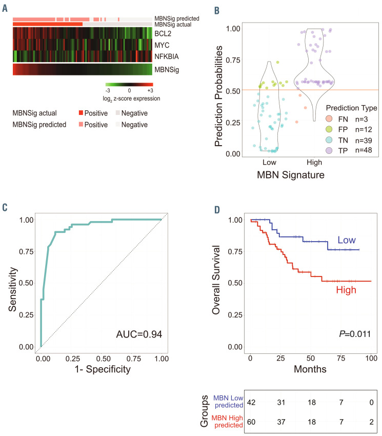
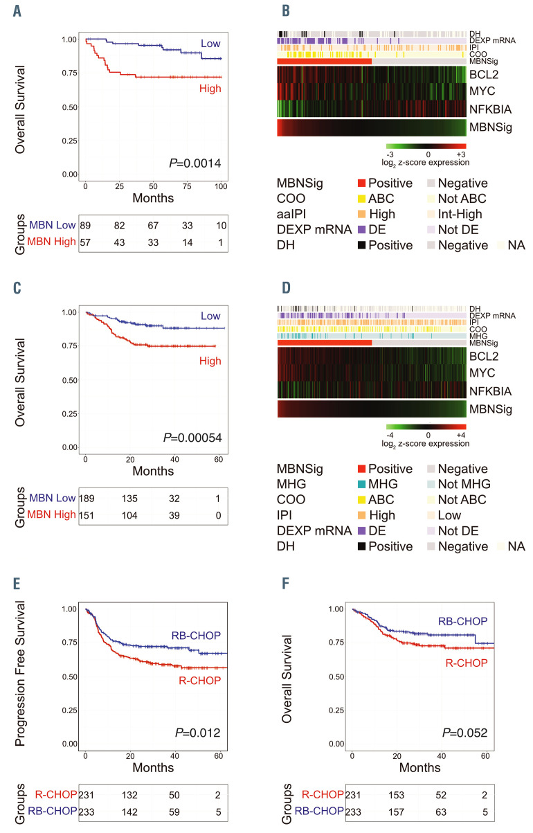
Recent randomized trials focused on gene expression-based determination of the cell of origin in diffuse large B-cell lymphoma could not show significant improvements by adding novel agents to standard chemoimmunotherapy. The aim of this study was the identification of a gene signature able to refine current prognostication algorithms and applicable to clinical practice. Here we used a targeted gene expression profiling panel combining the Lymph2Cx signature for cell of origin classification with additional targets including MYC, BCL-2 and NFKBIA, in 186 patients from 2 randomized trials (discovery cohort) (NCT00355199 and NCT00499018). Data were validated in 3 independent series (2 large public datasets and a real-life cohort). By integrating the cell of origin, MYC/BCL-2 double expressor status and NFKBIA expression, we defined a 3-gene signature combining MYC, BCL-2 and NFKBIA (MBN-signature), which outperformed the MYC/BCL-2 double expressor status in multivariate analysis, and allowed further risk stratification within the germinal center B-cell/unclassified subset. The high-risk (MBN Sig-high) subgroup identified the vast majority of double hit cases and a significant fraction of Activated B-Cell-derived diffuse large B-cell lymphomas. These results were validated in 3 independent series including a cohort from the REMoDL-B trial, where, in an exploratory ad hoc analysis, the addition of bortezomib in the MBN Sig-high subgroup provided a progression free survival advantage compared with standard chemoimmunotherapy. These data indicate that a simple 3-gene signature based on MYC, BCL-2 and NFKBIA could refine the prognostic stratification in diffuse large B-cell lymphoma, and might be the basis for future precision-therapy approaches.


Figures

Figure 1.
Figure 1.
Study algorithm. On the left, the discovery cohort is represented; 224 diffuse large B-cell lymphoma (DLBCL) patients enrolled in the DLCL04 (n= 130) and R-HDS0305 (n= 94) trials with available formalin-fixed, paraffin embedded (FFPE) tissue were initially considered in this analysis. Targeted gene expression profiling (TGEP) success rate was 92.4% (n=207), with 17 cases not yelding enough highquality mRNA to undergo successful GEP assessment. Only cases originally diagnosed as DLBCL non-otherwise specified (NOS) were considered. Therefore 21 cases classified in different DLBCL categories were excluded; 99 NOS-DLBCL FFPE patient samples from the DLCL04 trial and 87 samples from the R-HDS0305 trial were finally included in this study. On the right, the three validation cohorts: a cohort of 928 patients from Sha and coworkers (469 treated with R-CHOP; 459 with RBCHOP), a public gene expression dataset (Affymetrix Human Genome U133 Plus 2.0 Array), GSE10846, (https://www.ncbi.nlm.nih.gov/geo/query/acc.cgi?acc=GSE10846), including 233 patients treated with R-CHOP regimen (Lenz et al. 2008); an additional validation cohort including 102 consecutive DLBCL NOS cases with available FFPE tissue, treated with R-CHOP/R-CHOP-like regimens. RB-CHOP: R-CHOP plus bortezomib.
Figure 2.
Figure 2.
Integrating cell of origin with MYC/BCL-2 DEXP_mRNA status for prognostication in diffuse large Bcell lymphoma. (A) Recursive partitioning analysis integrating cell of origin (COO) classification and DEXP_mRNA status, allowing segregation of patients in three main prognostic subgroups (a low risk non-DEXP-mRNA GCB/U subset, and two high risk groups: MYC/BCL-2-DEXP-mRNA GCB/U and ABC). (B) Box plot graphs indicating the expression levels of the additional targets included in the panel (MYC, BCL-2, NFKBIA, STAT3, PIK3CA, PTEN) in the three main patients subgroups identified by the recursive partitioning analysis (non-DEXPmRNA GCB/U, MYC/BCL-2 DEXP_mRNA GCB/U, ABCderived diffuse large B-cell lymphoma [DLBCL]. P-value was calculated with Student ttest by comparing non DEXP_mRNA GCB/U group, selected as a reference, versus other groups. ABC: activated B-cells; GCB: germinal center B cells; GCB/U: GCB unclassified; DEXP_mRNA: double expressor GCB/U DLBCL. COO Nano: COO as determined by T-GEP with NanoString profiling
Figure 3.
Figure 3.
Survival curves according to MBN signature and multivariate analyses for overall survival. (A) Overall survival (OS) of the discovery cohort (RHDS0305+ DLCL04; n=186 patients) according to the MBN signature (MBN-Sig) showing significant differences in outcome between MBN-Sig low versus MBN-Sig high patient subsets. P-values were calculated with the log rank test. Frequencies of MBN-Sig high versus low cases in activated B-cells (ABC) and germinal center B cells/ unclassified (GCB/U) subsets in the discovery cohort are represented in the pie chart. (B) Forest plot depicting multivariate analyses for OS (discovery cohort). Only factors significantly asociated with OS in univariate analyses were considered. According to this analysis only the cell of origin (COO) as determined by NanoString-based targeted gene expression profiling (T-GEP) (COO_Nano), the MBN-Sig and the age adjusted international prognostic index (aaIPI) score retained statistical significance for OS, whereas MYC/BCL-2 DEXP_mRNA status, STAT3 and NFKBIA levels determined by T-GEP were not significantly associated with OS. HR: hazard ratio. (C) OS of the 469 patients treated with R-CHOP in the Sha’s cohort according to the MBN signature showing significant differences in outcome between MBN-Sig low versus MBN-Sig high patient subsets. P-values were calculated with the log rank test. Frequencies of MBN-Sig high versus low cases in ABC and GCB/U subsets in the Sha’s cohort are represented in the pie chart. (C) Forest plot depicting multivariate analyses for OS (Sha’s dataset), confirming the significant independent association with OS of the MBN-Sig in this large validation cohort.
Figure 4.
Figure 4.
Real-life applicability of the MBN signature. (A) Heatmap representing the three informative genes of the MBN signature (MBN-Sig) shown as rows and diffuse large B-cell lymphoma (DLBCL) tissue samples shown as columns in the real-life cohort of 102 patients, with the actual MBN-Sig and the predicted MBN-Sig class based on the application of a random forest (RF) model built on the discovery cohort on the top of the heatmap. (B) Violin plot showing the fractions of false predictions (false positive [FP], and false negative [FN]) as well as true predictions (true positive [TP], and true negative [TN]) in the real-life cohort by applying a three-gene RF model. (C) ROC curve of the real-life cohort using RF classifier. (D) Overall survival (OS) curve of the real-life cohort (n=102) based on the predicted MBN-Sig class. P-value was calculated with the log rank test.
Figure 5.
Figure 5.
The MBN signature identifies prognostically distinct subgroups including activated B cells and a fraction of germinal center B cells/ unclassified diffuse large B-cell lymphoma (DLBCL) enriched in double hit DLBCL cases, providing opportunities for precision therapies. (A) Overall survival (OS) of the germinal center B cells/ unclassified (GCB/U) subset in the discovery cohort (n=146 patients) according to the integration of the targeted gene expression profiling (T-GEP) panel (Lymph2Cx) with the MBN signature (MBN-Sig), distinguishing two risk categories according to the MBN-Sig. (B) Heatmap representing the three informative genes of the MBN-Sig shown as rows and diffuse large B-cell lymphoma (DLBCL) tissue samples shown as columns, in the discovery cohort (n=186 patients). (C) OS of the GCB/U subset in the Sha’s validation cohort (n=340 patients) according to the MBN-Sig, showing superimposable results compared to Figure 4C. (D) Heatmap representing the three informative genes of the MBN-Sig shown as rows and DLBCL tissue samples shown as columns, in the Sha’s cohort (n=469 patients treated with R-CHOP). (E) Progression-free survival (PFS) of patients treated with R-CHOP versus RB-CHOP in the MBN-Sig high subgroup (Full Sha’s cohort, n=928 patients; MBNSig high n=464 patients). (F) OS of patients treated with R-CHOP versus RB-CHOP in the MBN-Sig high subgroup (Full Sha’s cohort, n=928 patients; MBN-Sig high n=464 patients). In all panels the P-value was calculated with the log rank test. NA: not available; DH: double hit; DE: double expressor (based on DEXP_mRNA status); aaIPI: age adjusted international prognostic index; COO: cell of origin; IPI: international prognostic index); MHG: molecular high grade. RB-CHOP: R-CHOP plus Bortezomib.

References

    1. Alizadeh AA, Eisen MB, Davis RE, et al. . Distinct types of diffuse large B-cell lymphoma identified by gene expression profiling. Nature. 2000;403(6769):503-511. - PubMed
    1. Shipp MA, Ross KN, Tamayo P, et al. . Diffuse large B-cell lymphoma outcome prediction by gene-expression profiling and supervised machine learning. Nat Med. 2002;8(1):68-74. - PubMed
    1. Davis RE, Brown KD, Siebenlist U, Staudt LM. Constitutive nuclear factor kappaB activity is required for survival of activated B cell-like diffuse large B cell lymphoma cells. J Exp Med. 2001;194(12):1861-1874. - PMC - PubMed
    1. Hans CP, Weisenburger DD, Greiner TC, et al. . Confirmation of the molecular classification of diffuse large B-cell lymphoma by immunohistochemistry using a tissue microarray. Blood. 2004;103(1):275-282. - PubMed
    1. Meyer PN, Fu K, Greiner TC, et al. . Immunohistochemical methods for predicting cell of origin and survival in patients with diffuse large B-cell lymphoma treated with rituximab. J Clin Oncol. 2011; 29(2):200-207. - PMC - PubMed

Publication types

MeSH terms

Substances

Associated data