Review
. 2020 Aug 17;27(9):395-413.
doi: 10.1101/lm.052282.120. Print 2020 Sep.

Iron-responsive-like elements and neurodegenerative ferroptosis

Affiliations
Review

Iron-responsive-like elements and neurodegenerative ferroptosis

Jack T Rogers et al. Learn Mem. .

Abstract

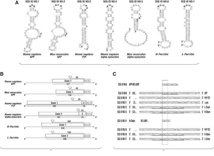
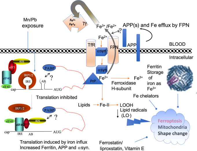
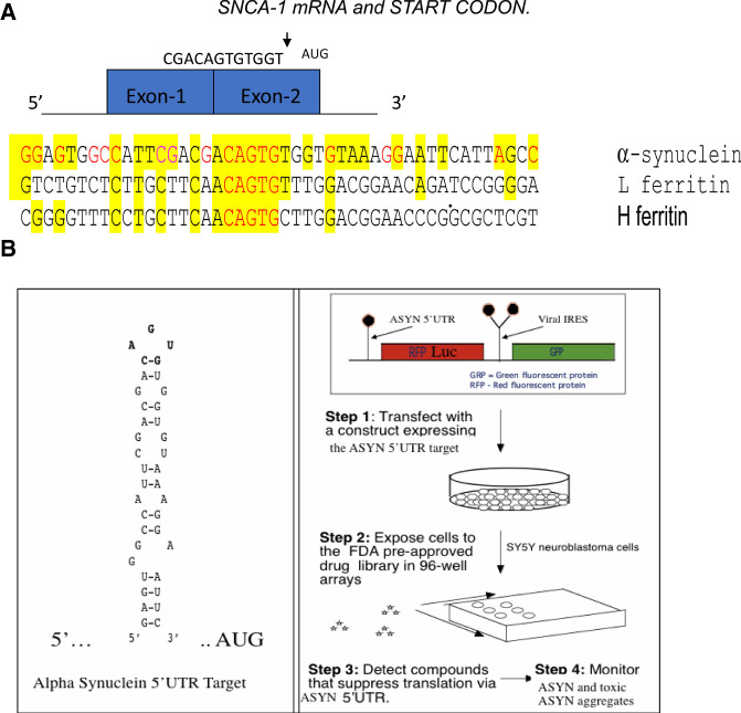
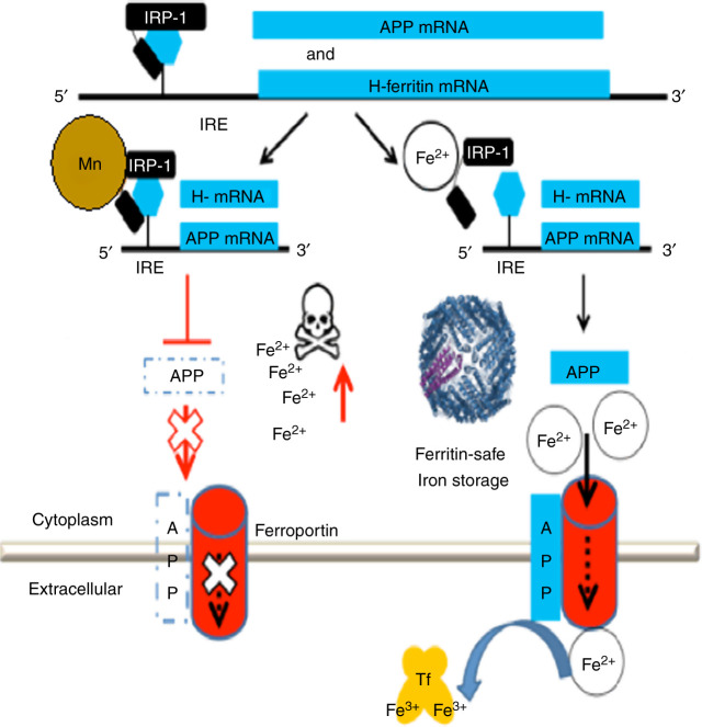
A set of common-acting iron-responsive 5'untranslated region (5'UTR) motifs can fold into RNA stem loops that appear significant to the biology of cognitive declines of Parkinson's disease dementia (PDD), Lewy body dementia (LDD), and Alzheimer's disease (AD). Neurodegenerative diseases exhibit perturbations of iron homeostasis in defined brain subregions over characteristic time intervals of progression. While misfolding of Aβ from the amyloid-precursor-protein (APP), alpha-synuclein, prion protein (PrP) each cause neuropathic protein inclusions in the brain subregions, iron-responsive-like element (IRE-like) RNA stem-loops reside in their transcripts. APP and αsyn have a role in iron transport while gene duplications elevate the expression of their products to cause rare familial cases of AD and PDD. Of note, IRE-like sequences are responsive to excesses of brain iron in a potential feedback loop to accelerate neuronal ferroptosis and cognitive declines as well as amyloidosis. This pathogenic feedback is consistent with the translational control of the iron storage protein ferritin. We discuss how the IRE-like RNA motifs in the 5'UTRs of APP, alpha-synuclein and PrP mRNAs represent uniquely folded drug targets for therapies to prevent perturbed iron homeostasis that accelerates AD, PD, PD dementia (PDD) and Lewy body dementia, thus preventing cognitive deficits. Inhibition of alpha-synuclein translation is an option to block manganese toxicity associated with early childhood cognitive problems and manganism while Pb toxicity is epigenetically associated with attention deficit and later-stage AD. Pathologies of heavy metal toxicity centered on an embargo of iron export may be treated with activators of APP and ferritin and inhibitors of alpha-synuclein translation.


Figures

Figure 1.
Figure 1.
RNA stem loops common to the 5′UTRs in the mRNAs for L- and H-ferritin and the key neurodegenerative transcripts for the amyloid precursor protein, alpha synuclein and Prion Protein. (A) IRE-like AGU/AGA tri-loops and potential iron-regulatory protein binding motifs in key neurodegenerative disease transcripts for human and mouse APP and SNCA mRNAs and in the human PrP and ferritin- L and H transcripts; further links between iron and AD and PD. (B) Organization of the positions of the IRE-like RNA stem loops in the 5′untranslated regions of human and mouse APP mRNAs and in the human PrP, SNCA and ferritin- L and H transcripts. (C) Alignment of the central AGU and AGA motifs in the 5′UTRs of human APP, αsyn and PrP transcripts relative to those for H- and H-ferritin.
Figure 2.
Figure 2.
A Model that merges the canonical IRE/IRP dependent pathways of translation of ferritin and APP to generate balanced iron homeostasis with a depiction of the reported roles of APP, αsyn, and PrP in iron transport (Singh et al. 2014; Baksi et al. 2016; Venkataramani et al. 2018; Tsatsanis et al. 2020). IRP1 and IRP2 are translation repressors in the absence of cellular iron and under conditions of iron chelation (Rogers et al. 2008). Iron influx releases IRPs from the IRE RNA stem loops in the 5′UTRs of L- and H-ferritin, Hif-2α and APP mRNAs to enhance the translation of these neuroprotective proteins (Cho et al. 2010; Goforth et al. 2010; Anderson et al. 2013; Chung et al. 2014). IRP2 in degraded from cells in conditions of iron influx while its deficiency has been associated with neurodegeneration (Zumbrennen-Bullough et al. 2014). Neurotoxic exposures to Mn and Pb repress IRP dependent translation of APP and ferritin H-chain (Venkataramani et al. 2018), a pathological path that we propose can enhance Fe levels to cause ferroptosis. Amyloidosis and α-synucleinopathy advance plaque and Lewy body formations while ferroptosis and neuronal viability appear to accelerate neurodegeneration with cognitive declines. There are testable links between by Nrf2 dependent gene expression of heme oxygenase and H-ferritin and glutathione production as a means to limit ferroptosis (Abdalkader et al. 2018; Song et al. 2020). Mn and Pb inhibition of APP can be predicted to promote ferroptosis and Fe buildup by interfering with APP/FPN complexes for Fe efflux while also toxically blocking ferritin translation, as associated with the production of cytotoxic reactive oxygen species (Rogers et al. 2016; Venkataramani et al. 2018; Fang et al. 2020), iron overload impairs epigenetic DNA methylation patterns to disrupt GABA neurotransmission (Ye et al. 2019). Ultimately the generation of highly dangerous lipid radicals resulting from iron overload can be scavenged by ferrostatin, the standard diagnostic inhibitor of ferroptosis, as well as by ferritin (Balla et al. 1992), activators of hepcidin (Yin et al. 2018), and by the application of nanochelators (Kang et al. 2019).
Figure 3.
Figure 3.
The 5′Untranslated region of alpha synuclein mRNA was the screening target for identifying αsyn translation blockers to address the need for Parkinson's disease and PDD and LBD therapies. The SNCA 5′UTR can be tested also to alleviate conditions of excess environmentally induced α-synucleinopathy (Friedlich et al. 2007; Rogers et al. 2011; Mikkilineni et al. 2012; Harischandra et al. 2019b). (A) The unique RNA target in the 5′UTR of the SNCA transcript. Depicted is the SNCA 5′UTR with its two exons immediately in front of the SNCA gene start codon (Olivares et al. 2009). Shown is the linear alignment of the SNCA 5′UTR with those for L- and H-ferritin mRNAs in the vicinity of their IRE RNA stem loops. (B) High-throughput screen for small molecule inhibitors of the RNA stem loop folded from the major neuronal neurodegenerative SNCA mRNA (MULTIFOLD, as described in Cho et al. 2010). Shown is a version of the transfection-based screen to identify SNCA 5′UTR directed translation inhibitors of a downstream luciferase reporter gene. During typical screens, inhibitors selectively maintained translation of a control GFP gene driven from an internal ribosome entry site (IRES) RNA structure. This RNA targeting technology was used to identify translation inhibitors of APP mRNA for AD therapy (Rogers et al. 2002a,b). The list of key HTS inhibitors of the SNCA 5′UTR are shown in PUBCHEM website as AID 1813. (Collaboration between Neurochemistry Laboratory at MGH and the Broad Inst. Cambridge, MA, https://pubchem.ncbi.nlm.nih.gov/bioassay/1813#section=Description
Figure 4.
Figure 4.
Model for manganese neurotoxicity invoking activation of APP and ferritin translation by iron-regulatory Proteins. (Right) In healthy individuals, APP facilitates FPN dependent iron export from neurons. (Left) Mn, like Pb exposure, interferes with APP and ferritin by blocking the translation of their mRNAs when causing super-repression by IRP1. Reduced FPN driven iron export by loss of APP(s) and the loss of Fe storage (ferritin) was shown to induce an embargo of iron inside the cell causing reactive oxygen species and REDOX active cell death/ferroptosis (Rogers et al. 2016; Venkataramani et al. 2018). Excess iron forms in magnetic nanoparticles throughout the amyloid plaque during AD (Plascencia-Villa et al. 2016) while ferritin was increased in brains and cerebrospinal fluid of AD patients (Ayton et al. 2015a; Ayton et al. 2016). Of note, amyloid form APP is protective in rodents burdened from acute bacterial infection/oxidative neurotoxicity (Kumar et al. 2016).

References

    1. Abdalkader M, Lampinen R, Kanninen KM, Malm TM, Liddell JR. 2018. Targeting Nrf2 to suppress ferroptosis and mitochondrial dysfunction in neurodegeneration. Front Neurosci 12: 466 10.3389/fnins.2018.00466 - DOI - PMC - PubMed
    1. Adamowicz DH, Roy S, Salmon DP, Galasko DR, Hansen LA, Masliah E, Gage FH. 2017. Hippocampal alpha-synuclein in dementia with Lewy bodies contributes to memory impairment and is consistent with spread of pathology. J Neurosci 37: 1675–1684. 10.1523/JNEUROSCI.3047-16.2016 - DOI - PMC - PubMed
    1. Ahn TB, Kim SY, Kim JY, Park SS, Lee DS, Min HJ, Kim YK, Kim SE, Kim JM, Kim HJ, et al. 2008. alpha-synuclein gene duplication is present in sporadic Parkinson disease. Neurology 70: 43–49. 10.1212/01.wnl.0000271080.53272.c7 - DOI - PubMed
    1. Allerson CR, Cazzola M, Rouault TA. 1999. Clinical severity and thermodynamic effects of iron-responsive element mutations in hereditary hyperferritinemia-cataract syndrome. J Biol Chem 274: 26439–26447. 10.1074/jbc.274.37.26439 - DOI - PubMed
    1. Alvarez-Fischer D, Henze C, Strenzke C, Westrich J, Ferger B, Hoglinger GU, Oertel WH, Hartmann A. 2008. Characterization of the striatal 6-OHDA model of Parkinson's disease in wild type and alpha-synuclein-deleted mice. Exp Neurol 210: 182–193. 10.1016/j.expneurol.2007.10.012 - DOI - PubMed

Publication types

MeSH terms