Comparative Study
. 2020 Sep 2;12(559):eabc3103.
doi: 10.1126/scitranslmed.abc3103. Epub 2020 Aug 17.

A comparison of four serological assays for detecting anti-SARS-CoV-2 antibodies in human serum samples from different populations

Ludivine Grzelak  1   2   3 Sarah Temmam  4 Cyril Planchais  5 Caroline Demeret  6   7 Laura Tondeur  8 Christèle Huon  4 Florence Guivel-Benhassine  1   2 Isabelle Staropoli  1   2 Maxime Chazal  9 Jeremy Dufloo  1   2   3 Delphine Planas  1   2 Julian Buchrieser  1   2 Maaran Michael Rajah  1   2   3 Remy Robinot  1   2 Françoise Porrot  1   2 Mélanie Albert  6   7   10 Kuang-Yu Chen  11 Bernadette Crescenzo-Chaigne  6   7 Flora Donati  6   7   10 François Anna  12 Philippe Souque  13 Marion Gransagne  14 Jacques Bellalou  15 Mireille Nowakowski  15 Marija Backovic  16 Lila Bouadma  17   18 Lucie Le Fevre  18 Quentin Le Hingrat  17   19 Diane Descamps  17   19 Annabelle Pourbaix  20 Cédric Laouénan  17   21 Jade Ghosn  17   20 Yazdan Yazdanpanah  17   20 Camille Besombes  8 Nathalie Jolly  22 Sandrine Pellerin-Fernandes  22 Olivia Cheny  22 Marie-Noëlle Ungeheuer  22 Guillaume Mellon  23 Pascal Morel  24 Simon Rolland  25   26 Felix A Rey  16 Sylvie Behillil  6   7   10 Vincent Enouf  6   7   10 Audrey Lemaitre  27 Marie-Aude Créach  28   29 Stephane Petres  15 Nicolas Escriou  14 Pierre Charneau  12   13 Arnaud Fontanet  8   30 Bruno Hoen  31 Timothée Bruel  1   2 Marc Eloit  32   33 Hugo Mouquet  5 Olivier Schwartz  34   2 Sylvie van der Werf  6   7   10
Affiliations
Comparative Study

A comparison of four serological assays for detecting anti-SARS-CoV-2 antibodies in human serum samples from different populations

Ludivine Grzelak et al. Sci Transl Med. .

Abstract

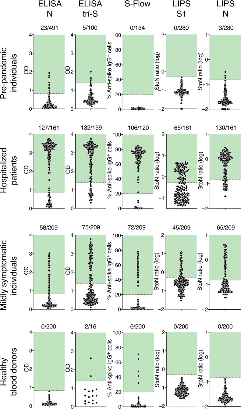
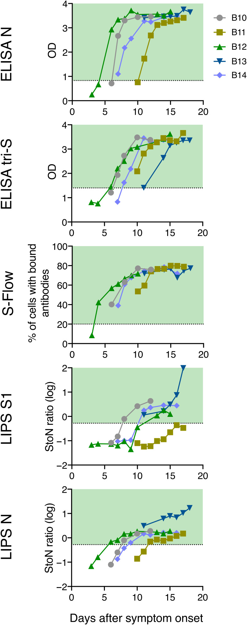
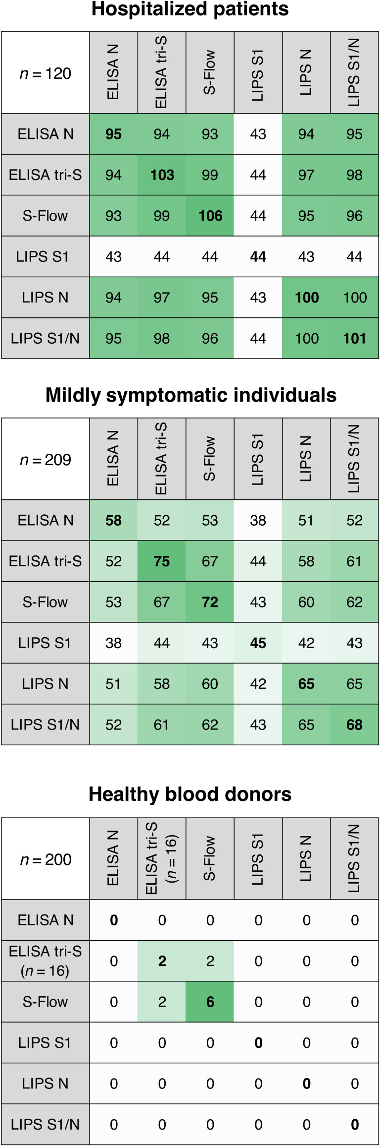
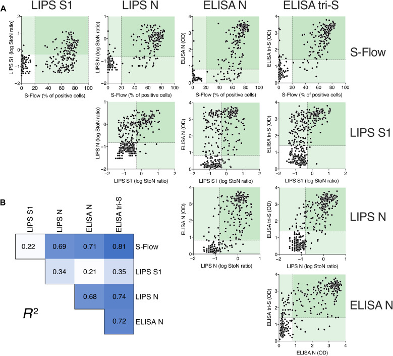
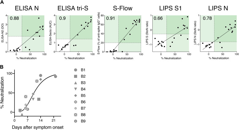
It is of paramount importance to evaluate the prevalence of both asymptomatic and symptomatic cases of SARS-CoV-2 infection and their differing antibody response profiles. Here, we performed a pilot study of four serological assays to assess the amounts of anti-SARS-CoV-2 antibodies in serum samples obtained from 491 healthy individuals before the SARS-CoV-2 pandemic, 51 individuals hospitalized with COVID-19, 209 suspected cases of COVID-19 with mild symptoms, and 200 healthy blood donors. We used two ELISA assays that recognized the full-length nucleoprotein (N) or trimeric spike (S) protein ectodomain of SARS-CoV-2. In addition, we developed the S-Flow assay that recognized the S protein expressed at the cell surface using flow cytometry, and the luciferase immunoprecipitation system (LIPS) assay that recognized diverse SARS-CoV-2 antigens including the S1 domain and the carboxyl-terminal domain of N by immunoprecipitation. We obtained similar results with the four serological assays. Differences in sensitivity were attributed to the technique and the antigen used. High anti-SARS-CoV-2 antibody titers were associated with neutralization activity, which was assessed using infectious SARS-CoV-2 or lentiviral-S pseudotype virus. In hospitalized patients with COVID-19, seroconversion and virus neutralization occurred between 5 and 14 days after symptom onset, confirming previous studies. Seropositivity was detected in 32% of mildly symptomatic individuals within 15 days of symptom onset and in 3% of healthy blood donors. The four antibody assays that we used enabled a broad evaluation of SARS-CoV-2 seroprevalence and antibody profiling in different subpopulations within one region.


Figures

Fig. 1
Fig. 1. Serological survey of SARS-Cov-2 antibodies in human serum samples.
Four serological assays were used to detect anti–SARS-Cov-2 antibodies in serum samples: (top row) individuals sampled between 2017 and 2019 (before pandemic), (second row) hospitalized cases with confirmed COVID-19, (third row) mildly symptomatic individuals from the Crépy-en-Vallois pandemic cluster with suspected COVID-19, and (bottom row) healthy blood donors. ELISA N and ELISA tri-S assays are conventional ELISAs using either N protein or the trimeric ectodomain of S protein as antigens. S-Flow is an assay detecting antibodies bound to cells expressing S protein by flow cytometry. The LIPS S1 and N assays detect either S1 or N protein fused to luciferase by immunoprecipitation. Pre-pandemic serum samples were used to determine the cutoff for each assay, which is indicated by a dotted line and a green area. The two ELISA assays were set to 95% specificity; the specificity of the S-Flow assay and LIPS assay was 99%. The number of positive samples is indicated. Each dot represents one sample. OD, optical density; StoN, signal-to-noise ratio.
Fig. 2
Fig. 2. Antibody detection in serum samples from five hospitalized patients with COVID-19.
The kinetics of seroconversion in serum samples from five hospitalized patients with COVID-19 (B10 to B14) were measured by four different serological assays. At least five longitudinal serum samples were collected for each patient up to 20 days after symptom onset. All patients were admitted to the intensive care unit. Each line represents one patient. Dotted lines and green areas indicate cutoff for positivity in the seroprevalence assays.
Fig. 3
Fig. 3. Comparison of positive serum samples.
The number of positive serum samples detected by each serological assay is shown for the three cohorts: hospitalized patients with COVID-19, mildly symptomatic individuals, and healthy blood donors. Correspondence of the positive results is shown among the four assays. For a given assay, each row indicates the number of positive samples that were also positive with the other three assays. Bold numbers indicate the number of positive samples for a given assay. The number of positive samples is color-coded: White corresponds to lower numbers, and green corresponds to higher numbers.
Fig. 4
Fig. 4. Correlations among the four serological assays.
To compare the four serological assays, results from serum samples from mildly symptomatic individuals and hospitalized patients with COVID-19 (n = 329) were pooled. (A) Results obtained with one assay were correlated with those of the other three assays. Dotted lines indicate assay cutoff values for positivity. Values in pale green areas are positive in one assay, and values in darker green areas are positive in two assays. Each dot represents one study participant. (B) Pearson correlation coefficient (R2) of each comparison is shown. R2 values are color-coded, with white corresponding to the lowest value and dark blue corresponding to the highest value. All correlations are significant (P < 0.0001).
Fig. 5
Fig. 5. Virus neutralizing activity in human serum samples.
(A) Virus-neutralizing activity (dilution 1:100) of 12 serum samples from the mildly symptomatic cohort of individuals with suspected COVID-19 (C1 to C12) and 9 serum samples from hospitalized patients with COVID-19 (B1 to B9). Virus-neutralizing activity was determined by the pseudovirus neutralization assay and compared to serology data obtained with the four serological assays. Numbers in the top left quadrant indicate the Spearman correlation coefficient, r. All correlations are significant (P < 0.0001). (B) Neutralization activity of serum samples (B1 to B9) from hospitalized patients with COVID-19 was plotted against days after symptom onset. The black line corresponds to a nonlinear fit of the data.

References

    1. Wu F., Zhao S., Yu B., Chen Y.-M., Wang W., Song Z.-G., Hu Y., Tao Z.-W., Tian J.-H., Pei Y.-Y., Yuan M.-L., Zhang Y.-L., Dai F.-H., Liu Y., Wang Q.-M., Zheng J.-J., Xu L., Holmes E. C., Zhang Y.-Z., A new coronavirus associated with human respiratory disease in China. Nature 579, 265–269 (2020). - PMC - PubMed
    1. Zhou P., Yang X.-L., Wang X.-G., Hu B., Zhang L., Zhang W., Si H.-R., Zhu Y., Li B., Huang C.-L., Chen H.-D., Chen J., Luo Y., Guo H., Jiang R.-D., Liu M.-Q., Chen Y., Shen X.-R., Wang X., Zheng X.-S., Zhao K., Chen Q.-J., Deng F., Liu L.-L., Yan B., Zhan F.-X., Wang Y.-Y., Xiao G.-F., Shi Z.-L., A pneumonia outbreak associated with a new coronavirus of probable bat origin. Nature 579, 270–273 (2020). - PMC - PubMed
    1. Corman V. M., Landt O., Kaiser M., Molenkamp R., Meijer A., Chu D. K. W., Bleicker T., Brünink S., Schneider J., Schmidt M. L., Mulders D. G. J. C., Haagmans B. L., van der Veer B., van den Brink S., Wijsman L., Goderski G., Romette J.-L., Ellis J., Zambon M., Peiris M., Goossens H., Reusken C., Koopmans M. P. G., Drosten C., Detection of 2019 novel coronavirus (2019-nCoV) by real-time RT-PCR. Euro Surveill. 25, 2000045 (2020). - PMC - PubMed
    1. Chu D. K. W., Pan Y., Cheng S. M. S., Hui K. P. Y., Krishnan P., Liu Y., Ng D. Y. M., Wan C. K. C., Yang P., Wang Q., Peiris M., Poon L. L. M., Molecular diagnosis of a Novel Coronavirus (2019-nCoV) causing an outbreak of pneumonia. Clin. Chem. 66, 549–555 (2020). - PMC - PubMed
    1. Pfefferle S., Reucher S., Nörz D., Lütgehetmann M., Evaluation of a quantitative RT-PCR assay for the detection of the emerging coronavirus SARS-CoV-2 using a high throughput system. Euro Surveill. 25, 2000152 (2020). - PMC - PubMed

Publication types

MeSH terms