. 2020 Aug 25;117(34):20794-20802.
doi: 10.1073/pnas.2008381117. Epub 2020 Aug 12.

Structural elucidation of the cis-prenyltransferase NgBR/DHDDS complex reveals insights in regulation of protein glycosylation

Affiliations

Structural elucidation of the cis-prenyltransferase NgBR/DHDDS complex reveals insights in regulation of protein glycosylation

Ban H Edani et al. Proc Natl Acad Sci U S A. .

Abstract

Cis-prenyltransferase (cis-PTase) catalyzes the rate-limiting step in the synthesis of glycosyl carrier lipids required for protein glycosylation in the lumen of endoplasmic reticulum. Here, we report the crystal structure of the human NgBR/DHDDS complex, which represents an atomic resolution structure for any heterodimeric cis-PTase. The crystal structure sheds light on how NgBR stabilizes DHDDS through dimerization, participates in the enzyme's active site through its C-terminal -RXG- motif, and how phospholipids markedly stimulate cis-PTase activity. Comparison of NgBR/DHDDS with homodimeric cis-PTase structures leads to a model where the elongating isoprene chain extends beyond the enzyme's active site tunnel, and an insert within the α3 helix helps to stabilize this energetically unfavorable state to enable long-chain synthesis to occur. These data provide unique insights into how heterodimeric cis-PTases have evolved from their ancestral, homodimeric forms to fulfill their function in long-chain polyprenol synthesis.

Keywords: cis-prenyltransferase; dolichol; glycosylation.


Conflict of interest statement

The authors declare no competing interest.

Figures

Fig. 1.
Fig. 1.
The catalytic core domain of human cis-PTase. (A) Comparing the domain structure of NgBR, DHDDS, and E. coli UPPS. The cis-PTase domain is colored yellow; N and C terminus of NgBR and DHDDS, gray; N terminus of E. coli UPPS, blue. (B) Purification of NgBR/DHDDS complex. (Left) Coomassie-stained SDS/PAGE showing purification steps. Lane 1, uncleaved 6HIS-SUMO-NgBR/DHDDS complex; lane 2, cleaved NgBR/DHDDS complex and SUMO; lane 3, NgBR/DHDDS complex after removing SUMO. (Right) Size exclusion chromatography profile of the purified complex after cleavage with SUMO protease. (C) Stimulation of cis-PTase activity of NgBR/DHDDS complex by PI. The values are means ± SD of eight independent measurements from two independent purifications.
Fig. 2.
Fig. 2.
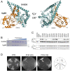
The overall structure of NgBR/DHDDS heterodimer. (A) Ribbon diagrams showing the front and back of the heteromeric complex. NgBR is colored in orange, and DHDDS is colored in deep teal. Mg2+ ion is shown as a gray sphere; IPP molecules occupying S1 and S2 sites are shown in red and green, respectively. (B) Limited proteolysis of the core domain was performed by incubating the protein with increasing amounts of thermolysin (0.005, 0.01, 0.02, 0.04, 0.08, 0.16 mg/mL). Untreated protein sample is denoted as “U.” In this gel, thermolysin comigrates with the full-length DHDDS. (C) Coimmunoprecipitation of NgBR/DHDDS mutations introduced at the complex interface. HEK293T cells were cotransfected with NgBR-HA and Flag-DHDDS cDNAs; cells were lysed 48 h posttransfection and immunoprecipitation performed using anti-flag magnetic beads. The lysate was analyzed by Western blotting. (D) Characterization of cis-PTase mutants in the C-terminal region of DHDDS using yeast complementation assay. The nus1Δ rer2Δ srt1Δ deletion strain expressing Giardia lamblia cis-PTase from URA3 plasmid was cotransformed with MET15 bearing WT NgBR and the LEU2 plasmid bearing either WT or mutant variants of DHDDS at the C terminus. Three variants were analyzed including a triple mutation (3A) corresponding to R306A, F313, L317, and two DHDDS truncation Δ256 and Δ289. The cells were streaked onto complete plates (YPD) or synthetic complete medium containing 1% FOA. The Ura3 protein, which is expressed from the URA3 marker converts FOA to toxic 5-fluorouracil, forcing the cells to lose the G. lamblia cis-PTase plasmid. Cell growth was monitored over time to assess phenotypic differences.
Fig. 3.
Fig. 3.
The active site of the NgBR/DHDDS complex. (A) Omit difference map, countered at 3.0 σ level, showing the two IPP molecules and Mg2+ ion bound at the active site. IPP1 is assigned to IPP molecule bound at S1 site and IPP2 to that at S2 site. The oxygen atoms are colored red, and phosphorus atoms are colored orange. The carbon atoms of IPP1 are colored salmon, and those of IPP2 are in green. (B) Detailed view of the -RXG- motif and the active site. The carbon atoms of NgBR -RXG- motif residues are colored orange and labeled, and those for DHDDS are colored cyan. Nitrogen atoms are colored blue and oxygen atoms are colored red. IPP1 and IPP2 are colored red and green, respectively. Mg2+ is shown as a gray sphere, and its coordination is indicated by the dashed lines. A coordinating water molecule is shown as a red sphere. (C) The K42E retinitis pigmentosa mutation in DHDDS. A cartoon representation indicating the locations of Lys-42 and Glu-234 relative to the P loop and bound substrates. DHDDS is colored in deep teal, IPP1 in red, IPP2 in green, and Mg2+ is shown as a gray sphere.
Fig. 4.
Fig. 4.
Missense NgBR mutations associated with Parkinson’s disease. (A) NgBR mutations related to Parkinson’s disease are shown as purple spheres and labeled. DHDDS is colored in gray and NgBR is colored in orange except for the N-terminal helix (α1), which is shown in green. (B) Detailed view showing the location of G91C disease mutant within NgBR colored orange except for α1 helix, shown in green. Gly-91 is involved in hydrophobic packing against Val-127. Lys-96 forms hydrogen bonds with Tyr-248 and Gln-289, which stabilize the C-terminal -RXG- motif. (C) cis-PTase activity was measured using purified WT and NgBR disease mutant, G91C. The mutant exhibits ∼40% reduction in cis-PTase activity compared to WT enzyme. The values are the mean ± SD of three independent measurements. (D) The majority of the mutated residues is solvent-exposed. The solvent accessible surface area (Å2) were calculated using GETAREA server.
Fig. 5.
Fig. 5.
DHDDS’s helix α0 functions as a membrane sensor. (A) A cartoon representation of DHDDS subunit colored in deep teal. The hydrophobic cavities of DHDDS before (Left) and after (Right) N-terminal loop deletion (residues 1–10) are shown in yellow. The N-terminal loop and α0 helix are shown in red; the side chains of the three exposed hydrophobic residues are shown and labeled. The hydrophobic cavity was generated using the 3V web server (54). (B) A cartoon representation of E. coli UPPs (PDB ID code 1X06) monomer colored in purple. The hydrophobic cavity is shown in yellow. Leu-137 involved in chain length control in UPPS is located at the end of the cavity. (C) Sequence alignment showing the conservation of three hydrophobic amino acids at the N-terminal helix α0 among DHDDS orthologs (highlighted in red). Conserved residues are highlighted in yellow. Proteins represented in this alignment are orthologs of human DHDDS cis-PTase subunit as follows: hDHDDS (human, UniProtKB Q86SQ9-1), XlDhdds (Xenopus laevis; UniProtKB Q7ZYJ5), DrDhdds (Danio rerio, UniProtKB Q6NXA2), CeDHDDS (Caenorhabditis elegans, UniProtKB Q5FC21), ScRer2 (S. cerevisiae, UniProtKB P35196), ScSrt1 (S. cerevisiae, UniProtKB Q03175), SpRer2 (Schizosaccharomyces pombe, UniProtKB O14171), TrRER2 (Trichoderma reesei, UniProtKB G0ZKV6), AfRer2 (Aspergillus fumigatus UniProtKB Q4WQ28), SlCPT3 (Solanum lycopersicum, UniProtKB K7WCI9), AtCPT3 (Arabidopsis thaliana, UniProtKB Q8S2T1), AtCPT4 (A. thaliana, UniProtKB Q8LAR7), AtCPT5 (A. thaliana, UniProtKB Q8LED0). (D) Phospholipid stimulation of WT and W12A/F15A/I19A triple mutant is shown. Stimulation was compared by measuring cis-PTase activity of purified WT and DHDDS triple mutant in the presence and absence of 1% PI. The values are the mean ± SD of five to eight independent measurements. (E) Reverse-phase TLC separation of dephosphorylated products from cis-PTase activity of WT and DHDDS triple mutation (W12A/F15A/I19A) denoted as MUT. Numbers correspond to the dominant polyprenols in each sample are shown at the bottom of the plate. The position of the polyprenol standards is shown on the left.
Fig. 6.
Fig. 6.
Mechanism of chain elongation by heteromeric cis-PTases. (A) Multiple amino acid sequence alignment comparing α3 helix between DHDDS orthologs and homodimeric cis-PTases. Highly conserved residues are highlighted in red, and less conserved ones are shown in yellow. Region corresponding to DHDDS EKE inset is highlighted in blue; the region is missing in homodimeric enzymes and a gap is present instead. Proteins represented in this alignment are: single subunit cis-PTs: EcUPPS (E. coli, UniProtKB P60472), MlDPPS (Micrococcus luteus, UniProtKB O82827), SaUPPS (Sulfolobus acidocaldarius, UniProtKB Q9HH76). Orthologs of human DHDDS cis-PTase subunit: hDHDDS (human, UniProtKB Q86SQ9-1), XlDhdds (Xenopus laevis, UniProtKB Q7ZYJ5), DrDhdds (D. rerio, UniProtKB Q6NXA2), CeDHDDS (C. elegans, UniProtKB Q5FC21), ScRer2 (S. cerevisiae, UniProtKB P35196), SpRer2 (S. pombe, UniProtKB O14171), SlCPT3 (Solanum lycopersicum, UniProtKB K7WCI9), AtCPT3 (A. thaliana, UniProtKB Q8S2T1), AtCPT4 (A. thaliana, UniProtKB Q8LAR7), AtCPT5 (A. thaliana, UniProtKB Q8LED0). (B) HPLC analysis of chain length of dolichol generated by WT and ΔEKE DHDDS mutant in yeast cells. The dolichol peaks were identified and labeled on the top of the chromatogram. The WT cells yielded the main compound Dol-20 compared to Dol-17 in ΔEKE mutant. The chain length and identity of lipids were confirmed by comparison with external standards of a polyprenol (Pren-10 to Pren-24) and dolichol (Dol-17 to Dol-23) mixtures. (C) Structural comparison between E. coli UPPS (PDB ID code 1X06) monomer and human DHDDS subunit. The EKE Inset within α3 helix of DHDDS creates a bigger bulge that may contribute to the stabilization of the enzyme:product complex during chain elongation. (D) Schematic diagram illustrating the proposed chain elongation mechanism for cis-PTases. Red arrow indicates the direction of product elongation. Exposed hydrophobic isoprene units may increasingly destabilize the enzyme:product complex by interacting with detergent micells (blue) or lipid bilayers (gray).

Similar articles

Cited by

References

    1. Takahashi S., Koyama T., Structure and function of cis-prenyl chain elongating enzymes. Chem. Rec. 6, 194–205 (2006). - PubMed
    1. Shimizu N., Koyama T., Ogura K., Molecular cloning, expression, and purification of undecaprenyl diphosphate synthase. No sequence similarity between E- and Z-prenyl diphosphate synthases. J. Biol. Chem. 273, 19476–19481 (1998). - PubMed
    1. Apfel C. M., Takács B., Fountoulakis M., Stieger M., Keck W., Use of genomics to identify bacterial undecaprenyl pyrophosphate synthetase: Cloning, expression, and characterization of the essential uppS gene. J. Bacteriol. 181, 483–492 (1999). - PMC - PubMed
    1. Sato M. et al. ., The yeast RER2 gene, identified by endoplasmic reticulum protein localization mutations, encodes cis-prenyltransferase, a key enzyme in dolichol synthesis. Mol. Cell Biol. 19, 471–483 (1999). - PMC - PubMed
    1. Endo S., Zhang Y. W., Takahashi S., Koyama T., Identification of human dehydrodolichyl diphosphate synthase gene. Biochim. Biophys. Acta 1625, 291–295 (2003). - PubMed

Publication types

MeSH terms