. 2020 Dec 1;130(12):6317-6324.
doi: 10.1172/JCI138416.

Antigen-specific B cell depletion for precision therapy of mucosal pemphigus vulgaris

Affiliations

Antigen-specific B cell depletion for precision therapy of mucosal pemphigus vulgaris

Jinmin Lee et al. J Clin Invest. .

Abstract

Desmoglein 3 chimeric autoantibody receptor T cells (DSG3-CAART) expressing the pemphigus vulgaris (PV) autoantigen DSG3 fused to CD137-CD3ζ signaling domains, represent a precision cellular immunotherapy approach for antigen-specific B cell depletion. Here, we present definitive preclinical studies enabling a first-in-human trial of DSG3-CAART for mucosal PV. DSG3-CAART specifically lysed human anti-DSG3 B cells from PV patients and demonstrated activity consistent with a threshold dose in vivo, resulting in decreased target cell burden, decreased serum and tissue-bound autoantibodies, and increased DSG3-CAART engraftment. In a PV active immune model with physiologic anti-DSG3 IgG levels, DSG3-CAART inhibited antibody responses against pathogenic DSG3 epitopes and autoantibody binding to epithelial tissues, leading to clinical and histologic resolution of blisters. DSG3 autoantibodies stimulated DSG3-CAART IFN-γ secretion and homotypic clustering, consistent with an activated phenotype. Toxicology screens using primary human cells and high-throughput membrane proteome arrays did not identify off-target cytotoxic interactions. These preclinical data guided the trial design for DSG3-CAART and may help inform CAART preclinical development for other antibody-mediated diseases.

Keywords: Autoimmune diseases; Autoimmunity; B cells; Immunotherapy; Therapeutics.


Conflict of interest statement

Conflict of interest: ASP and MCM possess equity, consult for, receive grants from, and have intellectual property (IP) with Cabaletta Bio. ASP, MCM, CTE, and JAF have IP with Novartis. MCM, CTE, and ASP have IP with Tmunity. DKL consults for Cabaletta Bio. CTE has equity and IP with Cabaletta Bio. JAF receives research support from Novartis, Tmunity, and Cabaletta Bio and is a consultant for L.E.K. Consulting and Guidepoint.

Figures

Figure 1
Figure 1. DSG3-CAART specifically depletes primary human anti-DSG3 IgG B cells from PV patients.
B cells purified from PV or healthy donor (HD) PBMCs were cocultured with DSG3-CAART, CART19, or NTD cells, and then stimulated with IL-2 and R848 before incubation on plates coated with rhDSG3 and anti–human IgG (anti-hIgG) to quantify anti-DSG3 and total IgG B cells, respectively. BSA served as a negative control antigen. ELISpot analysis shows that anti-DSG3 IgG B cells, but not total IgG B cells, are depleted by DSG3-CAART in PV patients 1 and 2 (columns 1 and 4). In PV patient 3, anti-DSG3 B cells were not detectable (column 7). CART19 eliminates all IgG B cells from PV patients and the HD (rows 3 and 6). Number of spots detected in each well is shown on the top right corner of each well. Quantification discussed in Methods.
Figure 2
Figure 2. DSG3-CAART demonstrates variable activity related to dose.
Luciferase-expressing anti-DSG3 hybridomas AK18, AK19, and AK23 (2 × 10⁵ total) were injected intravenously into NSG mice, followed 4 days later by injection with 3 × 10⁷, 1 × 10⁷, 3 × 10⁶ DSG3-CAART or 3 × 10⁷ NTD T cells. (A and B) Serial bioluminescence imaging indicates 24-fold, 302-fold, and 9-fold relative decreases in target cell burden in mice treated with 3 × 10⁷, 1 × 10⁷, and 3 × 10⁶ DSG3-CAART cells, respectively (black line, median). (C) Direct immunofluorescence staining of buccal mucosa with anti–human IgG indicates epithelial IgG binding (arrows) in 5 of 5 mice treated with NTD T cells and 4 of 5 mice treated with 3 × 10⁶ DSG3-CAART cells, compared with no mice treated with higher DSG3-CAART doses. Original magnification, ×20. (D) Anti-DSG3 ELISA performed on day 10 serum samples indicates that the 3 × 10⁷ and 1 × 10⁷ DSG3-CAART doses effectively abrogated serum anti-DSG3 IgG production relative to NTD or the 3 × 10⁶ DSG3-CAART dose. NS, nonsignificant. (E) Flow cytometric quantification of peripheral blood CD3+ T cells (left panel) and qPCR analysis of spleen (day 17, right panel) demonstrate dose-related DSG3-CAART engraftment. Average marking per human cell (mean ± standard deviation) reflects CAAR transgene copy number relative to p21 reference. NA, not applicable (not evaluated). Statistical analysis performed using Kruskal-Wallis (panels B and D) with post hoc Dunn’s test; ***P < 0.001, **P < 0.01, *P < 0.05.
Figure 3
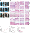
Figure 3. DSG3-CAART reverses rising serum antibody titers and improves blistering in a PV active immune model.
DSG3–/–DSG1tg/tg (DSG3-KO) mice were immunized with rhDSG3, followed by splenocyte transfer into syngeneic RAG2–/– mice. Twenty days after adoptive transfer of splenocytes, 10 and 5 mice were treated with DSG3-CAART or NTD T cells, followed by euthanasia on day 39 (5 mice in each treatment group) or day 69 (5 DSG3-CAART–treated mice), or earlier based on humane endpoints. (A and B) DSG3-CAART (images 6–15) improves hair loss, erosions, and histologic acantholysis (arrows), which persists in NTD-treated mice (images 1–5). Scale bars: 200 μm. (C) Mouse reagents were normalized for use in the human clinical DSG3 ELISA to calculate an anti-DSG3 antibody index value (RU/mL) for all mice with remaining serum samples. Serum anti-DSG3 antibody levels in the active immune model match or exceed those observed in human PV patients. (D) ELISA (mean OD with standard deviation) normalized to week 3 values indicates that DSG3-CAART treatment reduces serum anti-DSG3 antibody titers during the period of DSG3-CAART persistence. (E) T cell persistence by flow cytometry of peripheral blood, weeks 6–8 after splenocyte transfer.
Figure 4
Figure 4. High-throughput membrane proteome array (MPA) plus targeted cell screening did not identify off-target DSG3-CAART cytotoxic interactions.
(A) More than 5300 membrane proteins were expressed in HEK293T cells, and then screened with soluble Fc-tagged proteins to assess binding. Soluble Fc-tagged DSG3-CAAR ectodomain bound weakly to CLEC4M. Px44, anti-DSG3 positive control; FCGR1A, anti-Fc internal control. (B) MPA validation, indicating DSG3-CAAR-Fc binding to CLEC4M-overexpressing HEK293T relative to Fc-isotype control. (C) K562 cells were transduced with lentivectors encoding CLEC4M isoform v1 or v8, sorted to select for high CLEC4M expression, and then incubated with DSG3-CAART or CART19 cells. IFN-γ levels were undetectable in culture supernatants after 16 hours of coincubation of DSG3-CAART and CLEC4M-K562 cells, whereas IFN-γ was detected in positive control cocultures of DSG3-CAART with Nalm6 B cells expressing anti-DSG3 BCR PVB28, or CART19 cells cocultured with CD19+ Nalm6 cells. (D) qPCR verifies CLEC4M mRNA expression in pulmonary microvascular but not hepatic sinusoidal endothelial cells. HaCat keratinocytes, negative control. (E) Luminex cytokine analysis performed on DSG3-CAART and primary human cell coculture supernatants indicates no cytotoxic cytokine (IFN-γ, TNF-α) production. Red/blue × = below/above the range of detection. (F) xCELLigence cellular impedance assay of DSG3-CAART and primary human cell cocultures indicates no detectable cytolysis of CLEC4M-expressing pulmonary microvascular endothelial cells. DSG3-CAART induced changes in hepatic endothelial cell impedance only at a 50:1 effector-to-target ratio, a ratio not likely to be achieved in vivo.

References

    1. Maude SL, et al. Tisagenlecleucel in children and young adults with B-cell lymphoblastic leukemia. N Engl J Med. 2018;378(5):439–448. doi: 10.1056/NEJMoa1709866. - DOI - PMC - PubMed
    1. Schuster SJ, et al. Tisagenlecleucel in adult relapsed or refractory diffuse large B-cell lymphoma. N Engl J Med. 2019;380(1):45–56. doi: 10.1056/NEJMoa1804980. - DOI - PubMed
    1. Locke FL, et al. Long-term safety and activity of axicabtagene ciloleucel in refractory large B-cell lymphoma (ZUMA-1): a single-arm, multicentre, phase 1-2 trial. Lancet Oncol. 2019;20(1):31–42. doi: 10.1016/S1470-2045(18)30864-7. - DOI - PMC - PubMed
    1. Joly P, et al. First-line rituximab combined with short-term prednisone versus prednisone alone for the treatment of pemphigus (Ritux 3): a prospective, multicentre, parallel-group, open-label randomised trial. Lancet. 2017;389(10083):2031–2040. doi: 10.1016/S0140-6736(17)30070-3. - DOI - PubMed
    1. Kushner CJ, et al. Factors associated with complete remission after rituximab therapy for pemphigus. JAMA Dermatol. 2019;155(12):1404–1409. doi: 10.1001/jamadermatol.2019.3236. - DOI - PMC - PubMed

Publication types

MeSH terms