. 2020 Aug 20;16(8):e1008157.
doi: 10.1371/journal.pcbi.1008157. eCollection 2020 Aug.

Tumor vascular status controls oxygen delivery facilitated by infused polymerized hemoglobins with varying oxygen affinity

Affiliations

Tumor vascular status controls oxygen delivery facilitated by infused polymerized hemoglobins with varying oxygen affinity

Donald A Belcher et al. PLoS Comput Biol. .

Abstract

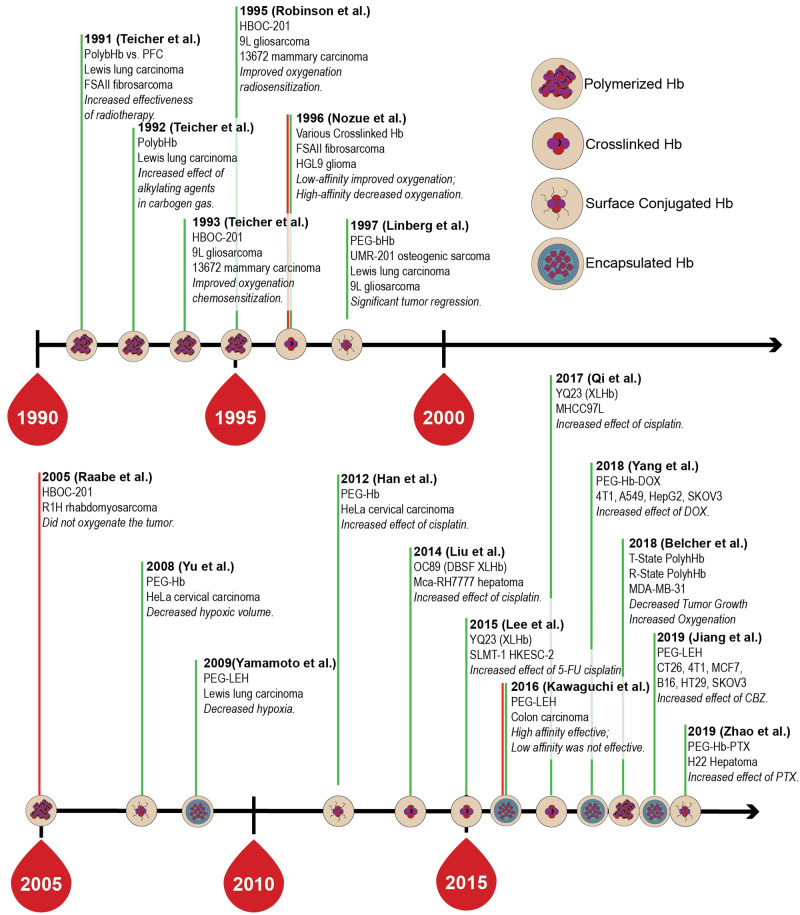
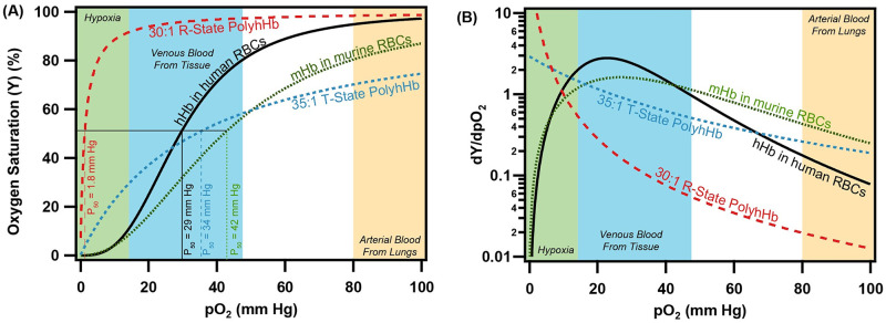
Oxygen (O2) delivery facilitated by hemoglobin (Hb)-based O2 carriers (HBOCs) is a promising strategy to increase the effectiveness of chemotherapeutics for treatment of solid tumors. However, the heterogeneous vascular structures present within tumors complicates evaluating the oxygenation potential of HBOCs within the tumor microenvironment. To account for spatial variations in the vasculature and tumor tissue that occur during tumor growth, we used a computational model to develop artificial tumor constructs. With these simulated tumors, we performed a polymerized human hemoglobin (hHb) (PolyhHb) enhanced oxygenation simulation accounting for differences in the physiologic characteristics of human and mouse blood. The results from this model were used to determine the potential effectiveness of different treatment options including a top load (low volume) and exchange (large volume) infusion of a tense quaternary state (T-State) PolyhHb, relaxed quaternary state (R-State) PolyhHb, and a non O2 carrying control. Principal component analysis (PCA) revealed correlations between the different regimes of effectiveness within the different simulated dosage options. In general, we found that infusion of T-State PolyhHb is more likely to decrease tissue hypoxia and modulate the metabolic rate of O2 consumption. Though the developed models are not a definitive descriptor of O2 carrier interaction in tumor capillary networks, we accounted for factors such as non-uniform vascular density and permeability that limit the applicability of O2 carriers during infusion. Finally, we have used these validated computational models to establish potential benchmarks to guide tumor treatment during translation of PolyhHb mediated therapies into clinical applications.


Conflict of interest statement

The authors have declared that no competing interests exist.

Figures

Fig 1
Fig 1. Time-line for the assessment of various HBOCs in the treatment of solid tumors.
Green lines indicate positive results, red lines indicate negative results.
Fig 2
Fig 2. OEC and O2 offloading plot for various O2 carrying species used in the simulations.
(A) The OEC is shown for human hemoglobin (hHb) in human RBCs, mouse hemoglobin (mHb) in mouse RBCs, 30:1 R-State PolyhHb, and 35:1 T-State PolyhHb. (B) The O2 offloading plot as a function of the pO2 is shown for the same species. Approximate pO2 regions for arterial and venous blood under normoxic conditions have been included on this graph for reference.
Fig 3
Fig 3. Model flow diagram.
At each time step, blood flow, distribution of nutrients/growth factors and vascular remodeling are computed. The tissue phase remodels at shorter time steps within the main loop. The resulting artificial tumors are transferred to the infusion model. Hemodilution and the HBOC enhanced viscosity modulates flow and vascular adaptation until the microvascular system is stable. After this, HBOC enhanced oxygenation is modeled.
Fig 4
Fig 4. Artificial mouse tumor growth and resulting biophysical properties after 40 days of growth.
(A) Visualization of cutaways, vessel (blue), tumor (green) and necrotic (red) volume fraction cross sections for artificial mouse tumor growth over 40 days. The tumor shown here was grown in a type A vascular bed. Also shown in this figure are the (B) radius, (C) rate of radial expansion, and (D) necrotic volume percentages for each vessel bed type (Type A-I). Shaded areas in these plots represent a 95% confidence interval across each type of vessel bed configuration. (E) Visualization of combined volume fraction cross sections for selected tumors from each of the vessel bed types (Type A-I). Comparison of (F) RBV and (G) CHb,tis between the tumor and host tissue in the artificial tumor constructs. The letter labels indicate the vessel configuration. The dashed line separates the tumor properties greater than and less than the host properties. For all tumor cross sections the scale bar is 1 mm.
Fig 5
Fig 5. PCA biplot for principal components 1 and 2 of the mouse and human tumor model.
In this figure, groupings are organized by organism type (human, mouse). Left: PC scores for tissue and O2 delivery for each of the simulated tumor treatments. Ellipses are drawn around each group with 68% of the normal probability. Right: Loading plots relating how each parameter influences the corresponding principal component. Green vectors indicate tumor properties while blue vectors indicate host tissue properties. Labels indicate the corresponding tumor property.
Fig 6
Fig 6. PCA biplot for principal components 1 and 2 for the analysis of the mouse model.
PC scores for tissue properties and O2 delivery for each of the simulated mouse tumor treatments for (A) principal component 1 and (C) principal component 3 as a function of principal component 2. Grouping in panel A is based on tumor vascular bed type. Grouping in panel B is based on dosing type. Ellipses are drawn around each group with 68% of the normal probability. Loading plots relating how each parameter influences the corresponding principal component is shown for (B) principal component 1 and (D) principal component 3 as a function of principal component 2. Loading vectors with magnitude less than 0.5 have been excluded from this plot. Green vectors indicate tumor properties while blue vectors indicate host tissue properties. Labels on each vector indicate the corresponding tumor property.
Fig 7
Fig 7. PCA biplot for principal components 1 and 2 for the analysis of the human model.
PC scores for tissue properties and O2 delivery for each of the simulated mouse tumor treatments for (A) principal component 1 and (C) principal component 3 as a function of principal component 2. Grouping in panel A is based on tumor vascular bed type. Grouping in panel B is based on dosing type. Ellipses are drawn around each group with 68% of the normal probability. Loading plots relating how each parameter influences the corresponding principal component is shown for (B) principal component 1 and (D) principal component 3 as a function of principal component 2. Loading vectors with magnitude less than 0.5 have been excluded from this plot. Green vectors indicate tumor properties while blue vectors indicate host tissue properties. Labels on each vector indicate the corresponding tumor property.
Fig 8
Fig 8. Changes in tumor vascular architecture and Hb/PolyhHb concentrations for the simulated PolyhHb enhanced infusion model.
Variations in the (A) RBV, (B) RBF, (C) CHb,tis, (D) Hb O2 saturation in RBC, (E) PolyhHb (HBOC) concentration, (F) PolyhHb (HBOC) O2 saturation, (G) blood pO2, and (H) MRO2 for the baseline, top-load, and exchange infusion of the control, 30:1 R-State PolyhHb, and 35:1 T-State PolyhHb.
Fig 9
Fig 9. Changes in OEF for the simulated PolyhHb enhanced infusion model.
Variations in (A) OEF, (B) Blood pO2, (C) OEFplas, (D) OEFHb O2 saturation in RBC, and (E) OEFHBOC (HBOC) concentration infusion of the control, 30:1 R-State PolyhHb, and 35:1 T-State PolyhHb.
Fig 10
Fig 10. Changes in tumor hypoxia for the simulated PolyhHb enhanced infusion model.
Variations in the (A) tumor tissue pO2, (B) boundary tissue pO2, (C) hypoxic fraction, and (D) boundary hypoxic fraction for infusion of the control, 30:1 R-State PolyhHb, and 35:1 T-State PolyhHb.
Fig 11
Fig 11. The effect of (A) host tissue Hb saturation, (B) tumor tissue Hb saturation, (C) tumor RBF and (D) percent decrease in total tumor hypoxic volume on the percent changes in the boundary hypoxic volume.
The baseline (unsupplemented) condition is depicted as the dashed line at zero. Letters labeling each data point indicate the vessel bed configuration for that artificial tumor construct. The non O2 carrying control is green, R-State PolyhHb is red, and T-State PolyhHb is blue.
Fig 12
Fig 12. The effect tumor (A) oxyHb concentration, (B) tissue blood saturation, (C) RBV, and (D)RBF on the percent changes in MRO2.
The baseline (unsupplemented) condition is depicted as the dashed line at zero. Letters labeling each data point indicate the vessel bed configuration for that artificial tumor construct.

References

    1. Nozue M, Lee I, Manning JM, Manning LR, Jain RK. Oxygenation in tumors by modified hemoglobins. Journal of Surgical Oncology. 1996;62(2):109–114. 10.1002/(SICI)1096-9098(199606)62:2<109::AID-JSO6>3.0.CO;2-C - DOI - PubMed
    1. Liu XB, Cheng Q, Geng W, Ling CC, Liu Y, Ng KTP, et al. Enhancement of cisplatin-based TACE by a hemoglobin-based oxygen carrier in an orthotopic rat HCC model. Artificial Cells, Nanomedicine and Biotechnology. 2014;42(4):229–236. 10.3109/21691401.2013.808647 - DOI - PubMed
    1. Lee NP, Chan KT, Choi MY, Lam HY, Tung LN, Tzang FC, et al. Oxygen carrier YQ23 can enhance the chemotherapeutic drug responses of chemoresistant esophageal tumor xenografts. Cancer Chemotherapy and Pharmacology. 2015;76(6):1199–1207. 10.1007/s00280-015-2897-2 - DOI - PubMed
    1. Qi X, Wong BL, Lau SH, Ng KTPP, Kwok SY, Kin-Wai Sun C, et al. A hemoglobin-based oxygen carrier sensitized Cisplatin based chemotherapy in hepatocellular carcinoma. Oncotarget. 2017;8(49):85311–85325. 10.18632/oncotarget.19672 - DOI - PMC - PubMed
    1. Robinson MF, Dupuis NP, Kusumoto T, Liu F, Menon K, Teicher Ba. Increased Tumor Oxygenation and Radiation Sensitivity in two Rat Tumors by A Hemoglobin-Based, Oxygen-Carrying Preparation. Artificial Cells, Blood Substitutes and Biotechnology. 1995;23(3):431–438. 10.3109/10731199509117959 - DOI - PubMed

Publication types