

COVID-19 severity scoring systems in radiological imaging - a review
- PMID: 32817769
- PMCID: PMC7425223
- DOI: 10.5114/pjr.2020.98009
COVID-19 severity scoring systems in radiological imaging - a review
Abstract
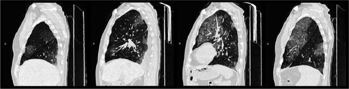
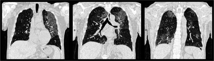
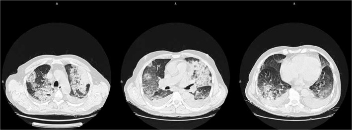
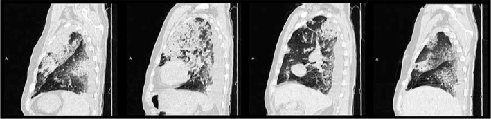
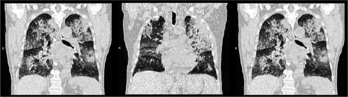
The current reference standard to make a definitive diagnosis of SARS-CoV-2 infection is the reverse transcription- polymerase chain reaction assay (rt-PCR). However, radiological imaging plays a crucial role in evaluating the course of COVID-19 and in choosing proper management of infected patients. Chest X-ray (CXR) is generally considered not to be sensitive for the detection of pulmonary abnormalities in the early stage of the disease. However, in the emergency setting CXR can be a useful diagnostic tool for monitoring the rapid progression of lung involvement in COVID-19, especially in patients admitted to intensive care units. The rapid course of SARS-CoV-2 infection and the severity and progression of lung aberrations require a method of radiological evaluation to implement and manage the appropriate treatment for infected patients. Computed tomography (CT) imaging is considered to be the most effective method for the detection of lung abnormalities, especially in the early stage of the disease. Moreover, serial chest CT imaging with different time intervals is also effective in estimating the evolution of the disease from initial diagnosis to discharge from hospital. Despite having low specificity in distinguishing abnormalities in viral infections, the high sensitivity of CT makes this method ideal for assessing the severity of the disease in patients with confirmed COVID-19. In this review, we present and discuss currently available scales that can be used to assess the severity of lung involvement in COVID-19 patients in everyday work, both for CXR and CT imaging.
Keywords: COVID-19; CT; CXR; SARS-CoV-2; X-ray; computed tomography.
Copyright © Polish Medical Society of Radiology 2020.
Conflict of interest statement
The authors report no conflict of interest.
Figures


References
-
- World Health Organization (WHO). Q&A on coronaviruses (COVID-19). Availabel at: https://www.who.int/emergencies/diseases/novel-coronavirus-2019/question... (Accessed: 24.02.2020).
-
- U.S. Centers for Disease Control and Prevention (CDC). How COVID-19 Spreads. 27 January 2020. Archived from the original on 28 January 2020. Retrieved 29 January 2020.
-
- Chinese Society of Radiology. Radiological diagnosis of new coronavirus infected pneumonitis: expert recommendation from the Chinese Society of Radiology (First edition). Chin J Radiol 2020; 54: E001.
Publication types
LinkOut - more resources
Full Text Sources
Medical
Miscellaneous