[Preprint]. 2020 Aug 17:2020.08.08.238469.
doi: 10.1101/2020.08.08.238469.

An ultra-potent synthetic nanobody neutralizes SARS-CoV-2 by locking Spike into an inactive conformation

Affiliations

An ultra-potent synthetic nanobody neutralizes SARS-CoV-2 by locking Spike into an inactive conformation

Michael Schoof et al. bioRxiv. .

Update in

  • An ultrapotent synthetic nanobody neutralizes SARS-CoV-2 by stabilizing inactive Spike.
    Schoof M, Faust B, Saunders RA, Sangwan S, Rezelj V, Hoppe N, Boone M, Billesbølle CB, Puchades C, Azumaya CM, Kratochvil HT, Zimanyi M, Deshpande I, Liang J, Dickinson S, Nguyen HC, Chio CM, Merz GE, Thompson MC, Diwanji D, Schaefer K, Anand AA, Dobzinski N, Zha BS, Simoneau CR, Leon K, White KM, Chio US, Gupta M, Jin M, Li F, Liu Y, Zhang K, Bulkley D, Sun M, Smith AM, Rizo AN, Moss F, Brilot AF, Pourmal S, Trenker R, Pospiech T, Gupta S, Barsi-Rhyne B, Belyy V, Barile-Hill AW, Nock S, Liu Y, Krogan NJ, Ralston CY, Swaney DL, García-Sastre A, Ott M, Vignuzzi M; QCRG Structural Biology Consortium; Walter P, Manglik A. Schoof M, et al. Science. 2020 Dec 18;370(6523):1473-1479. doi: 10.1126/science.abe3255. Epub 2020 Nov 5. Science. 2020. PMID: 33154106 Free PMC article.

Abstract

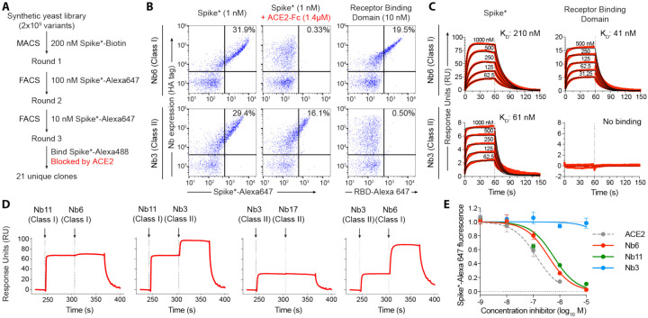
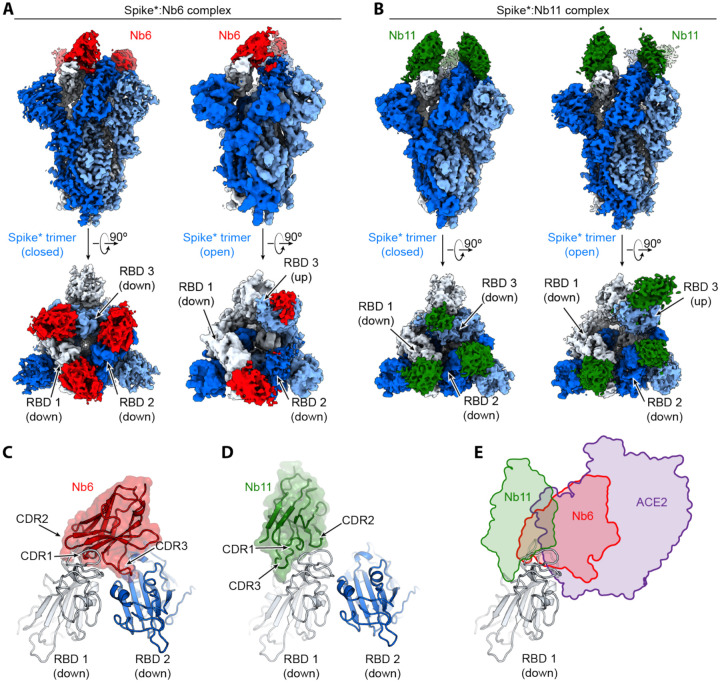
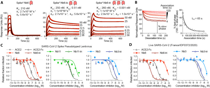
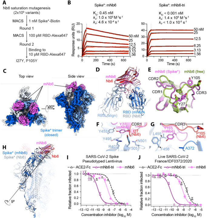
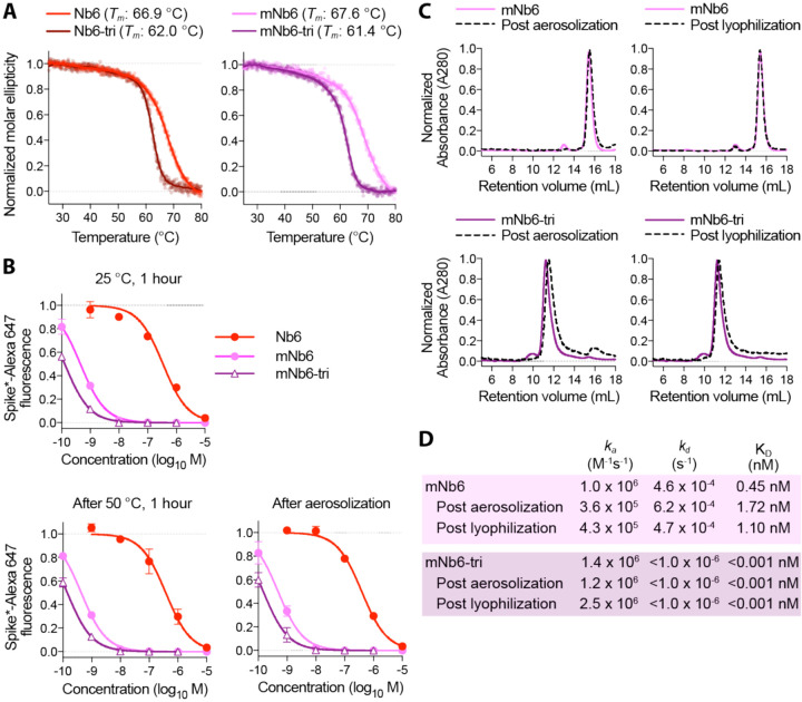
Without an effective prophylactic solution, infections from SARS-CoV-2 continue to rise worldwide with devastating health and economic costs. SARS-CoV-2 gains entry into host cells via an interaction between its Spike protein and the host cell receptor angiotensin converting enzyme 2 (ACE2). Disruption of this interaction confers potent neutralization of viral entry, providing an avenue for vaccine design and for therapeutic antibodies. Here, we develop single-domain antibodies (nanobodies) that potently disrupt the interaction between the SARS-CoV-2 Spike and ACE2. By screening a yeast surface-displayed library of synthetic nanobody sequences, we identified a panel of nanobodies that bind to multiple epitopes on Spike and block ACE2 interaction via two distinct mechanisms. Cryogenic electron microscopy (cryo-EM) revealed that one exceptionally stable nanobody, Nb6, binds Spike in a fully inactive conformation with its receptor binding domains (RBDs) locked into their inaccessible down-state, incapable of binding ACE2. Affinity maturation and structure-guided design of multivalency yielded a trivalent nanobody, mNb6-tri, with femtomolar affinity for SARS-CoV-2 Spike and picomolar neutralization of SARS-CoV-2 infection. mNb6-tri retains stability and function after aerosolization, lyophilization, and heat treatment. These properties may enable aerosol-mediated delivery of this potent neutralizer directly to the airway epithelia, promising to yield a widely deployable, patient-friendly prophylactic and/or early infection therapeutic agent to stem the worst pandemic in a century.


Conflict of interest statement

Competing Interests M.Schoof, B.Faust, R.A.Saunders, N.Hoppe, P.Walter, and A.Manglik are inventors on a provisional patent describing anti-Spike nanobodies described in this manuscript.

Figures

Figure 1.
Figure 1.. Discovery of two distinct classes of anti-Spike nanobodies.
A, Selection strategy for identification of anti-Spike nanobodies that disrupt Spike-ACE2 interactions using magnetic bead selections (MACS) or fluorescence activated cell sorting (FACS). B, Flow cytometry of yeast displaying Nb6 (a Class I nanobody) or Nb3 (a Class II nanobody). Nb6 binds Spike*-Alexa 647 and receptor binding domain (RBD-Alexa 647). Nb6 binding to Spike* is completely disrupted by an excess (1.4 μM) of ACE2-Fc. Nb3 binds Spike*, but not the RBD. Nb3 binding to Spike* is partially decreased by ACE2-Fc. C, SPR of Nb6 and Nb3 binding to either Spike* or RBD. Red traces are raw data and global kinetic fits are shown in black. Nb3 shows no binding to RBD. D, SPR experiments with immobilized Spike* show that Class I and Class II nanobodies can bind Spike* simultaneously. By contrast, two Class I nanobodies or Class II nanobodies do not bind simultaneously. E, Nanobody inhibition of 1 nM Spike*-Alexa 647 binding to ACE2 expressing HEK293T cells. n = 3 (ACE2, Nb3) or 5 (Nb6, Nb11) biological replicates. All error bars represent s.e.m.
Figure 2.
Figure 2.. Cryo-EM structures of Nb6 and Nb11 bound to Spike.
A, Cryo-EM maps of Spike*-Nb6 complex in either closed (left) or open (right) Spike* conformation. B, Cryo-EM maps of Spike*-Nb11 complex in either closed (left) or open (right) Spike* conformation. The top views show receptor binding domain (RBD) up- or down-states. C, Nb6 straddles the interface of two down-state RBDs, with CDR3 reaching over to an adjacent RBD. D, Nb11 binds a single RBD in the down-state (displayed) or similarly in the up-state. No cross-RBD contacts are made by Nb11 in either RBD up- or down-state. E, Comparison of RBD epitopes engaged by ACE2 (purple), Nb6 (red), or Nb11 (green). Both Nb11 and Nb6 directly compete with ACE2 binding.
Figure 3.
Figure 3.. Multivalency improves nanobody affinity and inhibitory efficacy.
A, SPR of Nb6 and multivalent variants. Red traces show raw data and black lines show global kinetic fit for Nb6 and independent fits for association and dissociation phases for Nb6-bi and Nb6-tri. B, Dissociation phase SPR traces for Nb6-tri after variable association time ranging from 4 to 520 s. Curves were normalized to maximal signal at the beginning of the dissociation phase. Percent fast phase is plotted as a function of association time (right) with a single exponential fit. n = 3 independent biological replicates. C, Inhibition of pseudotyped lentivirus infection of ACE2 expressing HEK293T cells. n = 3 biological replicates for all but Nb11-tri (n = 2) D, Inhibition of live SARS-CoV-2 virus. Representative biological replicate with n = 3 (right panel) or 4 (left panel) technical replicates per concentration. n = 3 biological replicates for all but Nb3 and Nb3-tri (n = 2). All error bars represent s.e.m.
Figure 4.
Figure 4.. Affinity maturation of Nb6 yields a picomolar SARS-CoV-2 neutralizing molecule.
A, A saturation mutagenesis library of Nb6 was subjected to two rounds of selection to identify consensus mutations I27Y and P105Y. B, SPR of mNb6 and mNb6-tri binding to immobilized Spike*. Red traces show raw data and black lines show global kinetic fit. No dissociation was observed for mNb6-tri over 10 minutes. C, Cryo-EM structure of Spike*-mNb6 complex. D, Comparison of receptor binding domain (RBD) engagement by Nb6 and mNb6. One RBD was used to align both structures (RBD align), demonstrating changes in Nb6 and mNb6 position and the adjacent RBD. E, Comparison of mNb6 complementarity determining regions in either the cryo-EM structure of the Spike*-mNb6 complex or an X-ray crystal structure of mNb6 alone. F, CDR1 of Nb6 and mNb6 binding to the RBD. As compared to I27 in Nb6, Y27 of mNb6 hydrogen bonds to Y453 and optimizes pi-pi and pi-cation interactions with the RBD. G, CDR3 of Nb6 and mNb6 binding to the RBD demonstrating a large conformational rearrangement of the entire loop in mNb6. H, Comparison of closed Spike* bound to mNb6 and Nb6. Rotational axis for RBD movement is highlighted. I, Inhibition of pseudotyped lentivirus infection of ACE2 expressing HEK293T cells by mNb6 and mNb6-tri. n = 3 biological replicates J, mNb6 and mNb6-tri inhibit SARS-CoV-2 infection of VeroE6 cells in a plaque assay. Representative biological replicate with n = 4 technical replicates per concentration. n = 3 biological replicates for all samples. All error bars represent s.e.m.
Figure 5.
Figure 5.. Nb6 and its derivates are robust proteins.
A, Thermal denaturation of nanobodies assessed by circular dichroism measurement of molar ellipticity at 204 nm. Apparent melting temperatures (Tm) for each nanobody are indicated. B, Nanobody inhibition of 1 nM Spike*-Alexa 647 binding to ACE2 expressing HEK293T cells after incubation at either 25 °C or 50 °C for 1 hour or after aerosolization. C, Size exclusion chromatography of nanobodies after lyophilization or aerosolization. D, Summary table of SPR kinetics data and affinities for aerosolized or lyophilized mNb6 and mNb6-tri.

References

    1. Ksiazek T. G. et al., A novel coronavirus associated with severe acute respiratory syndrome. N Engl J Med 348, 1953–1966 (2003). - PubMed
    1. Zaki A. M., van Boheemen S., Bestebroer T. M., Osterhaus A. D., Fouchier R. A., Isolation of a novel coronavirus from a man with pneumonia in Saudi Arabia. N Engl J Med 367, 1814–1820 (2012). - PubMed
    1. Zhou P. et al., A pneumonia outbreak associated with a new coronavirus of probable bat origin. Nature 579, 270–273 (2020). - PMC - PubMed
    1. Chan J. F. et al., A familial cluster of pneumonia associated with the 2019 novel coronavirus indicating person-to-person transmission: a study of a family cluster. Lancet 395, 514–523 (2020). - PMC - PubMed
    1. Huang C. et al., Clinical features of patients infected with 2019 novel coronavirus in Wuhan, China. Lancet 395, 497–506 (2020). - PMC - PubMed

Publication types