. 2020 Nov 5;27(5):840-851.e6.
doi: 10.1016/j.stem.2020.07.020. Epub 2020 Aug 19.

Next-Generation Surrogate Wnts Support Organoid Growth and Deconvolute Frizzled Pleiotropy In Vivo

Affiliations

Next-Generation Surrogate Wnts Support Organoid Growth and Deconvolute Frizzled Pleiotropy In Vivo

Yi Miao et al. Cell Stem Cell. .

Abstract

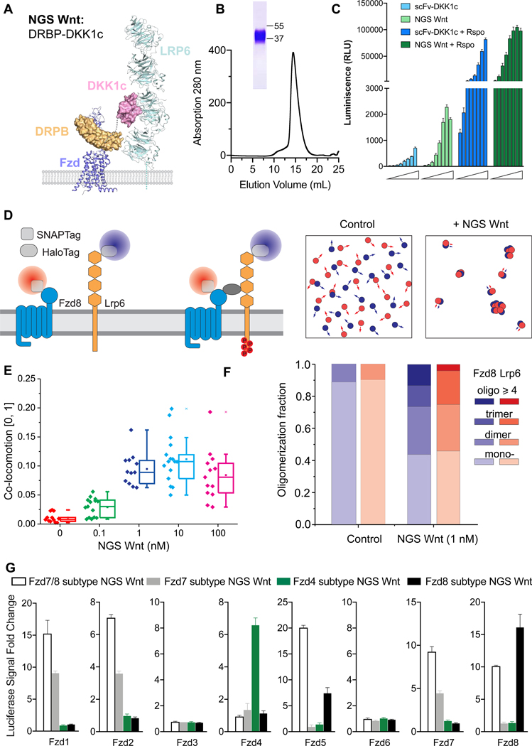
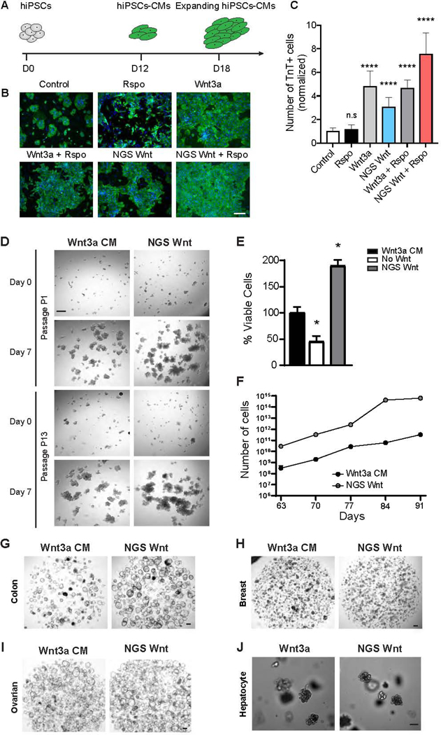
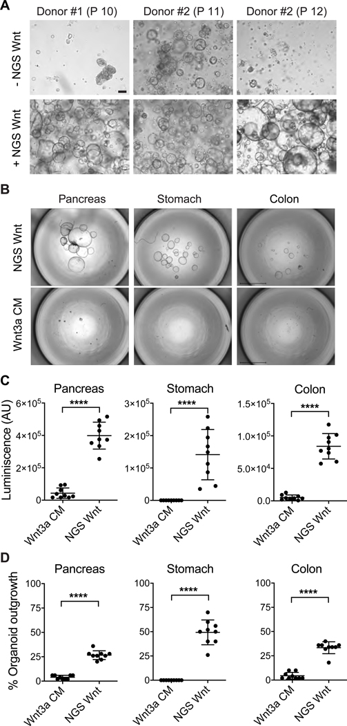
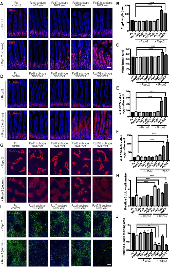
Modulation of Wnt signaling has untapped potential in regenerative medicine due to its essential functions in stem cell homeostasis. However, Wnt lipidation and Wnt-Frizzled (Fzd) cross-reactivity have hindered translational Wnt applications. Here, we designed and engineered water-soluble, Fzd subtype-specific "next-generation surrogate" (NGS) Wnts that hetero-dimerize Fzd and Lrp6. NGS Wnt supports long-term expansion of multiple different types of organoids, including kidney, colon, hepatocyte, ovarian, and breast. NGS Wnts are superior to Wnt3a conditioned media in organoid expansion and single-cell organoid outgrowth. Administration of Fzd subtype-specific NGS Wnt in vivo reveals that adult intestinal crypt proliferation can be promoted by agonism of Fzd5 and/or Fzd8 receptors, while a broad spectrum of Fzd receptors can induce liver zonation. Thus, NGS Wnts offer a unified organoid expansion protocol and a laboratory "tool kit" for dissecting the functions of Fzd subtypes in stem cell biology.

Keywords: DARPin; Frizzled; Wnt; canonical Wnt signaling; organoids; protein engineering; regenerative medicine; stem cell; surrogate Wnt.


Conflict of interest statement

Declaration of Interests Y.M., L.T.D., D.B., and K.C.G. are inventors on patent applications submitted by Leland Stanford Junior University that cover the use of NGS Wnt. H.C. is an inventor on several patents related to organoid technology. W.H. works at U-Protein Express BV, a contract research organization that produces recombinant proteins and antibodies as commercial activity. K.C.G., C.J.K., C.Y.J., H.C. and R.N. are founders of Surrozen, Inc.

Figures

Figure 1.
Figure 1.. Design concept of NGS Wnt.
(A) Design schematic of NGS Wnt. Fzd subtype (purple) specific binder DRPB (orange) is fused with Lrp5/6 (cyan) binder DKK1c (pink) using a flexible Gly-Ser linker (grey dash line). Figure is adapted from PDBs (3S8V, 3S94, 6BD4 and 6NDZ). (B) Size exclusion chromatography of Fzd7/8 subtype NGS Wnt. The band on SDS-PAGE gel represents Fzd7/8 subtype NGS Wnt. (C) NGS Wnt (156 pM to 20 nM) induces higher β-catenin signaling change than scFv-DKK1c (2 nM to 250 nM) with or without Rspo (25 nM). Data represent mean and S.E., N = 3 technical replicates from representative experiment. (D) Schematic for characterizing NGS Wnt induced heterodimer-/oligomerization ofFzd8 and Lrp6 by single molecule imaging in live cells. (E) Co-locomotion Lrp6 and Fzd8 measured within 20 min after addition of NGS Wnt at indicated concentrations. N > 12 for each condition. Error bar represents mean and S.E. (F) Quantification of Lrp6 and Fzd8 receptor oligomer fractions. Control refers tountreated samples. More than 2800 individual complex intensities were examined for each receptor. (G) Fzd subtype NGS Wnts specifically induce β-catenin signaling in through targeted Fzd receptors. All data represents mean and S.E., N = 3 technical replicates from representative experiment. See also Figures S1, S2 and Video S1 and S2.
Figure 2.
Figure 2.. NGS Wnt promotes hiPSC-CM expansion and supports proliferation of multiple organoids.
(A) Experimental diagram of hiPSC-CM expansion. At day 12 (D12), hiPSC-derived CMs were treated with Wnt activators and expansion was monitored at day 18 (D18). (B) Representative immunofluorescence images for cardiac troponin T (TnT) (green) and DAPI (blue) in hiPSC-CMs treated with indicated Wnt activators or control. Scale bar represents 100 μm. (C) Quantification of the TnT positive cell number with the indicated Wnt activators orcontrol. **** indicates P<0.0001 by unpaired t-test. Data were from 3 independent experiments and 2 replicates. Results are expressed as mean and S.D.. (D) Representative brightfield images of human cystic fibrosis colon organoid cultures in either Wnt3a CM or NGS Wnt. N=5. Scale bar represents 100 μm. (E) Quantification of viable cells (as percentage) of human cystic fibrosis colon organoids after first passage (7 days) cultured in Wnt3a CM, no Wnt source or NGS Wnt. * indicates P<0.05. (F) Growth curves of human cystic fibrosis colon organoids were analyzed from passage P9 to P13. Cell number counts are expressed as mean and S.D. of three independent assays. * indicates P<0.05. (G-J) Representative brightfield images of human colon, ovarian, breast and mouse hepatocyte organoids expanded with Wnt3a CM/Wnt3a or NGS Wnt at 0.5 nM. N=10 for colon and N=3 for other organoid types. Scale bar represents 100 μm. See also Figures S3 and S4
Figure 3.
Figure 3.. NGS Wnt increases human tubuloid life span and improves organoid outgrowth efficiency from single cells.
(A) Representative brightfield images of human tubuloids without/with NGS Wnt at indicated passage number. Scale bar represents 100 μm. (B) Representative images of human pancreas, stomach and colon organoids at 2 weeks after seeding 50 single cells, cultured with either Wnt3a CM or NGS Wnt. N=1 for individual organoid type. Scale bars represent 2 mm. (C-D) Quantification of organoid outgrowth by CellTiter-Glo assay (C) and organoid counting (D). Mean and S.E. for 9 technical replicates are displayed. **** indicates P<0.0001. See also Figure S4.
Figure 4.
Figure 4.. NGS Wnts reveal functionally relevant Fzd receptor profiles in adult intestinal crypt and liver homeostasis.
(A-F) Analysis of representative jejunum cross-sectional immunofluorescence images for (A) Ki67 (red) and (D) CD44v6 (red) following adenovirus administration. DAPI staining is represented in blue. Crypt length (B), villus length (C), Ki67+ cells/crypt-villus unit (E), CD44v6+ cells/crypt-villus unit (F) were measured in each condition shown in (A) and (D). Scale bars represent 50 μm. (G-J) Analysis of representative liver cross-sectional immunofluorescence images for GLUL (G) and E-Cadherin (E-Cad) (I) following adenovirus administration. (H) The number of GLUL positive hepatocytes around the central vein was measured in each condition shown in (G). (J) The relative E-Cadherin+ cells within a fixed area was measured in each condition shown in (I). Scale bars represent 100 μm. Data are expressed as mean ± S.E. N=4 animals per group and experiments were repeated twice. Statistical analysis was performed using one-way ANOVA followed by Tukey’s multiple comparison test(**P< 0.01; ****P< 0.0001).

References

    1. Bandaranayake AD, Correnti C, Ryu BY, Brault M, Strong RK, and Rawlings DJ (2011). Daedalus: a robust, turnkey platform for rapid production of decigram quantities of active recombinant proteins in human cell lines using novel lentiviral vectors. Nucleic Acids Research 39, 11. - PMC - PubMed
    1. Barker N, and Clevers H. (2006). Mining the Wnt pathway for cancer therapeutics. Nat Rev Drug Discov 5, 997–1014. - PubMed
    1. Bartfeld S, Bayram T, van de Wetering M, Huch M, Begthel H, Kujala P, Vries R, Peters PJ, and Clevers H. (2015). In Vitro Expansion of Human Gastric Epithelial Stem Cells and Their Responses to Bacterial Infection. Gastroenterology 148, 126–136. - PMC - PubMed
    1. Benhamouche S, Decaens T, Godard C, Chambrey R, Rickman DS, Moinard C, Vasseur-Cognet M, Kuo CJ, Kahn A, Perret C, et al. (2006). Apc tumor suppressor gene is the “zonation-keeper” of mouse liver. Developmental cell 10, 759–770. - PubMed
    1. Boj SF, Hwang CI, Baker LA, Chio IIC, Engle DD, Corbo V, Jager M, Ponz-Sarvise M, Tiriac H, Spector MS, et al. (2015). Organoid Models of Human and Mouse Ductal Pancreatic Cancer. Cell 160, 324–338. - PMC - PubMed

Publication types

Substances