. 2020 Aug 20;8(1):141.
doi: 10.1186/s40478-020-01015-3.

Assessment of radial glia in the frontal lobe of fetuses with Down syndrome

Affiliations

Assessment of radial glia in the frontal lobe of fetuses with Down syndrome

Ana A Baburamani et al. Acta Neuropathol Commun. .

Abstract

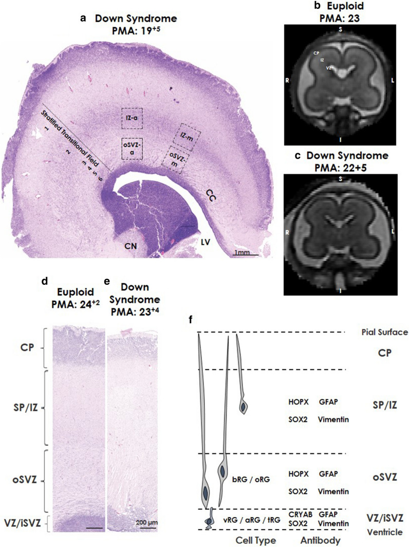
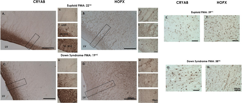
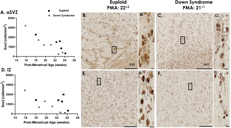
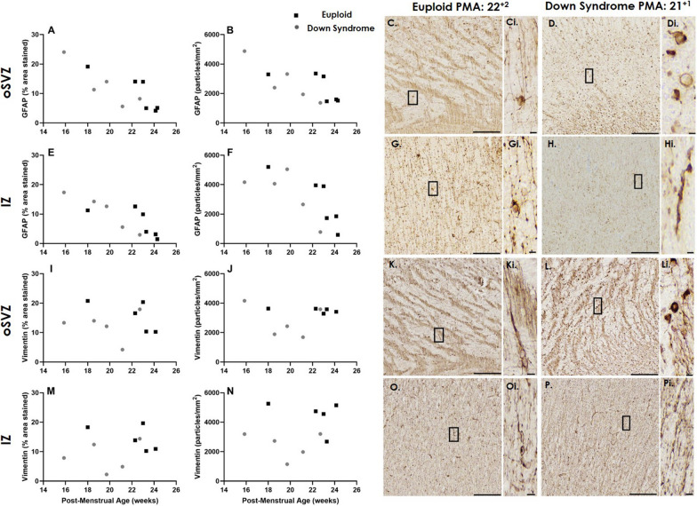
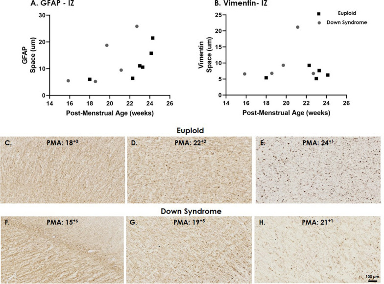
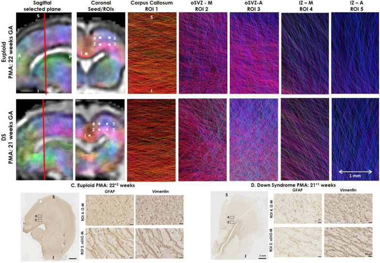
Down syndrome (DS) occurs with triplication of human chromosome 21 and is associated with deviations in cortical development evidenced by simplified gyral appearance and reduced cortical surface area. Radial glia are neuronal and glial progenitors that also create a scaffolding structure essential for migrating neurons to reach cortical targets and therefore play a critical role in cortical development. The aim of this study was to characterise radial glial expression pattern and morphology in the frontal lobe of the developing human fetal brain with DS and age-matched controls. Secondly, we investigated whether microstructural information from in vivo magnetic resonance imaging (MRI) could reflect histological findings from human brain tissue samples. Immunohistochemistry was performed on paraffin-embedded human post-mortem brain tissue from nine fetuses and neonates with DS (15-39 gestational weeks (GW)) and nine euploid age-matched brains (18-39 GW). Radial glia markers CRYAB, HOPX, SOX2, GFAP and Vimentin were assessed in the Ventricular Zone, Subventricular Zone and Intermediate Zone. In vivo diffusion MRI was used to assess microstructure in these regions in one DS (21 GW) and one control (22 GW) fetal brain. We found a significant reduction in radial glial progenitor SOX2 and subtle deviations in radial glia expression (GFAP and Vimentin) prior to 24 GW in DS. In vivo, fetal MRI demonstrates underlying radial projections consistent with immunohistopathology. Radial glial alterations may contribute to the subsequent simplified gyral patterns and decreased cortical volumes observed in the DS brain. Recent advances in fetal MRI acquisition and analysis could provide non-invasive imaging-based biomarkers of early developmental deviations.

Keywords: Cortical development; Diffusion MRI; Down syndrome; Fetal brain; Radial glia; SOX2.


Conflict of interest statement

The authors declare that they have no competing interests.

Figures

Fig. 1
Fig. 1
Coronal section of regions of interest. Photomicrographs of Haemotoxlin and Eosin (H&E) stained (a) coronal section from the frontal lobe of a Down syndrome case at 19+5 weeks PMA showing the stratified transitional field (1–6) and regions of interest. b, c Coronal T2-weighted MRI images from (b) control, 23 weeks PMA and (c) Down syndrome, 22+5 weeks PMA, highlighting different regions based on T2-weighted signal intensities. Higher magnification of H&E stained frontal lobe coronal sections from (d) age-matched Euploid case at 24+2 weeks PMA, and (e) Down syndrome case at 23+4 weeks PMA. F Schematic of regions of interest and associated protein labels of radial glia. Scale bars indicated (a) 1 mm, (b, c) 200 µm. Abbreviations: –a anterior, CN caudate nucleus, CC corpus callosum, CP cortical plate, IZ intermediate zone, LV lateral ventricle, -m medial, RG radial glia (aRG; apical, bRG basal, oRG outer, vRG; ventral, tRG; truncated), SVZ subventricular zone (iSVZ; inner, oSVZ; outer), PMA post-menstrual age, STF stratified transitional field, SP subplate, VZ ventricular zone
Fig. 2
Fig. 2
CRYAB and HOPX in the developing brain. Photomicrographs of CRYAB (apical/ventral/truncated radial glia), and HOPX (basal/outer radial glia) in coronal frontal lobe sections in mid-gestation from (A, B) age-matched euploid, 22+2 weeks PMA and (C, D) Down syndrome 19+5 weeks PMA. (EH) In late-gestation CRYAB and HOPX staining is cellular in both Down syndrome (38+4 weeks PMA) and euploid (39+1 weeks PMA) cases. Scale bars indicate (AD) 500 µm, (i–iii) 50 µm, (EH) 100 µm. Abbreviations: LV; lateral ventricle, SVZ; subventricular zone (iSVZ; inner, oSVZ; outer), PMA; post-menstrual age, VZ; ventricular zone
Fig. 3
Fig. 3
SOX2 cells in the oSVZ and IZ in mid-gestation. The number of SOX2 positive cells in the (A) oSVZ and (D) IZ across mid-gestation in age-matched euploid (black squares) and Down syndrome (grey circles) cases. Photomicrographs of SOX2 staining in the (B, C) iSVZ/oSVZ and (E, F) IZ in (B, E) euploid, 22+2 weeks PMA and Down syndrome, 21+1 weeks PMA. Scale bars indicate (B, C, E, F) 150 µm, (i) 5 µm. Abbreviations: IZ; intermediate zone, SVZ; subventricular zone (iSVZ; inner, oSVZ; outer), PMA; post-menstrual age
Fig. 4
Fig. 4
Amount and Number of GFAP and Vimentin in the oSVZ and IZ during mid-gestation. The amount (% area stained) and number (particles/mm2) of (A, B, E, F) GFAP and (I, J, M, N) Vimentin in the (A, B, I, J) oSVZ and (E, F, M, N) IZ during mid-gestation in age-matched euploid (black squares) and Down syndrome (grey circles) cases. Photomicrographs of (C, D, G, H) GFAP and (K, L, O, P) Vimentin in the (C, D, K, L) oSVZ and (G, H, O, P) IZ, in (C, G, k, O) Euploid and (D, H, L, P) Down syndrome. IZ intermediate zone, oSVZ outer subventricular zone, PMA post-menstrual age. Scale bars indicate (C, D, G, H, K, L, O, P) 150 µm, (i) 5 µm
Fig. 5
Fig. 5
Space between radial glial fibres in the IZ. The average space between (A) GFAP and (B) Vimentin labelled radial glial fibres in the IZ across mid-gestation in age-matched euploid (black squares) and Down syndrome (grey circles) cases. Photomicrographs of GFAP staining across mid-gestation in (C–E) euploid and (F–H) Down syndrome cases. Scale bars indicate (C–H) 100 µm. IZ intermediate zone, PMA post-menstrual age
Fig. 6
Fig. 6
Fetal diffusion MRI and immunohistochemistry in the fetal brain. Upper panels show a (A) Euploid (22 GW) and (B) DS (21 GW) fetal brain, following in vivo fetal MRI in sagittal plane, and 5 seed/regions of interest (ROI), approximately 1 × 1 × 1 voxel, (2 mm), corresponding to immunohistological ROIs assessed and the corpus callosum. The colour-coding of the tractography is based on the standard red, green, blue code to indicate directionality; red for left–right, green for anterior–posterior and blue for dorsal–ventral. The lower panel reflect GFAP and Vimentin labelled radial glia, with images rotated in a comparable plane to the fetal MRI images in the (C) Euploid and (D) DS brain, showing direction of fibres in comparable ROI 2 (oSVZ-M) and ROI 4 (IZ-M) stained with GFAP and Vimentin. S superior, I inferior. Scale bar indicates 2 mm on low magnification and 50 µm on high magnification photomicrographs

References

    1. Altman J, Bayer SA. Regional differences in the stratified transitional field and the honeycomb matrix of the developing human cerebral cortex. J Neurocytol. 2002;31:613–632. doi: 10.1023/a:1025787427576. - DOI - PubMed
    1. Baburamani AA, Patkee PA, Arichi T, Rutherford MA. New approaches to studying early brain development in Down syndrome. Dev Med Child Neurol. 2019;61:867–879. doi: 10.1111/dmcn.14260. - DOI - PMC - PubMed
    1. Beattie R, Hippenmeyer S. Mechanisms of radial glia progenitor cell lineage progression. FEBS Lett. 2017;591:3993–4008. doi: 10.1002/1873-3468.12906. - DOI - PMC - PubMed
    1. Borrell V. Recent advances in understanding neocortical development. F1000 Research. 2019 doi: 10.12688/f1000research.20332.1. - DOI - PMC - PubMed
    1. Borrell V, Reillo I. Emerging roles of neural stem cells in cerebral cortex development and evolution. Dev Neurobiol. 2012;72:955–971. doi: 10.1002/dneu.22013. - DOI - PubMed

Publication types