Review
. 2020 Sep;41(9):758-770.
doi: 10.1016/j.it.2020.07.004. Epub 2020 Aug 17.

Astrocyte Reactivity: Subtypes, States, and Functions in CNS Innate Immunity

Affiliations
Review

Astrocyte Reactivity: Subtypes, States, and Functions in CNS Innate Immunity

Michael V Sofroniew. Trends Immunol. 2020 Sep.

Abstract

Astrocytes are neural parenchymal cells that ubiquitously tile the central nervous system (CNS). In addition to playing essential roles in healthy tissue, astrocytes exhibit an evolutionarily ancient response to all CNS insults, referred to as astrocyte reactivity. Long regarded as passive and homogeneous, astrocyte reactivity is being revealed as a heterogeneous and functionally powerful component of mammalian CNS innate immunity. Nevertheless, concepts about what astrocyte reactivity comprises and what it does are incomplete and sometimes controversial. This review discusses the goal of differentiating reactive astrocyte subtypes and states based on composite pictures of molecular expression, cell morphology, cellular interactions, proliferative state, normal functions, and disease-induced dysfunctions. A working model and conceptual framework is presented for characterizing the diversity of astrocyte reactivity.


Figures

Figure. 1.
Figure. 1.. Mammalian astrocytes respond to diverse reactivity-inducing triggers and produce diverse molecular effectors.
A. Astrocyte reactivity can be triggered by a wide variety of molecules that can derive from diverse sources, including any cell type in CNS tissue, as well as from microbial pathogens, circulating inflammatory cells, serum proteins, peripheral metabolic disorders or environmental toxins. B. Reactive astrocytes can exhibit diverse functional responses to these triggers and can elaborate a wide variety of effector molecules that can influence many different cell types in a context-specific manner. BBB blood-brain barrier, bv blood vessel, OPC oligodendrocyte progenitor cell. Protein abbreviations are per international guidelines (https://www.genecards.org/). Small molecule abbreviations: Aβ amyloid beta, Glut glutamate, NE norepinephrine, NO nitric oxide, ROS reactive oxygen species.
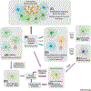
Figure 2.
Figure 2.. Key Figure – Working model of diverse astrocyte responses to CNS disorders.
A. In healthy tissue, astrocytes exhibit regional and local heterogeneity in gene expression and function. Diverse starting conditions can influence subsequent reactivity responses. B. Different forms of astrocyte reactivity are triggered by diverse non-cell-autonomous signals emanating from diverse CNS insults. C. Proliferative astrocyte reactivity occurs in response to tissue damage caused e.g. by traumatic or ischemic cell degeneration, blood-brain barrier leak, infection or autoimmune leukocytic inflammation. Newly proliferated astrocytes (red nuclei) form limitans borders that permanently separate and corral areas of damage, inflammation and non-neural scar tissue from adjacent viable neural tissue. D. Non-proliferative astrocyte reactivity can exhibit diverse states with different changes in gene expression and functions that are context dependent as determined by astrocyte starting conditions and incoming reactivity triggers. Non-proliferative reactive astrocytes maintain but modify their interactions with surrounding cells in preserved tissue architecture. E. Non-proliferative astrocyte reactivity can resolve over time if acute triggers recede. F. Astrocyte reactivity can become chronic if triggers persist. G. Chronic astrocyte reactivity can lead to loss- or gain-of-functions resulting in disease states with dysfunctional reactivity that can exacerbate tissue pathologies and worsen disorder outcome. H. Genetic mutations and polymorphisms can lead to cell-autonomous dysfunctions in astrocytes that lead to non-reactive disease states. Such disease states can in the absence of astrocyte reactivity, cause or contribute to tissue dysfunctions that in turn lead to production of astrocyte reactivity triggers. The ensuing dysfunctional astrocyte reactivity exhibits gain- or loss-of-functions that can contribute to further tissue pathology in a vicious cycle. Details and literature references are in the main text.
Figure 3.
Figure 3.. Astrocytes in healthy human and mouse tissue express multiple DAMP and PAMP receptors.
Pink boxes indicate significant receptor gene expression. DAMP and PAMP receptor information taken from reference [90]. CP, caudate putamen, Cx cerebral cortex, Hp hippocampus, Hu, human, Ms mouse, SC spinal cord. Expression data taken from references [–36, 57] ) and their associated open access databases: a http://www.brainrnaseq.org/ b http://astrocyternaseq.org/ c https://astrocyte.rnaseq.sofroniewlab.neurobio.ucla.edu./addgene?query=Clec9a

References

    1. Sofroniew MV and Vinters HV (2010) Astrocytes: biology and pathology. Acta Neuropathol 119 (1), 7–35. - PMC - PubMed
    1. Verkhratsky A and Nedergaard M (2018) Physiology of Astroglia. Physiol Rev 98 (1), 239–389. - PMC - PubMed
    1. Magistretti PJ and Allaman I (2018) Lactate in the brain: from metabolic end-product to signalling molecule. Nat Rev Neurosci 19 (4), 235–249. - PubMed
    1. MacVicar BA and Newman EA (2015) Astrocyte regulation of blood flow in the brain. Cold Spring Harb Perspect Biol 7 (5). - PMC - PubMed
    1. Plog BA and Nedergaard M (2018) The Glymphatic System in Central Nervous System Health and Disease: Past, Present, and Future. Annu Rev Pathol 13, 379–394. - PMC - PubMed

Publication types