. 2020 Oct 16;147(20):dev192997.
doi: 10.1242/dev.192997.

Loss of the seipin gene perturbs eggshell formation in Caenorhabditiselegans

Affiliations

Loss of the seipin gene perturbs eggshell formation in Caenorhabditiselegans

Xiaofei Bai et al. Development. .

Abstract

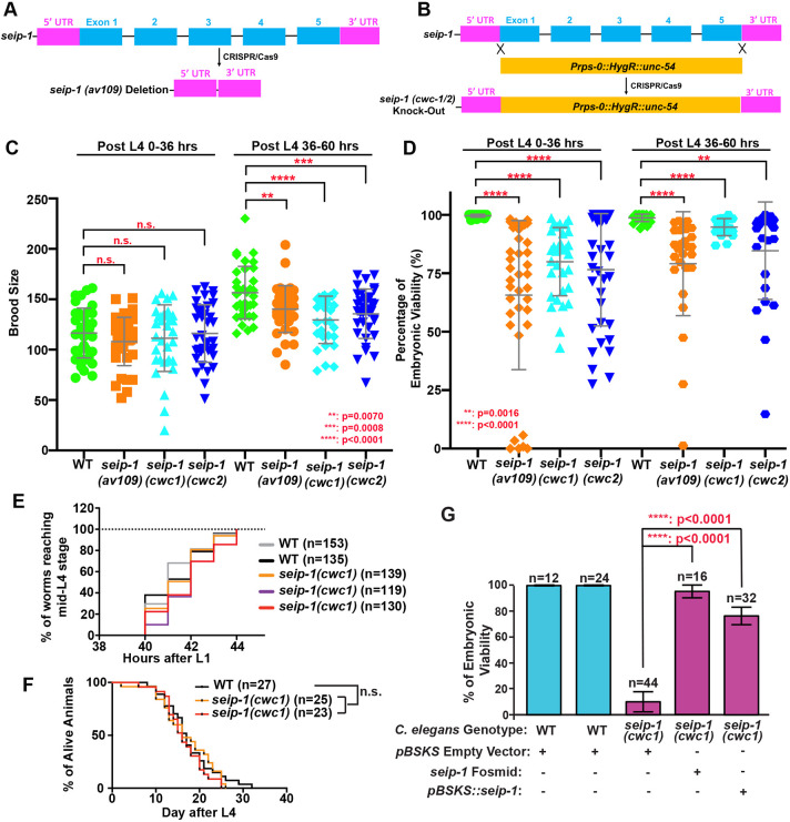
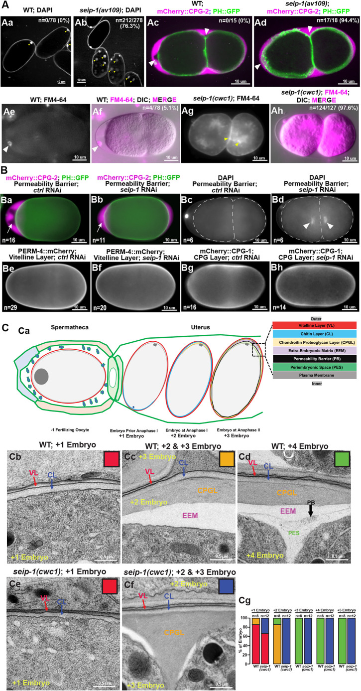
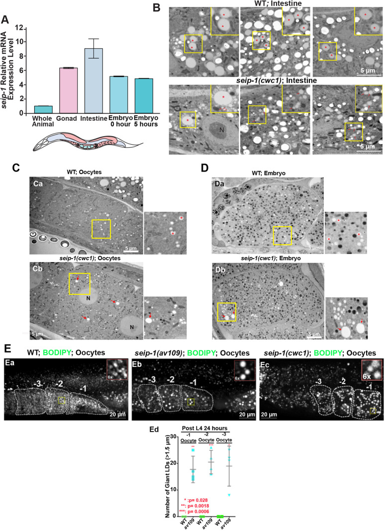
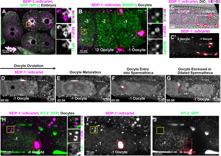
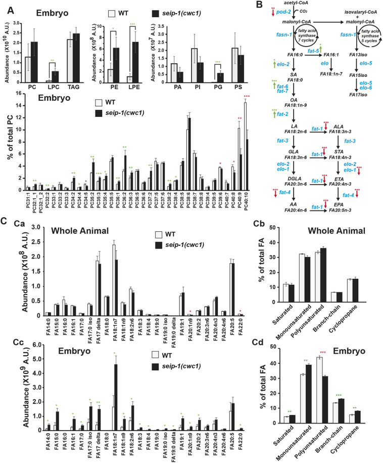
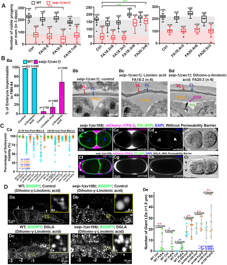
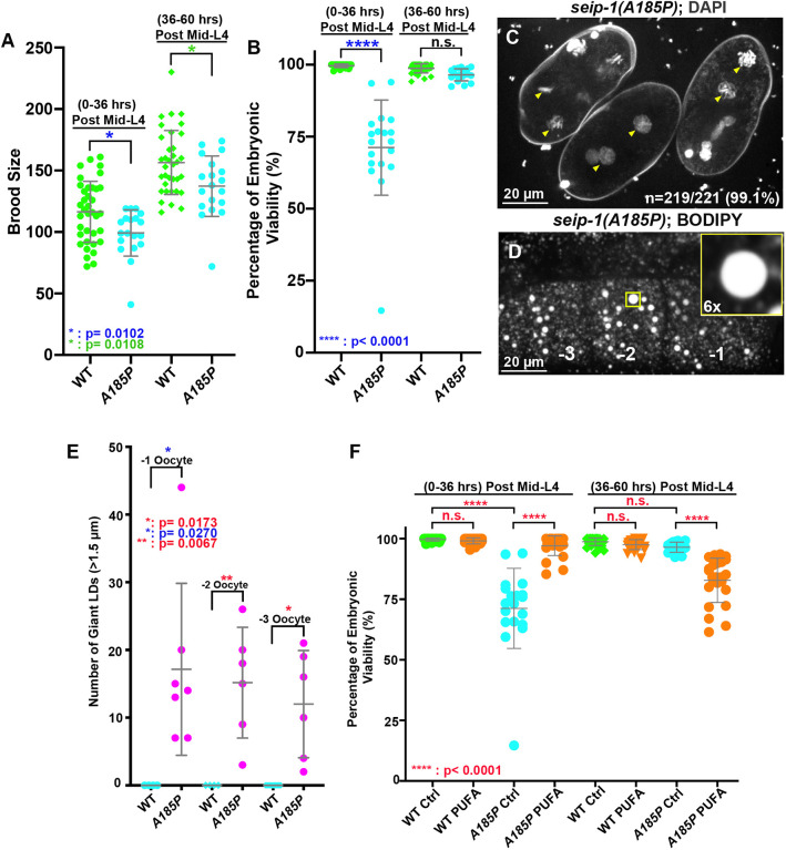
Seipin, an evolutionary conserved protein, plays pivotal roles during lipid droplet (LD) biogenesis and is associated with various human diseases with unclear mechanisms. Here, we analyzed Caenorhabditis elegans mutants deleted of the sole SEIPIN gene, seip-1 Homozygous seip-1 mutants displayed penetrant embryonic lethality, which is caused by the disruption of the lipid-rich permeability barrier, the innermost layer of the C. elegans embryonic eggshell. In C. elegans oocytes and embryos, SEIP-1 is associated with LDs and is crucial for controlling LD size and lipid homeostasis. The seip-1 deletion mutants reduced the ratio of polyunsaturated fatty acids (PUFAs) in their embryonic fatty acid pool. Interestingly, dietary supplementation of selected n-6 PUFAs rescued the embryonic lethality and defective permeability barrier. Accordingly, we propose that SEIP-1 may maternally regulate LD biogenesis and lipid homeostasis to orchestrate the formation of the permeability barrier for eggshell synthesis during embryogenesis. A lipodystrophy allele of seip-1 resulted in embryonic lethality as well and could be rescued by PUFA supplementation. These experiments support a great potential for using C. elegans to model SEIPIN-associated human diseases.

Keywords: Eggshell; Fatty acid; Lipid droplet; PUFAs; Permeability barrier; Seipin.


Conflict of interest statement

Competing interestsThe authors declare no competing or financial interests.

Figures

Fig. 1.
Fig. 1.
Deletion of the seip-1 gene causes hatching defects. (A) Diagram of deleted regions in the seip-1(av109) mutant or (B) replacement of the seip-1 gene with a hygromycin-resistant gene (HygR) cassette in seip-1(cwc1) and seip-1(cwc2) mutants. (C) Brood size was compared between wild-type (WT) and seip-1 mutants over two time periods post L4. (D) The percentage of viable embryos was reduced in the seip-1 deletion animals. (E) The developmental time required for synchronized L1 larvae to reach L4 and (F) the longevity of viable animals in the population were similar to wild-type animals. (G) Expression of the seip-1 containing fosmid and plasmid in the seip-1(cwc1) mutant significantly rescues the embryonic lethality. Data are mean±s.d. Statistical significance was determined using an unpaired two-tailed Student's t-test. n.s., not significant.
Fig. 2.
Fig. 2.
Deletion of the seip-1 gene causes severe eggshell formation defects. (A) DAPI staining of zygotic chromatin (yellow arrows, Ab) was apparent in seip-1 embryos, whereas only the first polar body (yellow arrows, Aa) stained in wild-type (WT) embryos (n=number of embryos with zygotic chromatin stained with DAPI/number of embryos imaged). mCherry::CPG-2 was used to visualize the integrity of the permeability barrier. In wild type, the inner layer of the eggshell prevents diffusion of mCherry::CPG-2 beyond the space between the outer layers of the eggshell and the embryo surface (white arrowheads, Ac). However, in seip-1(av109) embryos, mCherry::CPG-2 penetrates inside the entire space between the eggshell and the embryo surface (white arrowheads, Ad) (n=number of embryos with mCherry::CPG-2 penetration/number of embryos imaged). Only the plasma membrane surrounding the first polar body (white arrowheads) was stained by FM4-64 (magenta) in wild-type embryos (Ae,Af), whereas staining was observed at the plasma membrane and cytosol (yellow arrowheads) in seip-1(cwc1) mutant embryos (Ag,Ah) (n=number of embryos with FM4-64 penetration/number of embryos imaged). (B) Depletion of SEIP-1 by RNAi resulted in defective permeability barrier formation (Ba,Bb), allowing mCherry::CPG-2 (white arrows in Ba and Bb) to fill the space between the outer layers of the eggshell and the embryo surface (n=number of embryos with mCherry::CPG-2 penetration), and DAPI (white arrowheads, Bd) to penetrate inside the embryos to stain zygotic chromatin (n=number of embryos with DAPI penetration). The eggshell proper appears mainly intact when SEIP-1 was depleted by RNAi, as markers for the vitelline layer (PERM-4::mCherry, Be,Bf) and the CPG layer (mCherry::CPG-1, Bg,Bh) are properly localized compared with embryos treated by control RNAi (n=the number of embryos examined). (C) Diagram of the sequential steps during eggshell assembly (Ca). At anaphase of meiosis II, the permeability barrier is formed between the outer eggshell and the plasma membrane. TEM micrographs of high-pressure frozen embryos demonstrate that the vitelline layer (VL), chitin layer (CL) and the chondroitin proteoglycan layer (CPGL) are easily observed in both wild-type and seip-1(cwc1) embryos that have completed meiosis I (Cb,Cc,Ce). A clear edge of the permeability barrier (PB, black arrow) was visualized in every wild-type embryo that completed meiosis II (Cd). EEM defines the space between the chondroitin proteoglycan layer and the permeability barrier, whereas the periembryonic space (PES) bridges the permeability barrier and the embryo proper. However, no permeability barrier was found at any stage of the seip-1(cwc1) embryo (Ce,Cf). The white patches in Cf show the severely compromised EEM. Quantification of the embryos with the forming eggshell in both WT and seip-1(cwc1) embryos is presented in Cg. The embryos containing different components of the eggshell were categorized into four groups, which are represented by the colored squares inserted at the top right of each panel (Cb-Cf). Red, embryos containing the vitelline layer and chitin layer; blue, embryos with the vitelline layer, chitin layer and chondroitin proteoglycan layer; orange, embryos with four layers, including the vitelline layer, chitin layer, chondroitin proteoglycan layer and EEM; green, embryos with all five eggshell layers (vitelline layer, chitin layer, chondroitin proteoglycan layer, EEM and permeability barrier). n=the number of embryos examined.
Fig. 3.
Fig. 3.
SEIP-1 depletion alters LD size in the oocytes and embryos. (A) qRT-PCR was performed using RNAs isolated from whole animals, dissected tissues or embryos immediately prepared (0 h) from gravid adults or embryos allowed to develop for 5 h after dissection from gravid adults. seip-1 is highly expressed in the C. elegans germline, embryos and intestine. (B) TEM micrographs of high-pressure frozen animals displayed normal LDs in wild-type and abnormal LDs in seip-1(cwc1) intestinal cells. Red asterisks and arrows mark the LDs. A 1.6× magnification of the regions marked with a yellow square are also shown in upper right insets in each panel. (C,D) TEM micrographs of high-pressure frozen animals show normal LDs (red asterisks) in wild-type oocytes (C) and embryos (D), as well as enlarged (red arrows) and tiny LDs in the seip-1(cwc1) oocytes and embryos. A 2.5× magnification of the regions marked with a yellow square are also shown to the right of each panel. (E) BODIPY-stained LDs in the −1 to −3 oocytes of wild-type (Ea) and seip-1 (Eb,Ec) animals. A 6× magnification of the yellow squares are shown on the top right corner of each panel. Quantification of the enlarged LDs (diameter >1.5 μm) inside the −1 to −3 oocytes of both wild-type (wt; green) and seip-1 mutants (blue) is presented in Ed. Data are mean±s.d. Statistical significance was determined using an unpaired two-tailed Student's t-test. N, nucleus.
Fig. 4.
Fig. 4.
SEIP-1 localization in C. elegans oocytes and embryos. (A) SEIP-1::mScarlet observed in embryos. Right inserts represent the magnified area of the yellow square, showing that SEIP-1::mScarlet is adjacent to the ER labeled by GFP::SP12. (B) In the oocytes, SEIP-1::mScarlet is either adjacent to or surrounds the LDs stained by BODIPY. Right inserts represent the magnified area of the yellow square, showing that SEIP-1::mScarlet localizes to the surface of the BODIPY stained LDs. −1, −2 and −3 indicate oocyte from the most proximal to the distal to the spermathecal. (B-C′) SEIP-1::mScarlet localizes to the pseudocoelomic space (red squares in panel B; red circles in panels C,C′) surrounding the ovulating oocytes (-1 oocyte). (D-G) Representative images of SEIP-1::mScarlet localization during ovulation and fertilization. A strong SEIP-1::mScarlet signal was observed in the fifth sheath cell (red arrows) that surrounds the −1 oocyte (D) until the oocyte is ovulated and enters the spermatheca (E-G). (H-J) A vitellogenin reporter (VIT-2::GFP) transgene was used to monitor yolk lipoprotein localization, showing no colocalization with SEIP-1::mScarlet either in the LDs (yellow squares) or the pseudocoelomic space (red squares). Amplified images of the yellow square are shown to the right of H.
Fig. 5.
Fig. 5.
Lipidomic perturbations and fatty acid imbalances were observed in the seip-1 mutant embryos. (A) Lipidomic analysis of the early embryos indicated that phospholipid levels were broadly affected in seip-1(cwc1) mutant embryos compared with wild-type embryos. Detailed phosphatidylcholine (PC) species in the lipidome of wild-type and the seip-1(cwc1) mutant were compared based on their ratio normalized to the total PC level. Green and red asterisks indicate those values that are significantly increased or reduced, respectively, in the seip-1(cwc1) mutant compared with wild type. (B) The de novo fatty acid synthesis pathway of C. elegans. Enzymes (blue fonts) that catalyze each of the biochemical steps (black fonts) are indicated. Expression of the de novo fatty acid synthetic genes were either upregulated (elo-2, fat-2, fat-5 and fat-6, green arrows) or downregulated (pod-2, elo-1, fat-1, fat-4, red arrows) in seip-1(cwc1) mutant embryos (see also Fig. S6). Green and red arrows indicate that gene expression is significantly increased or reduced in seip-1(cwc1) mutant embryos compared with wild type. (C) The absolute abundance of different fatty acid (FA) species (Ca) or the fatty acid species in subgroups (Cb) were not affected in seip-1(cwc1) whole animals measured by GC/MS. However, the absolute abundance of most fatty acid species was increased in seip-1(cwc1) embryos (Cc). Fatty acids were divided into subgroups and compared based on the ratio normalized to the total fatty acid level. The percentages of MUFAs, branch-chain and cyclopropane were increased in the isolated seip-1 mutant embryos (Cd). In contrast, the percentage of PUFAs was significantly decreased in seip-1 mutant embryos. Green and red asterisks indicate those values that are significantly increased or reduced, respectively, in the seip-1(cwc1) mutant compared with wild type. Data are mean±s.d. Statistical significance was determined using an unpaired two-tailed Student's t-test. *P<0.05; **P<0.01; ***P<0.001. A.U., arbitrary units.
Fig. 6.
Fig. 6.
Dietary supplementation of PUFAs rescued the permeability barrier defects but not the abnormal LDs in seip-1 mutants. (A) The total number of progeny of wild-type (WT) and seip-1 animals after feeding with NGM supplemented with 300 µM of the indicated fatty acids for 3 days were counted and compared. n=the number of L4 larvae used for the experiment. ***P<0.001. The boxes extend from the 25th to 75th percentiles. The whiskers extend from minimum to maximum. (B) Dietary supplementation of DGLA (FA18:3n6), but not LA (FA18:2n6), reduced the population of penetrant seip-1 embryos stained with FM4-64 (Ba). TEM micrographs of high-pressure frozen embryos showing the permeability barrier (black arrow) formed after seip-1 mutant animals were fed with DGLA, but not formed when fed with LA (Bb-Bd). n=number of embryos showing representative eggshell layers/number of embryos imaged. (C) The percentage of viable embryos was significantly rescued in the DGLA-fed seip-1 deletion animals (wild type, green; mutant, blue; DGLA-fed, orange) (Ca). The penetration of DAPI, the expression pattern of CPG2::mCherry, and DIC images, demonstrated that the permeability barrier was defective in seip-1 mutants embryos (Cb-Ci), which can be restored after the diet of seip-1 mutants animals was supplemented with DGLA (Cf-Ci). The GFP::PH transgene was used to indicate the plasma membranes. n=number of embryos with abnormal DAPI penetration and abnormal CPG2::mCherry pattern/number of embryos imaged. The permeability barrier can be easily seen in panel Cf (white arrowheads) and panel Ci (black arrowheads). (D) Enlarged LDs were frequently observed in the −1 to −3 oocytes of both wild-type (Dc) and seip-1(av109) (Dd) animals fed with DGLA. Right top insets represent 6× amplified images of the inlaid yellow squares (Da-Dd). Quantification of enlarged LDs (diameter >1.5 μm) in −1 to −3 oocytes of both PUFA-fed wild-type and seip-1(av109) animals is shown in De. Data are mean±s.d. Statistical significance was determined using an unpaired two-tailed Student's t-test. n.s., not significant.
Fig. 7.
Fig. 7.
A seipin patient-specific allele disrupts eggshell formation and displays enlarged LDs in C. elegans. (A) A conserved patient-specific allele seip-1(A185P) was generated and resulted in reduced brood sizes. (B) seip-1(A185P) causes embryonic lethality in the day 1 (post L4 0-36 h) adult animals. However, embryonic viability of seip-1(A185P) is not affected in the day 2 and 3 (post L4 36-60 h) animals. (C) DAPI staining of zygotic chromatin (yellow arrowheads) in seip-1(A185P) mutant embryos is indicative of a defective permeability barrier. (D) seip-1(A185P) exhibits enlarged LDs in the −1 to −3 oocytes marked by BODIPY. The panel inset shows a 6× enlarged view of a single enlarged LD indicated by the yellow box in the main panel. (E) Quantification of enlarged LDs (diameter >1.5 μm) in −1 to −3 oocytes of seip-1(A185P) animals. (F) The percentage of viable embryos was significantly rescued in the day 1 (post L4 0-36 h) DGLA (PUFA)-fed seip-1(A185P) animals compared with the control. In contrast, the percentage of viable embryos was reduced in the day 2 and 3 animals (post L4 36-60 h) fed with PUFAs. Data are mean±s.d. Statistical significance was determined using an unpaired two-tailed Student's t-test. n.s., not significant.

References

    1. Benenati G., Penkov S., Müller-Reichert T., Entchev E. V. and Kurzchalia T. V. (2009). Two cytochrome P450s in Caenorhabditis elegans are essential for the organization of eggshell, correct execution of meiosis and the polarization of embryo. Mech. Dev. 126, 382-393. 10.1016/j.mod.2009.02.001 - DOI - PubMed
    1. Cao Z., Hao Y., Fung C. W., Lee Y. Y., Wang P. F., Li X. S., Xie K., Lam W. J., Qiu Y. F., Tang B. Z. et al. (2019). Dietary fatty acids promote lipid droplet diversity through seipin enrichment in an ER subdomain. Nat. Commun. 10. 10.1038/s41467-019-10835-4 - DOI - PMC - PubMed
    1. Cartwright B. R. and Goodman J. M. (2012). Thematic Review Series: Lipid Droplet Synthesis and Metabolism: from Yeast to Man Seipin: from human disease to molecular mechanism. J. Lipid Res. 53, 1042-1055. 10.1194/jlr.R023754 - DOI - PMC - PubMed
    1. Chen W., Yechoor V. K., Chang B. H., Li M. V., March K. L. and Chan L. (2009). The human lipodystrophy gene product Berardinelli-Seip congenital lipodystrophy 2/seipin plays a key role in adipocyte differentiation. Endocrinology 150, 4552-4561. 10.1210/en.2009-0236 - DOI - PMC - PubMed
    1. Cutler R. G., Thompson K. W., Camandola S., Mack K. T. and Mattson M. P. (2014). Sphingolipid metabolism regulates development and lifespan in Caenorhabditis elegans. Mech. Ageing Dev. 143-144, 9-18. 10.1016/j.mad.2014.11.002 - DOI

Publication types

MeSH terms

Substances