An official website of the United States government
The .gov means it’s official.
Federal government websites often end in .gov or .mil. Before
sharing sensitive information, make sure you’re on a federal
government site.
The site is secure.
The https:// ensures that you are connecting to the
official website and that any information you provide is encrypted
and transmitted securely.
1 Graduate Program in Chemical and Physical Biology, Vanderbilt University, Nashville, Tennessee, USA.
2 Department of Medicine, Veterans Affairs Medical Center, San Francisco, California, USA; Department of Medicine, University of California, San Francisco, California, USA.
3 Department of Pharmacology, Vanderbilt University, Nashville, Tennessee, USA.
4 Department of Chemistry, University of California, Davis, California, USA.
5 Department of Medicine, Veterans Affairs Medical Center, San Francisco, California, USA.
6 Department of Pharmacology, Vanderbilt University, Nashville, Tennessee, USA; Department of Biochemistry, Vanderbilt University, Nashville, Tennessee, USA; Center for Structural Biology, Vanderbilt University, Nashville, Tennessee, USA. Electronic address: tina.iverson@vanderbilt.edu.
Sialic acid-binding immunoglobulin-like lectins (Siglec)-like domains of streptococcal serine-rich repeat (SRR) adhesins recognize sialylated glycans on human salivary, platelet, and plasma glycoproteins via a YTRY sequence motif. The SRR adhesin from Streptococcus sanguinis strain SK1 has tandem sialoglycan-binding domains and has previously been shown to bind sialoglycans with high affinity. However, both domains contain substitutions within the canonical YTRY motif, making it unclear how they interact with host receptors. To identify how the S. sanguinis strain SK1 SRR adhesin affects interactions with sialylated glycans and glycoproteins, we determined high-resolution crystal structures of the binding domains alone and with purified trisaccharides. These structural studies determined that the ligands still bind at the noncanonical binding motif, but with fewer hydrogen-bonding interactions to the protein than is observed in structures of other Siglec-like adhesins. Complementary biochemical studies identified that each of the two binding domains has a different selectivity profile. Interestingly, the binding of SK1 to platelets and plasma glycoproteins identified that the interaction to some host targets is dominated by the contribution of one binding domain, whereas the binding to other host receptors is mediated by both binding domains. These results provide insight into outstanding questions concerning the roles of tandem domains in targeting host receptors and suggest mechanisms for how pathogens can adapt to the availability of a range of related but nonidentical host receptors. They further suggest that the definition of the YTRY motif should be changed to ϕTRX, a more rigorous description of this sialic acid-recognition motif given recent findings.
Conflict of interest—The authors declare that they have no conflicts of interest with the contents of this article.
Figures
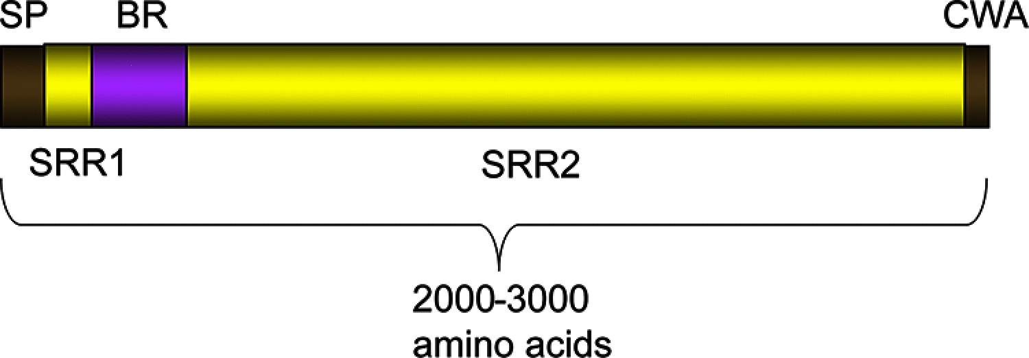
Figure 1.
General organization of SRR adhesin…
Figure 1.
General organization of SRR adhesin proteins. SRR adhesins initiate with a ∼90-amino acid…
Figure 1.
General organization of SRR adhesin proteins. SRR adhesins initiate with a ∼90-amino acid N-terminal signal peptide (SP) that facilitates trafficking to a specialized glycoprotein transporter known as the accessory Sec system. The serine-rich repeat regions (SRR1 and SRR2) are extensively O-glycosylated in the bacterial cytoplasm prior to transport. The ligand-binding region (BR) varies depending upon the organism and contains structural modules that are highly diverse in sequence, fold, and function, which may provide binding specificity for different bacterial strains (6). The C-terminal cell wall anchor (CWA) includes an LPXTG sequence motif that covalently links the cell wall peptidoglycan.
Figure 2.
The sialoglycan-binding motif of Siglec-like…
Figure 2.
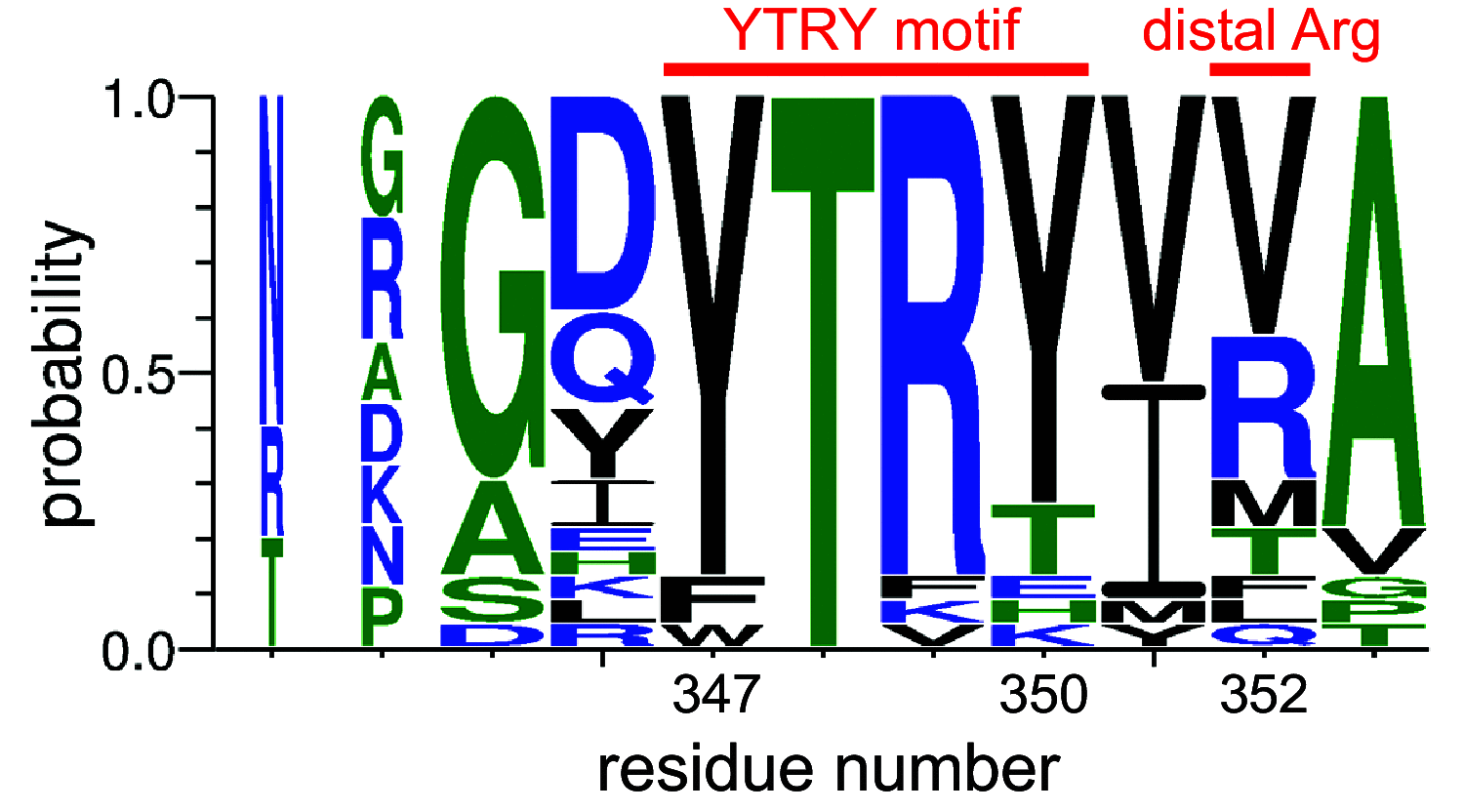
The sialoglycan-binding motif of Siglec-like SRR adhesins. The conservation of residues is indicated…
Figure 2.
The sialoglycan-binding motif of Siglec-like SRR adhesins. The conservation of residues is indicated by letter size with the larger letters representing a more strongly conserved residue. The positions of the YTRY motif (positions 347–350) and the distal Arg (position 352) are notated with a red line above the letters. The numbering reflects the residue positions within SK1Siglec1. The letters colored blue, green, and black indicate charged, nonpolar, and polar residues, respectively. The adhesins included in the alignment are WP_125444382.1 from S. gordonii strain M99, WP_046165954.1 from S. gordonii strain 72-40, WP_080889728.1 from S. gordonii strain G9B, WP_046165954.1 from Streptococcus sp. strain 1236FAA, WP_009659981.1 from Streptococcus sp. strain AS14, WP_002906900.1 from S. sanguinis strain SK115, WP_125439128.1 from S. sanguinis strain SK150, WP_125444035.1 from S. sanguinis strain SK678, WP_081102781.1 from S. gordonii strain Challis, WP_045635027.1 from S. gordonii strain UB10712, WP_080555651.1 from S. sanguinis strain SK1, WP_011836739.1 from S. sanguinis strain SK36, WP_080555852.1 from S. sanguinis strain SK408, WP_000466180.1 from S. sanguinis strain SK140, WP_046165954.1 from S. sanguinis strain PS478, WP_087941957.1 from S. sanguinis strain SK1056, WP_080557024.1 from S. sanguinis strain SK330, WP_080560819.1 from S. sanguinis strain SK355, WP_080555460.1 from S. sanguinis strain SK405, WP_061600538.1 from S. gordonii strain SK49, and WP_000466181.1 from S. oralis strain SF100. Although previously termed the YTRY motif, this binding sequence motif is formally defined as ϕTRX, where ϕ represents Trp, Phe, or Thr, and X represents Tyr, Thr, Glu, His, or Lys.
Figure 3.
Structure of the binding region…
Figure 3.
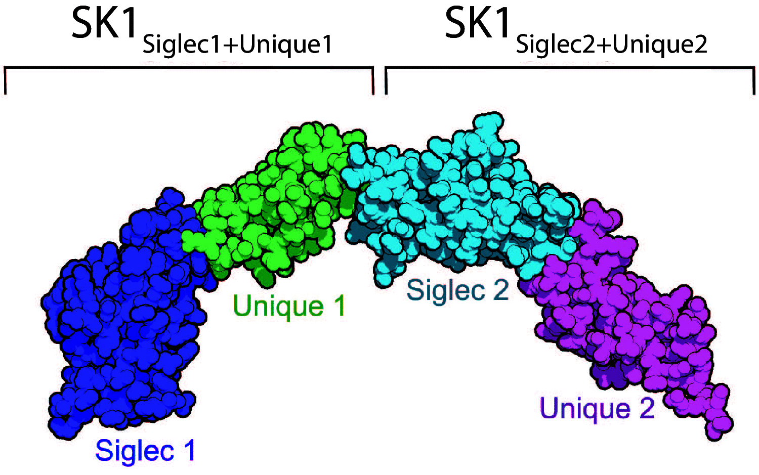
Structure of the binding region of unliganded SK1. SK1 BR has four domains…
Figure 3.
Structure of the binding region of unliganded SK1. SK1BR has four domains in the order SK1Siglec1, SK1Unique1, SK1Siglec2, and SK1Unique2 depicted from left to right and colored by domain. The domain repeats, named SK1Siglec1+Unique1 and SK1Siglec2+Unique2, are homologous but not identical.
Figure 4.
Structural comparison of the tandem…
Figure 4.
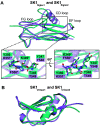
Structural comparison of the tandem Siglec and Unique domains. A , and …
Figure 4.
Structural comparison of the tandem Siglec and Unique domains.A, and B, cartoon diagrams of unliganded SK1BR. SK1Siglec1 and SK1Unique1 are colored in teal, and the SK1Siglec2 and SK1Unique2 are colored in lavender. A, 98 eight atoms were aligned, and 26 were rejected after 5 cycles to output an RMS deviation of 1.058 Å. The noncanonical binding motifs of SK1Siglec1 and SK1Siglec2 are shown as sticks, and residues that deviate from the canonical YTRY motif definition are noted with an asterisk. B, 72 were aligned, and 3 were rejected after 2 cycles to output an RMS deviation of 0.639 Å.
Figure 5.
Ligand electron density. A–B …
Figure 5.
Ligand electron density. A–B , SK1 Siglec1 is shown in teal cartoon. …
Figure 5.
Ligand electron density.A–B, SK1Siglec1 is shown in teal cartoon. C–D, SK1Siglec2 is shown in lavender cartoon. A, C, sTa and B, D, 3′sLn are shown as yellow and green sticks, respectively. Oxygen atoms are colored in red, and nitrogen atoms are in blue. Ligands were manually placed in Coot after refinement of the protein and prior to solvent placement. The ligands were then refined with rigid-body and real space refinement in Coot prior to solvent placement and final structure refinements. |Fo| − |Fc| electron density maps were calculated from coordinates that had been refined in Phenix (40) for three rounds after the removal of the sialoglycans from the model. Maps are contoured at 3σ and are shown in dark gray mesh.
Figure 6.
SK1 interactions with ligands. The…
Figure 6.
SK1 interactions with ligands. The structures of the Siglec domains of A–B ,…
Figure 6.
SK1 interactions with ligands. The structures of the Siglec domains of A–B, SK1Siglec1+Unique1, C–D, SK1Siglec2+Unique2, and E, Hsa (PDB entry 6EFD) (18) are shown in cartoon in teal, lavender, and orange, respectively. The adhesin residues that hydrogen bond with the ligands are shown as sticks. Hydrogen bonds between the adhesins and ligands are shown as dark gray dashed lines. The ligands sTa and 3´sLn are shown as yellow and green sticks, respectively. Oxygen and nitrogen atoms are colored red and blue, respectively.
Figure 7.
Temperature factor analysis of Siglec…
Figure 7.
Temperature factor analysis of Siglec domains and bound ligands. A–D , SK1 …
Figure 7.
Temperature factor analysis of Siglec domains and bound ligands.A–D, SK1Siglec1 is shown in teal cartoon, and SK1Siglec2 is shown in lavender cartoon. The sTa liganded structures are shown in A and C, and the 3´sLn liganded structures are shown in B and D. Both sTa and 3´sLn are shown as sticks and colored by temperature factor, where blue represents a low temperature factor, and red represents a high temperature factor as depicted by the scale in the bottom right corner of each panel. The scale values are in Å2.
Figure 8.
Siglec domain colored by temperature…
Figure 8.
Siglec domain colored by temperature factor. A–B , SK1 unliganded, ( C–D …
Figure 8.
Siglec domain colored by temperature factor.A–B, SK1 unliganded, (C–D) sta-bound, and E–F 3′sLn-bound are shown in cartoon and colored by temperature factor, where blue represents a low temperature factor, and red represents a high temperature factor. The color bars in the bottom right corner of each panel indicate the ranges of B factors in Å2. sTa and 3´sLn are shown as sticks. The oxygen and nitrogen atoms are colored red and blue, respectively.
Figure 9.
Comparison of bacterial sialic acid–binding…
Figure 9.
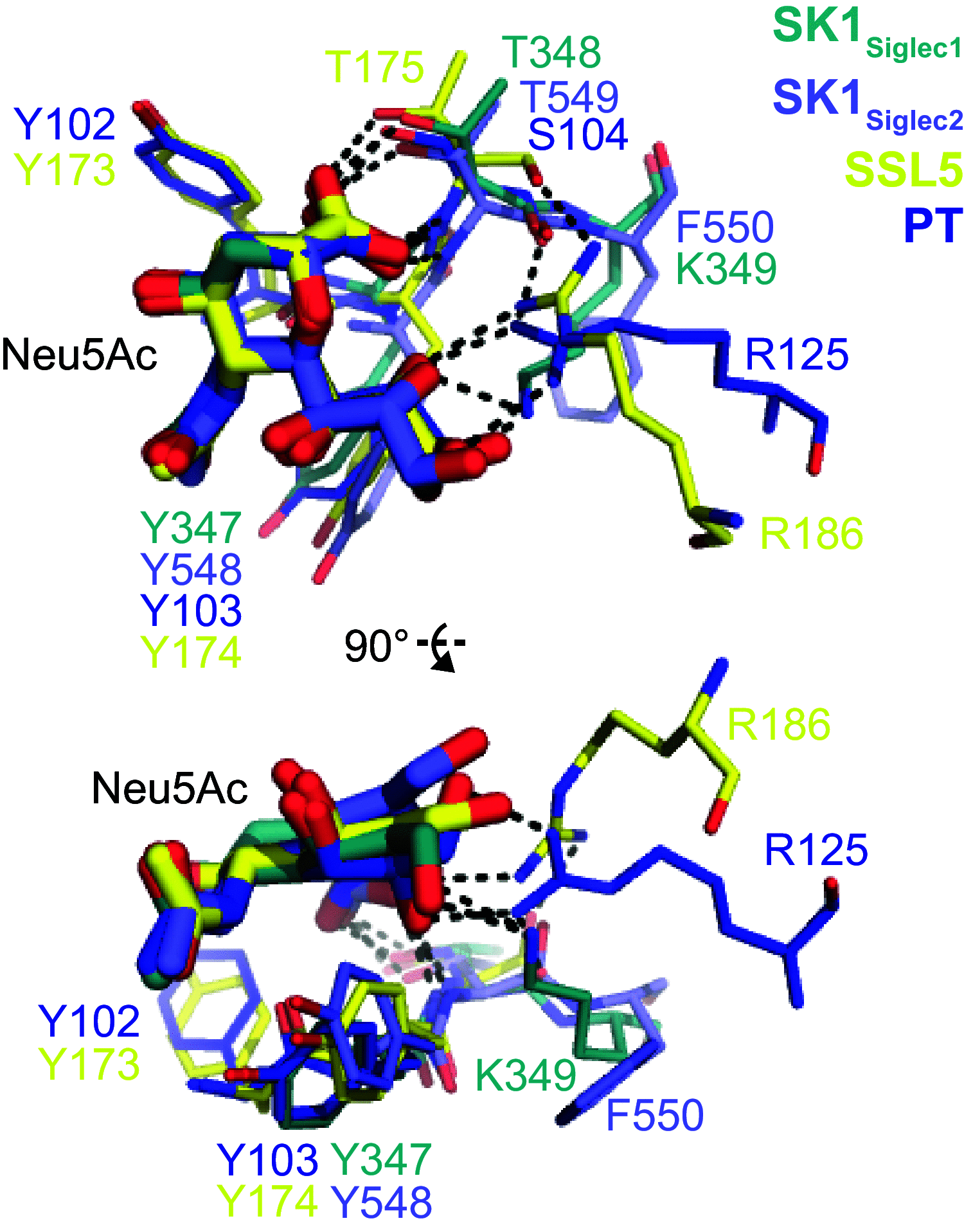
Comparison of bacterial sialic acid–binding pockets. Shown here is an overlay of the…
Figure 9.
Comparison of bacterial sialic acid–binding pockets. Shown here is an overlay of the two Siglec domains of SK1BR with staphylococcal superantigen-like protein 5 (SSL5; PDB entry 2R61) and pertussis toxin (PT; PDB entry 1PTO) (48), two proteins in which the sialic acid recognition motif has previously been identified (22).
Figure 10.
Binding of SK1 BR and…
Figure 10.
Binding of SK1 BR and split variants to glycans and glycoproteins. A …
Figure 10.
Binding of SK1BR and split variants to glycans and glycoproteins.A, biotin-glycan binding to immobilized GST-tagged SK1BR and the split binding modules (n = 4 technical replicates). The asterisk indicates binding that was significantly greater than the level of binding to all other glycans in the set of six (p < 0.05 using a two-way analysis of variance with Tukey's correction for multiple comparisons). B, binding of biotinylayed sTa or 3´sLn to immobilized GST-tagged SK1BR and split constructs (n = 3 technical replicates). C, binding of GST-tagged SK1BR and split constructs to glycoproteins in human plasma (lane 1), platelet lysate (lane 2), or submandibular sublingual saliva (lane 3). D, binding of GST-tagged SK1BR and SK1BR deletion constructs to immobilized human platelets (n = 3 technical replicates). In A, B, and D, mean values ± standard deviation are indicated. In cases where error bars are not evident, the deviations were smaller than the size of the symbol used for the data point. Background values for GST alone were not subtracted but are shown in B and D.
Figure 11.
SRR adhesins binding pocket size…
Figure 11.
SRR adhesins binding pocket size comparison. A–E , the adhesins are shown…
Figure 11.
SRR adhesins binding pocket size comparison.A–E, the adhesins are shown in surface representations (16–18). The top image of the binding site is rotated 80° around the z axis and 70° around the x axis. The binding pocket of each is colored in gray, and the portions of the CD, EF, and FG loops that create the walls of the binding pocket are colored in teal, lavender, cyan, orange, and magenta for SK1Siglec1, SK1Siglec2, SrpA, Hsa, and GspB, respectively. The Arg distal to the YTRY motif is colored blue. Sialyl T antigen is shown as yellow sticks bound to each adhesin. F, multiple sequence alignment of the above adhesins is shown. The YTRY motif is outlined in red, and the distal Arg is outlined in blue.
Figure 12.
Model of target-specific effects of…
Figure 12.
Model of target-specific effects of SK1. A , the distance between the…
Figure 12.
Model of target-specific effects of SK1.A, the distance between the two binding sites is 67 Å, measured from the Cα atoms of Tyr-347, the first residue in the YTRY motif of SK1Siglec1, and Arg-553, the distal arginine residue of SK1Siglec2. The length of the binging site in SK1Siglec1 is 14 Å, measured from the Cα atoms of Gly-344 and Lys-349. The length of the binding site in SK1Siglec2 is 24 Å, measured from the Cα atoms of Gly-545 to Arg-553. B–E, note that the glycans shown here are only meant to serve as a hypothetical glycan structure and are not meant to represent a specific, defined glycan target. B, multivalent binding of a branched glycan. Given the distance between the two binding sites, SK1BR could bind the same branched glycan with both SK1Siglec1 and SK1Siglec2. C, multivalent binding of a patch of clustered glycans. Each binding site of SK1BR binds a separate glycan structure. D, SK1Siglec1 dominant binding. E, SK1Siglec2 dominant binding. Given the openness of the binding site and the presence of the distal Arg, SK1Siglec2 may bind a short saccharide, two short saccharides, or a longer hexasaccharide.
Ramboarina S., Garnett J. A., Zhou M., Li Y., Peng Z., Taylor J. D., Lee W. C., Bodey A., Murray J. W., Alguel Y., Bergeron J., Bardiaux B., Sawyer E., Isaacson R., Tagliaferri C., et al. (2010) Structural insights into serine-rich fimbriae from Gram-positive bacteria. J. Biol. Chem. 285, 32446–32457 10.1074/jbc.M110.128165
-
DOI
-
PMC
-
PubMed
Deng L., Bensing B. A., Thamadilok S., Yu H., Lau K., Chen X., Ruhl S., Sullam P. M., and Varki A. (2014) Oral streptococci utilize a Siglec-like domain of serine-rich repeat adhesins to preferentially target platelet sialoglycans in human blood. PLoS Pathog. 10, e1004540 10.1371/journal.ppat.1004540
-
DOI
-
PMC
-
PubMed
Zheng W., Tan M. F., Old L. A., Paterson I. C., Jakubovics N. S., and Choo S. W. (2017) Distinct biological potential of Streptococcus gordonii and Streptococcus sanguinis revealed by comparative genome analysis. Sci. Rep. 7, 2949 10.1038/s41598-017-02399-4
-
DOI
-
PMC
-
PubMed
Ronis A., Brockman K., Singh A. K., Gaytán M. O., Wong A., McGrath S., Owen C. D., Magrini V., Wilson R. K., van der Linden M., and King S. J. (2019) Streptococcus oralis subsp. dentisani produces monolateral serine-rich repeat protein fibrils, one of which contributes to saliva binding via sialic acid. Infect. Immun. 87, e00406–19 10.1128/IAI.00406-19
-
DOI
-
PMC
-
PubMed
Denapaite D., Brückner R., Nuhn M., Reichmann P., Henrich B., Maurer P., Schähle Y., Selbmann P., Zimmermann W., Wambutt R., and Hakenbeck R. (2010) The genome of Streptococcus mitis B6: what is a commensal? PLoS One 5, e9426 10.1371/journal.pone.0009426
-
DOI
-
PMC
-
PubMed