. 2020 Sep 11;369(6509):1359-1365.
doi: 10.1126/science.abb5317. Epub 2020 Aug 20.

Structural basis of transcription-translation coupling

Affiliations

Structural basis of transcription-translation coupling

Chengyuan Wang et al. Science. .

Abstract

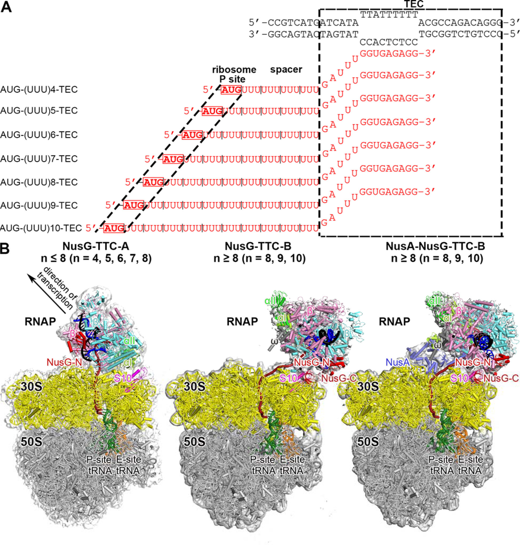
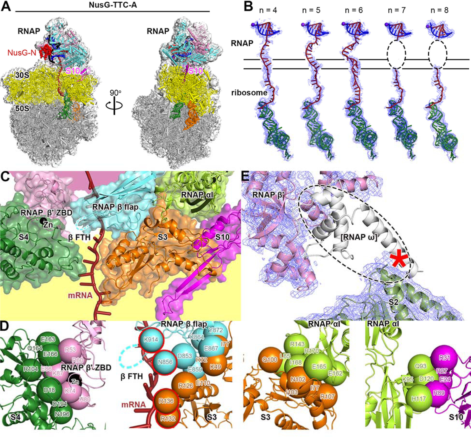
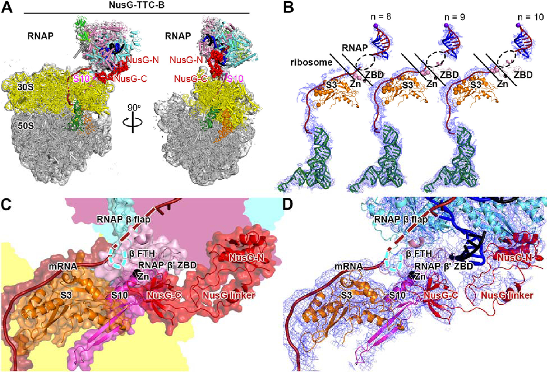
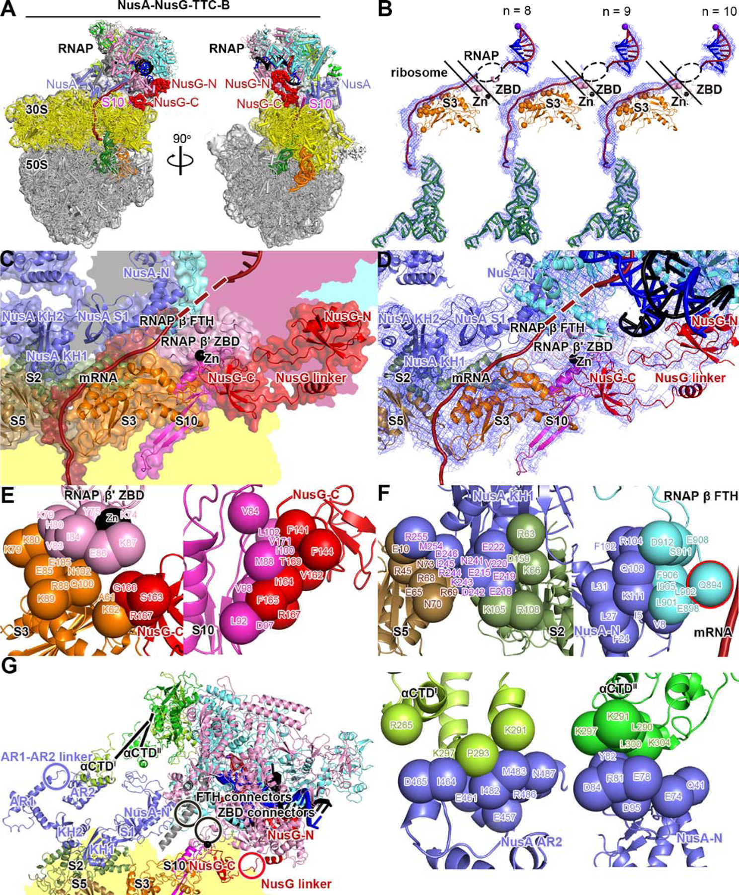
In bacteria, transcription and translation are coupled processes in which the movement of RNA polymerase (RNAP)-synthesizing messenger RNA (mRNA) is coordinated with the movement of the first ribosome-translating mRNA. Coupling is modulated by the transcription factors NusG (which is thought to bridge RNAP and the ribosome) and NusA. Here, we report cryo-electron microscopy structures of Escherichia coli transcription-translation complexes (TTCs) containing different-length mRNA spacers between RNAP and the ribosome active-center P site. Structures of TTCs containing short spacers show a state incompatible with NusG bridging and NusA binding (TTC-A, previously termed "expressome"). Structures of TTCs containing longer spacers reveal a new state compatible with NusG bridging and NusA binding (TTC-B) and reveal how NusG bridges and NusA binds. We propose that TTC-B mediates NusG- and NusA-dependent transcription-translation coupling.


Figures

Fig. 1.
Fig. 1.. Structure determination: transcription-translation complexes (TTCs)
(A) Nucleic-acid scaffolds. Each scaffold comprises nontemplate- and template-strand oligodeoxyribonucleotides (black) and one of seven oligoribonucleotides having spacer length n of 4, 5, 6, 7, 8, 9, or 10 codons (red), corresponding to mRNA. Dashed black box labeled “TEC,” portion of nucleic-acid scaffold that forms TEC upon addition of RNAP (10 nt nontemplate- and template-strand ssDNA segments forming “transcription bubble,” 10 nt of mRNA engaged with template-strand DNA as RNA-DNA “hybrid,” and 5 nt of mRNA, on diagonal, in RNAP RNA-exit channel); dashed black lines labeled “ribosome P-site,” mRNA AUG codon intended to occupy ribosome active-center P site upon addition of ribosome and tRNAfMet; “spacer,” mRNA spacer between TEC and AUG codon in ribosome active-center P site. (B) Cryo-EM structures of NusG-TTC-A (obtained with spacer lengths of 4–8 codons), NusG-TTC-B (obtained with spacer lengths of 8–10 codons), and NusA-NusG-TTC-B (obtained with spacer lengths of 8–10 codons). Structures shown are NusG-TTC-A (3.7 Å; n = 4; Table S1), NusG-TTC-B (4.7 Å; n = 9; Table S1), and NusA-NusG-TTC-B2 (3.5 Å; n = 8; Table S1). Images show EM density (gray surface) and fit (ribbons) for TEC, NusG and NusA (at top; direction of transcription, defined by downstream dsDNA, indicated by arrow in left panel and directly toward viewer in center and right panels) and for ribosome 30S and 50S subunits and P- and E-site tRNAs (at bottom). RNAP β’, β, αI, αII, and ω subunits are in pink, cyan, light green, and dark green, and gray; 30S subunit, 50S subunit, P-site tRNA, E-site tRNA are in yellow, gray, green, and orange; DNA nontemplate strand, DNA template strand, and mRNA are in black, blue, and brick-red (brick-red dashed line where modelled). NusG, NusA, and ribosomal protein S10 are in red, light blue, and magenta. Ribosome L7/L12 stalk omitted for clarity in this and all subsequent images.
Fig. 2.
Fig. 2.. Cryo-EM structure of NusG-TTC-A
(A) Structure of NusG-TTC-A (3.7 Å; n = 4; Table S1). Two orthogonal views. Colors as in Fig. 1B. (B) Accommodation of mRNA spacer lengths of 4, 5, 6, 7 and 8 codons in NusG-TTC-A. EM density, blue mesh; mRNA, brick-red (disordered mRNA nucleotides indicated by dashed oval); template-strand DNA in RNA-DNA hybrid, blue; RNAP active-center catalytic Mg2+, purple sphere; tRNA in ribosome P site, green. Upper and lower black horizontal lines indicate edges of RNAP and ribosome. (C) RNAP-ribosome interface in NusG-TTC-A (n = 4; identical interface for n = 5, 6, 7, or 8), showing RNAP β’ zinc binding domain, (ZBD, pink; Zn2+ ion as black sphere), RNAP β flap, cyan, RNAP β flap tip helix (β FTH; disordered residues indicated by cyan dashed line), and RNAP αI (green) interacting with ribosomal proteins S4 (forest green), S3 (orange), and S10 (magenta) and with mRNA (brick red). Portions of RNAP β’ and ribosome 30S not involved in interactions are shaded pink and yellow, respectively. (D) RNAP-ribosome interactions involving RNAP β’ ZBD and S4 (subpanel 1), RNAP β flap and S3 (subpanel 2; β FTH, dashed line; β and S3 residues that interact with mRNA, cyan and orange spheres with red outlines; mRNA, brick-red), RNAP αI and S3 (subpanel 3), and RNAP αI and S10 (subpanel 4). Other colors as in (C). (E) Absence of EM density for RNAP ω subunit. EM density, blue mesh; atomic models for RNAP β’ and S2, pink ribbon and forest-green ribbon, respectively; location of missing EM density for ω, dashed oval; ω in TEC in absence of ribosome (PDB 6P19; 17), white ribbon.
Fig. 3.
Fig. 3.. Cryo-EM structure of NusG-TTC-B
(A) Structure of NusG-TTC-B (4.7 Å; n = 9; Table S1). Views and colors as in Fig. 2A. (B) Accommodation of mRNA spacer lengths of 8, 9, and 10 codons in NusG-TTC-B. EM density, blue mesh; mRNA, brick-red (disordered mRNA nucleotides indicated by dashed oval); template-strand DNA in RNA-DNA hybrid, blue; RNAP active-center catalytic Mg2+, purple sphere; tRNA in ribosome P site, green; ribosomal protein S3, orange (positively charged residues positioned to contact mRNA as orange spheres); RNAP β’ zinc binding domain (ZBD, pink; Zn2+ ion as black sphere; positively charged residues positioned to contact mRNA as pink spheres). Upper and lower black diagonal lines indicate edges of RNAP and ribosome. (C) RNAP-ribosome interface and NusG bridging in NusG-TTC-B (n = 9; identical interface for n = 8, 9, or 10). RNAP β’ zinc binding domain, (ZBD, pink; Zn2+ ion as black sphere) interacts with ribosomal protein S3 (orange) and mRNA (brick red). NusG (red) bridges RNAP and ribosome, with NusG-N interacting with RNAP and NusG-C interacting with ribosomal protein S10 (magenta). Portions of RNAP β’, β, and ribosome 30S not involved in interactions are shaded pink, cyan, and yellow, respectively. (D) As C, showing cryo-EM density as blue mesh.
Fig. 4.
Fig. 4.. Cryo-EM structure of NusA-NusG-TTC-B
(A) Structure of NusA-NusG-TTC-B (NusA-NusG-TTC-B2; 3.5 Å; n = 9; Table S1). NusA, light blue. Views and other colors as in Figs. 2A and 3A. (B) Accommodation of mRNA spacer lengths of 8, 9, and 10 codons in NusA-NusG-TTC-B. Views and colors as in Fig 3B. (C) RNAP-ribosome interface, NusG bridging, and NusA binding in NusA-NusG-TTC-B (n = 9; identical interface for n = 8, 9, or 10). RNAP β’ zinc binding domain, (ZBD, pink; Zn2+ ion as black sphere) interacts with ribosomal protein S3 (orange) and mRNA (brick red). NusG (red) bridges RNAP and ribosome, with NusG-N interacting with RNAP and NusG-C interacting with ribosomal protein S10 (magenta). NusA (light blue) KH1 domain interacts with ribosomal proteins S5 and S2 (brown and forest green). Portions of RNAP β’, β, ω, and ribosome 30S not involved in interactions are shaded pink, cyan, gray, and yellow, respectively. (D) As C, showing cryo-EM density as blue mesh. (E) RNAP-ribosome interactions involving RNAP β’ ZBD and S3 (subpanel 1) and NusG-ribosome interactions involving NusG-C and S10 (subpanel 2). (F) NusA-ribosome interactions involving NusA KH1 and S5 and S2 (subpanel 1) and NusA-RNAP interactions involving NusA-N and RNAP β FTH (subpanel 2; β FTH residue that interacts with mRNA, cyan sphere with red outline; mRNA, brick-red), NusA AR2 and RNAP αCTDI (subpanel 3), and NusA-N and RNAP αCTDII (subpanel 4). (G) Points of flexibility in NusA-NusG-TTC-B (NusA “coupling pantograph”): flexible linkage in NusA structure (AR1-AR2 linker; light blue circle), three flexible linkages between NusA and RNAP (αCTDI linker, αCTDII linker, and β FTH connectors; black lines and black circle), flexible linkage between RNAP and ribosome (β’ ZBD connectors; black circle), and flexible NusG bridging of RNAP and ribosome (NusG linker; red circle).

Comment in

Similar articles

Cited by

References

    1. Miller O, Hamkalo B, Thomas C, Visualization of bacterial genes in action, Science 169, 392–395 (1970). - PubMed
    1. Burmann B, Schweimer K, Luo X, Wahl M, Stitt B, Gottesman M, Rösch P, A NusE-NusG complex links transcription and translation, Science 328, 501–504 (2010). - PubMed
    1. Proshkin S, Rahmouni A, Mironov A, Nudler E, Cooperation between translating ribosomes and RNA polymerase in transcription elongation., Science 328, 504–508 (2010). - PMC - PubMed
    1. Castro-Roa D, Zenkin N, In vitro experimental system for analysis of transcription-translation coupling, Nucl. Acids Res 40, e45 (2012). - PMC - PubMed
    1. McGary K, Nudler E, RNA polymerase and the ribosome: the close relationship, Curr. Opin. Microbiol 16, 112–117 (2013). - PMC - PubMed

Publication types

MeSH terms