. 2020 Sep 25;369(6511):1633-1637.
doi: 10.1126/science.abb9818. Epub 2020 Aug 20.

Succination inactivates gasdermin D and blocks pyroptosis

Affiliations

Succination inactivates gasdermin D and blocks pyroptosis

Fiachra Humphries et al. Science. .

Abstract

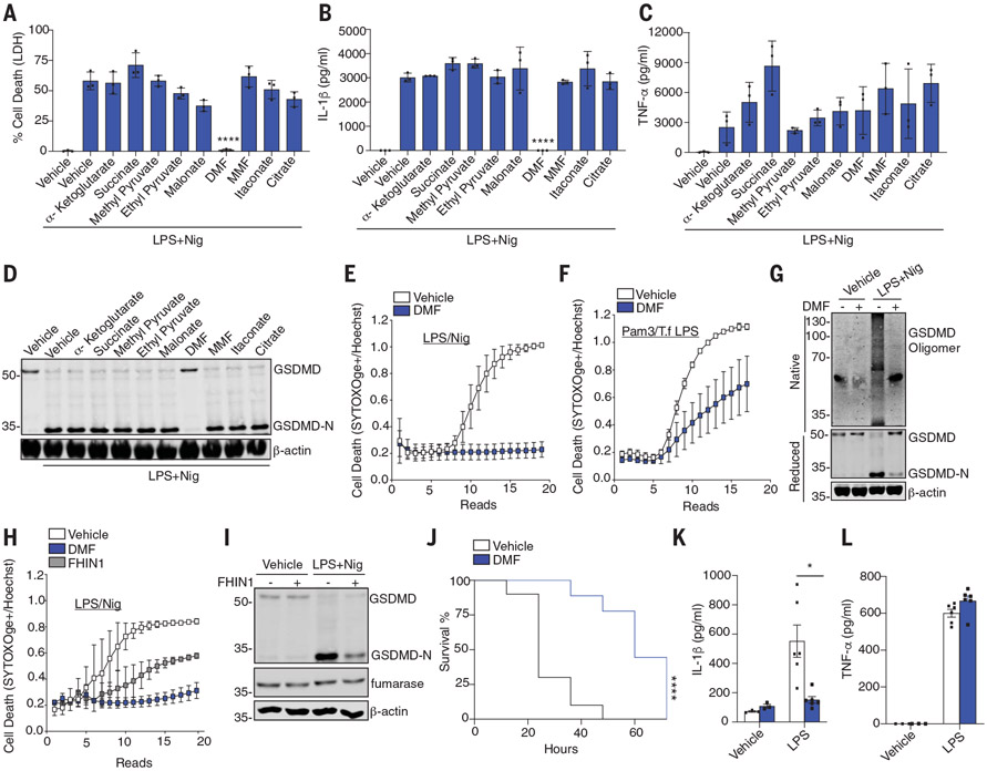
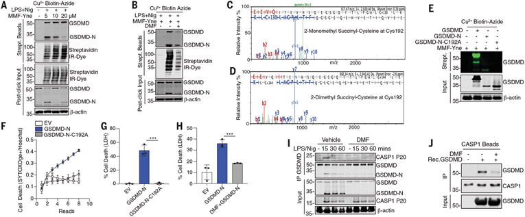
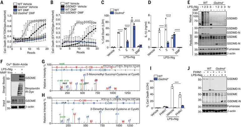
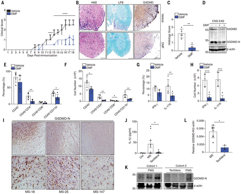
Activated macrophages undergo a metabolic switch to aerobic glycolysis, accumulating Krebs' cycle intermediates that alter transcription of immune response genes. We extended these observations by defining fumarate as an inhibitor of pyroptotic cell death. We found that dimethyl fumarate (DMF) delivered to cells or endogenous fumarate reacts with gasdermin D (GSDMD) at critical cysteine residues to form S-(2-succinyl)-cysteine. GSDMD succination prevents its interaction with caspases, limiting its processing, oligomerization, and capacity to induce cell death. In mice, the administration of DMF protects against lipopolysaccharide shock and alleviates familial Mediterranean fever and experimental autoimmune encephalitis by targeting GSDMD. Collectively, these findings identify GSDMD as a target of fumarate and reveal a mechanism of action for fumarate-based therapeutics that include DMF, for the treatment of multiple sclerosis.


Figures

Fig. 1.
Fig. 1.. DMF inhibits pyroptosis.
(A to F) BMDM treated as indicated and LDH, IL-1β, or TNF-α measured with [(A) to (C)] enzyme-linked immunosorbent assay (ELISA), (D) GSDMD/β-actin by immunoblotting, or [(E) and (F)] permeability of cells to SYTOX Orange. (G) GSDMD oligomerization in native and reduced cell lysates treated as above. (H and I) Time course of permeabilization to SYTOX Orange or GSDMD-N formation in cells treated as indicated. (J) Survival rates of WT mice after 50 mg/kg of LPS (n = 10 mice) or LPS+ DMF (n = 9 mice). (K and L) Serum IL-1β or TNF-α levels of WT mice 5 hours after intraperitoneal injection with 5 mg/kg LPS (n = 6 mice) or phosphate-buffered saline (PBS) (n = 3 mice). (A) to (C) are pooled from three independent experiments. (D) to (I) are representative from three independent experiments. In (J) to (L), data points indicate individual mice. *P < 0.05; **P < 0.01; ***P < 0.001; ****P < 0.0001) [(J), Mantel–Cox survival analysis; (A) to (C) and (K), two-way analysis of variance (ANOVA)]. Error bars indicate means ± SEM.
Fig. 2.
Fig. 2.. Succination of GSDMD inhibits pyroptosis.
(A and B) Immunoblot of GSDMD in streptavidin pulldown from clicked lysates treated as indicated. (C and D) Representative mass spectrometry spectra of 2-monomethyl and 2-dimethylsuccination of GSDMD immunoprecipitated from DMF-treated BMDMs. (E) Immunoblot of GSDMD in clicked lysates from transfected HEK293T cells. (F to H) Cell death of HEK293T transfected as indicated. (I) Immunoblots of CASP1p20 and GSDMD in BMDMs. (J) Immunoblots of an in vitro binding assay of succinated and nonsuccinated GSDMD incubated with caspase 1 beads. [(A), (B), (F), (J), and (K)] Representative images from three independent experiments. [(C) to (E)] Representative mass spectrometry spectra from two independent experiments. [(H) and (I)] Pooled data from three independent experiments. ***P < 0.001 (one-way ANOVA). Error bars indicate means ± SEM.
Fig. 3.
Fig. 3.. DMF targets GSDMD and GSDME.
(A and B) Kinetic cell death of (A) WT and Gsdmd−/− BMDMs or (B) WT and GSDMD−/− THP1 cells treated as indicated. (C and D) LDH and IL-1β release from WT and Gsdmd−/− BMDMs. (E) Immunoblot of GSDMD and GSDME in native and reduced cell lysates from BMDMs. (F) Immunoblot of GSDME in streptavidin pulldown from clicked lysates. (G and H) Representative mass spectrometry spectra of succinated GSDME. (I) Immunoblot analysis of GSDMD and GSDME from WT and Gsdmd−/− BMDMs. [(A) and (B)] Representative of three independent experiments. [(C), (D), and (I)] Pooled data from three independent experiments. [(E), (F), and (J)] Representative images from three independent experiments. ***P < 0.0001; ****P < 0.00001 (two-way ANOVA). Error bars indicate means ± SEM.
Fig. 4.
Fig. 4.. Succination of GSDMD alleviates EAE and MS.
(A) Clinical scores of WT mice administered vehicle or DMF daily after EAE induction, respectively (n = 10 mice). (B) Representative hematoxylin and eosin (H&E), Luxol fast blue (LFB), and anti-GSDMD staining and (C) pathology evaluation of spinal cord sections from mice, showing inflammatory cell infiltration and demyelination, respectively. Scale bar, 200 μm. (D) Immunoblot of GSDMD in spinal cord tissue. (E and F) Flow cytometry analysis of CD45+ leukocytes, CD45+CD4+ T cells, CD45+CD8+ T cells, and CD45+CD11b+ monocytes that infiltrated the spinal cords and brains of the mice in (A) (n = 5 mice). (G and H) TH1 (IFN-γ+) and TH17 (IL-17A+) cells from CD4+ T cells (n = 5 mice). (I) Immunohistochemistry staining of GSDMD-N in post mortem lesions from MS patients. Scale bars, 100 μm (top), 25 μm (bottom). (J) IL-1β levels in serum from healthy controls (n = 6 donors), MS patients (MS; n = 9 donors), and MS patients receiving Tecfidera delayed release capsules (n = 9 donors). (K) Immunoblot of GSDMD-N and (L) densitometry analysis of GSDMD-N in PBMCs from [(J) and (K)] MS (n = 8 donors) or PMS Tecfidera (n = 3 donors). [(A), (C), and (E) to (H)] Pooled data from two independent EAE experiments. *P < 0.05; **P < 0.01; ***P < 0.001 [(A) and (C), Mann–Whitney U test; (E) to (H), multiple t test]. (A) Box and whisker plot. [(C), (E) to (H), (J), and (L)] Error bars indicate means ± SEM.

Comment in

References

    1. Shi J et al., Nature 526, 660–665 (2015). - PubMed
    1. Kayagaki N et al., Nature 526, 666–671 (2015). - PubMed
    1. Mills EL et al., Nature 556,113–117 (2018). - PMC - PubMed
    1. Liao S-T et al., Nat. Commun 10, 5091 (2019). - PMC - PubMed
    1. Lampropoulou V et al. Cell Metab. 24, 158–166 (2016). - PMC - PubMed

Publication types

MeSH terms