

Involvement of p53, p21, and Caspase-3 in Apoptosis of Coronary Artery Smooth Muscle Cells in a Kawasaki Vasculitis Mouse Model
- PMID: 32820144
- PMCID: PMC7456161
- DOI: 10.12659/MSM.922429
Involvement of p53, p21, and Caspase-3 in Apoptosis of Coronary Artery Smooth Muscle Cells in a Kawasaki Vasculitis Mouse Model
Abstract
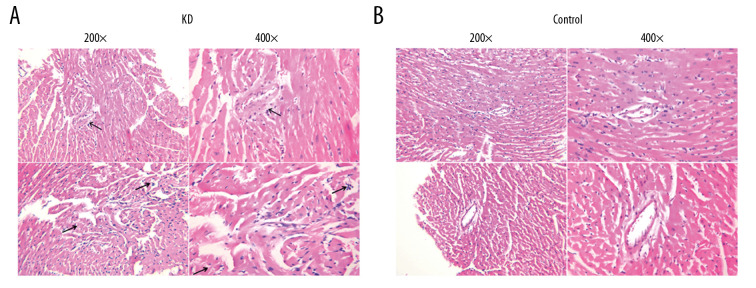
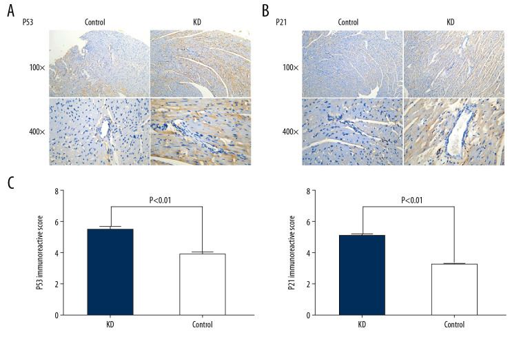
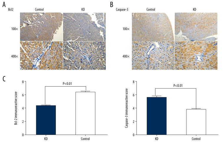
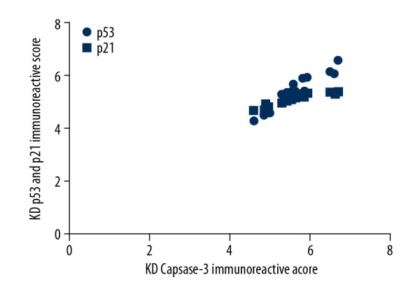
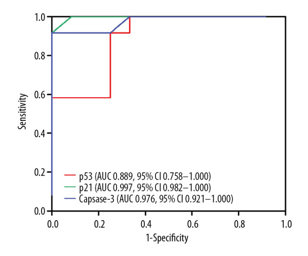
BACKGROUND Overexpression of p53, p21, and caspase-3 promotes apoptosis of vascular smooth muscle cells. However, the mechanisms that lead to apoptosis of coronary artery smooth muscle cells (CASMCs) is unclear in Kawasaki disease (KD). This study investigated involvement of p53, p21, and caspase-3 in the apoptosis of CASMCs from a Kawasaki vasculitis mouse model. MATERIAL AND METHODS The Kawasaki vasculitis mouse model with coronary artery lesions was generated via administration of Lactobacillus casei cell wall extract. In 2 groups of mice (healthy control and KD vasculitis mice), the levels of p53, p21, and caspase-3 protein in the root of the coronary artery were evaluated via immunohistochemistry. Receiver operating characteristic curves were plotted for determination of area under the curve, 95% confidence interval, sensitivity, specificity, and cutoff values for the ability of p53, p21, and caspase-3 expression to predict CASMC apoptosis and coronary artery lesion formation in KD vasculitis mice. RESULTS Compared with healthy mice, KD vasculitis mice had a significantly higher apoptosis index and upregulated p53, p21, and caspase-3 expression. Also, the immunoreactive score for caspase-3 was positively correlated with the immunoreactivity scores for p53 and p21. The optimal cutoff values for p53, p21, and caspase-3 expression for predicting the presence of coronary artery lesions were 4.15, 4.18, and 4.22, respectively. CONCLUSIONS Upregulated levels of p53, p21, and caspase-3 promoted apoptosis of CASMCs in KD vasculitis mice. Thus, the levels of p53, p21, and caspase-3 may serve as valuable predictors of coronary artery lesion formation in KD.
Conflict of interest statement
None.
Figures
Similar articles
-
Reduced Platelet miR-223 Induction in Kawasaki Disease Leads to Severe Coronary Artery Pathology Through a miR-223/PDGFRβ Vascular Smooth Muscle Cell Axis.Circ Res. 2020 Sep 11;127(7):855-873. doi: 10.1161/CIRCRESAHA.120.316951. Epub 2020 Jun 29. Circ Res. 2020. PMID: 32597702 Free PMC article.
-
[Role and mechanisms of CHI3L1 in coronary artery lesions in a mouse model of Kawasaki disease-like vasculitis].Zhongguo Dang Dai Er Ke Za Zhi. 2023 Dec 15;25(12):1227-1233. doi: 10.7499/j.issn.1008-8830.2309080. Zhongguo Dang Dai Er Ke Za Zhi. 2023. PMID: 38112139 Free PMC article. Chinese.
-
IL-1 Signaling Is Critically Required in Stromal Cells in Kawasaki Disease Vasculitis Mouse Model: Role of Both IL-1α and IL-1β.Arterioscler Thromb Vasc Biol. 2015 Dec;35(12):2605-16. doi: 10.1161/ATVBAHA.115.306475. Epub 2015 Oct 29. Arterioscler Thromb Vasc Biol. 2015. PMID: 26515418 Free PMC article.
-
Animal models of vasculitis. Lessons we can learn to improve our understanding of Kawasaki disease.Rheum Dis Clin North Am. 1988 Aug;14(2):479-87. Rheum Dis Clin North Am. 1988. PMID: 3140306 Review.
-
An update on understanding the pathophysiology in Kawasaki disease: Possible role of immune complexes in coronary artery lesion revisited.Int J Rheum Dis. 2023 Aug;26(8):1453-1463. doi: 10.1111/1756-185X.14816. Epub 2023 Jul 11. Int J Rheum Dis. 2023. PMID: 37431726 Review.
Cited by
-
Serum TP53 Protein Level as a Sensitive Biomarker for the Diagnosis of Myocardial Damage in Children.Med Sci Monit. 2022 Jun 25;28:e936248. doi: 10.12659/MSM.936248. Med Sci Monit. 2022. PMID: 35751366 Free PMC article.
-
The role of p21 in cellular senescence and aging-related diseases.Mol Cells. 2024 Nov;47(11):100113. doi: 10.1016/j.mocell.2024.100113. Epub 2024 Sep 19. Mol Cells. 2024. PMID: 39304134 Free PMC article. Review.
-
Mechanisms of cordycepin in the treatment of pulmonary arterial hypertension in rats based on metabonomics and transcriptomics.Sci Rep. 2024 May 30;14(1):12431. doi: 10.1038/s41598-024-62163-3. Sci Rep. 2024. PMID: 38816406 Free PMC article.
References
-
- Yorifuji T, Tsukahara H, Doi H. Early childhood exposure to maternal smoking and Kawasaki disease: A longitudinal survey in Japan. Sci Total Environ. 2019;655:141–46. - PubMed
-
- Denby KJ, Clark DE, Markham LW. Management of Kawasaki disease in adults. Heart. 2017;103:1760–69. - PubMed
-
- Pinna GS, Kafetzis DA, Tselkas OI, et al. Kawasaki disease: An overview. Curr Opin Infect Dis. 2008;21:263–70. - PubMed
-
- Nakamura Y, Yashiro M, Uehara R, et al. Increasing incidence of Kawasaki disease in Japan: Nationwide survey. Pediatr Int. 2008;50:287–90. - PubMed
MeSH terms
Substances
LinkOut - more resources
Full Text Sources
Medical
Research Materials
Miscellaneous
