

Generation of interconnected vesicles in a liposomal cell model
- PMID: 32820180
- PMCID: PMC7441142
- DOI: 10.1038/s41598-020-70562-5
Generation of interconnected vesicles in a liposomal cell model
Abstract
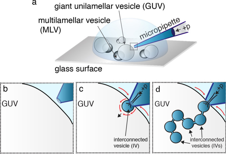
We introduce an experimental method based upon a glass micropipette microinjection technique for generating a multitude of interconnected vesicles (IVs) in the interior of a single giant unilamellar phospholipid vesicle (GUV) serving as a cell model system. The GUV membrane, consisting of a mixture of soybean polar lipid extract and anionic phosphatidylserine, is adhered to a multilamellar lipid vesicle that functions as a lipid reservoir. Continuous IV formation was achieved by bringing a micropipette in direct contact with the outer GUV surface and subjecting it to a localized stream of a Ca2+ solution from the micropipette tip. IVs are rapidly and sequentially generated and inserted into the GUV interior and encapsulate portions of the micropipette fluid content. The IVs remain connected to the GUV membrane and are interlinked by short lipid nanotubes and resemble beads on a string. The vesicle chain-growth from the GUV membrane is maintained for as long as there is the supply of membrane material and Ca2+ solution, and the size of the individual IVs is controlled by the diameter of the micropipette tip. We also demonstrate that the IVs can be co-loaded with high concentrations of neurotransmitter and protein molecules and displaying a steep calcium ion concentration gradient across the membrane. These characteristics are analogous to native secretory vesicles and could, therefore, serve as a model system for studying secretory mechanisms in biological systems.
Conflict of interest statement
The authors declare no competing interests.
Figures


Similar articles
-
DNA-induced endocytosis upon local microinjection to giant unilamellar cationic vesicles.Eur Biophys J. 1999;28(2):142-50. doi: 10.1007/s002490050193. Eur Biophys J. 1999. PMID: 10028239
-
Pulling Membrane Nanotubes from Giant Unilamellar Vesicles.J Vis Exp. 2017 Dec 7;(130):56086. doi: 10.3791/56086. J Vis Exp. 2017. PMID: 29286431 Free PMC article.
-
Artificial exocytotic system that secretes intravesicular contents upon Ca2+ influx.Langmuir. 2010 Sep 21;26(18):14788-92. doi: 10.1021/la102737e. Langmuir. 2010. PMID: 20722459
-
Protein Reconstitution Inside Giant Unilamellar Vesicles.Annu Rev Biophys. 2021 May 6;50:525-548. doi: 10.1146/annurev-biophys-100620-114132. Epub 2021 Mar 5. Annu Rev Biophys. 2021. PMID: 33667121 Review.
-
Elementary Processes and Mechanisms of Interactions of Antimicrobial Peptides with Membranes-Single Giant Unilamellar Vesicle Studies.Adv Exp Med Biol. 2019;1117:17-32. doi: 10.1007/978-981-13-3588-4_3. Adv Exp Med Biol. 2019. PMID: 30980351 Review.
Cited by
-
Calcium-induced compaction and clustering of vesicles tracked with molecular resolution.Biophys J. 2023 Jul 11;122(13):2646-2654. doi: 10.1016/j.bpj.2023.05.019. Epub 2023 May 22. Biophys J. 2023. PMID: 37218132 Free PMC article.
-
Identifying and Manipulating Giant Vesicles: Review of Recent Approaches.Micromachines (Basel). 2022 Apr 19;13(5):644. doi: 10.3390/mi13050644. Micromachines (Basel). 2022. PMID: 35630111 Free PMC article. Review.
References
-
- Walde P, Cosentino K, Engel H, Stano P. Giant vesicles: preparations and applications. ChemBioChem. 2010;11:848–865. - PubMed
-
- Bahrami A, et al. Wrapping of nanoparticles by membranes. Adv. Colloid Interface Sci. 2014;208:214–224. - PubMed
-
- Zimmerberg J, McLaughlin S. Membrane curvature: How BAR domains bend bilayers. Curr. Biol. 2004;14:R250–R252. - PubMed
Publication types
MeSH terms
Substances
LinkOut - more resources
Full Text Sources
Miscellaneous
