. 2020 Dec;57(12):4989-4999.
doi: 10.1007/s12035-020-02078-y. Epub 2020 Aug 20.

Thallium-201 Imaging in Intact Olfactory Sensory Neurons with Reduced Pre-Synaptic Inhibition In Vivo

Affiliations

Thallium-201 Imaging in Intact Olfactory Sensory Neurons with Reduced Pre-Synaptic Inhibition In Vivo

Hideaki Shiga et al. Mol Neurobiol. 2020 Dec.

Abstract

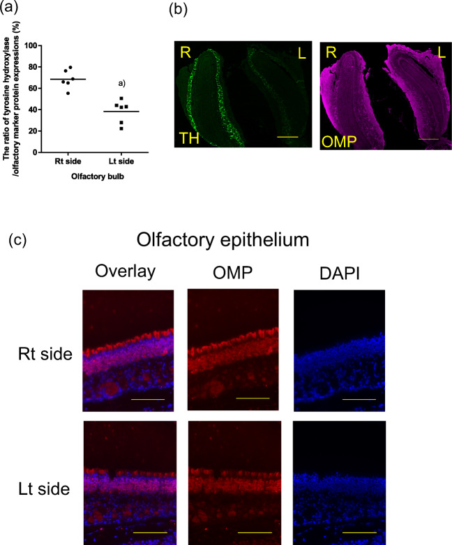
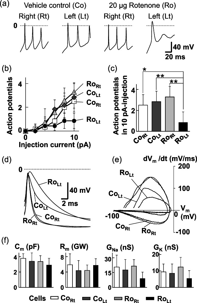
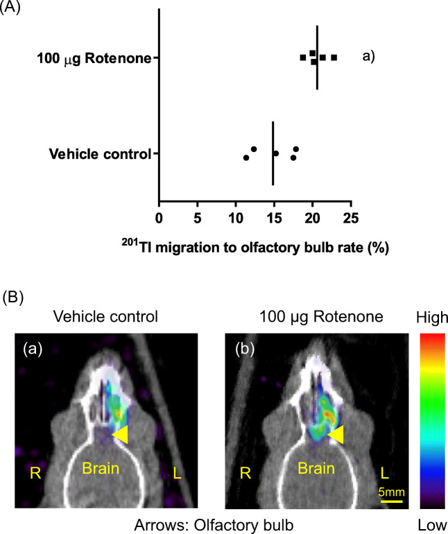
In this study, we determined whether the 201Tl (thallium-201)-based olfactory imaging is affected if olfactory sensory neurons received reduced pre-synaptic inhibition signals from dopaminergic interneurons in the olfactory bulb in vivo. The thallium-201 migration rate to the olfactory bulb and the number of action potentials of olfactory sensory neurons were assessed 3 h following left side nasal administration of rotenone, a mitochondrial respiratory chain complex I inhibitor that decreases the number of dopaminergic interneurons without damaging the olfactory sensory neurons in the olfactory bulb, in mice (6-7 animals per group). The migration rate of thallium-201 to the olfactory bulb was significantly increased following intranasal administration of thallium-201 and rotenone (10 μg rotenone, p = 0.0012; 20 μg rotenone, p = 0.0012), compared with that in control mice. The number of action potentials was significantly reduced in the olfactory sensory neurons in the rotenone treated side of 20 μg rotenone-treated mice, compared with that in control mice (p = 0.0029). The migration rate of thallium-201 to the olfactory bulb assessed with SPECT-CT was significantly increased in rats 24 h after the left intranasal administration of thallium-201 and 100 μg rotenone, compared with that in control rats (p = 0.008, 5 rats per group). Our results suggest that thallium-201 migration to the olfactory bulb is increased in intact olfactory sensory neurons with reduced pre-synaptic inhibition from dopaminergic interneurons in olfactory bulb glomeruli.

Keywords: Action potential; Dopaminergic interneuron; Olfactory dysfunction; Olfactory transport; Rotenone; Tyrosine hydroxylase.


Conflict of interest statement

The authors declare that they have no conflict of interest.

Figures

Fig. 1
Fig. 1
The thallium-201 migration rate to the olfactory bulb was significantly increased in normal mice 3 h after the left intranasal administration of 201Tl and rotenone ([a] 10 μg rotenone, p = 0.0012, N = 6; [b] 20 μg rotenone, p = 0.0012, N = 6) compared with the rate after treatment with 201Tl and the vehicle control (1% DMSO in PBS, N = 6). The bars indicate the mean
Fig. 2
Fig. 2
(a) The ratio of tyrosine hydroxylase (TH) expression divided by the olfactory marker protein (OMP) expressions in the left olfactory bulb was significantly decreased 3 h after the left intranasal administration of 20 μg rotenone, compared with the ratio in the right olfactory bulb in the mice ([a] p = 0.0002, N = 6/group). The bars indicate mean. (b) Representative axial images of TH (green) and OMP (magenta) expressions determined with immunohistochemical staining in the olfactory bulb of mice treated with 20 μg rotenone. R, right side; L, left side. TH expression was mostly observed in the glomerular layer of the olfactory bulb. The bars indicate 500 μm. (c) Representative images of olfactory marker protein (OMP; red) and DAPI (blue) expression determined with immunohistochemical staining in the olfactory epithelium of mice 3 h after the left intranasal administration of 20 μg rotenone. Scale bars indicate 100 μm. Rt, right; Lt, left
Fig. 3
Fig. 3
Excitability of olfactory sensory neurons 3 h after the left intranasal administration of 20 μg rotenone or the vehicle control (1% DMSO) in normal mice. (a) Typical firing of olfactory sensory neurons after intranasal administration of 20 μg rotenone or vehicle control. Action potentials were elicited by 10-pA current injections. (b) Relationships between number of action potentials and amplitude of injection currents during 100-ms step pulses. Squares and diamonds: olfactory sensory neurons on the right and left olfactory epithelium in the control mice (CoRt, N = 6 and CoLt, N = 6), respectively; triangles and circles: olfactory sensory neurons on the right and left olfactory epithelium in 20 μg rotenone-treated mice (RoRt, N = 7 and RoLt, N = 7), respectively. (c) Significant reduction of the number of action potentials by the intranasal administration of rotenone. Data points are shown in Fig. 3b at 10 pA. **p < 0.01 and *p < 0.05 by Scheffe’s multiple comparisons following one-way ANOVA. (d) Different form of action potentials in ensemble average: CoRt (N = 3), CoLt (N = 6), RoRt (N = 6), RoLt (N = 4). (e) Phase-plane plot for the action potentials shown in Fig. 3d. The ensemble average of the time derivative of the action potential (dVm/dt) is plotted to the average value of Vm of each time point. The time derivative of the membrane potentials shows current density through voltage-gated channels (Iionic/Cm = − dVm/dt). (f) Comparison of membrane properties between CoRt (N = 7), CoLt (N = 8), RoRt (N = 9), and RoLt (N = 8). Cm, membrane capacitance; Rm, membrane resistance; GNa and GK, conductance for voltage-gated Na+ and K+ currents, respectively. No statistical significance was shown by one-way ANOVA in all comparison
Fig. 4
Fig. 4
Clustering of olfactory sensory neurons 3 h after the left intranasal administration of 20 μg rotenone or the vehicle control (1% DMSO) in normal mice. (a) Dendrogram showing olfactory sensory neurons clustered based on the values of GNa and GK. Examined cells comprising of CoRt (N = 7), CoLt (N = 8), RoRt (N = 9), and RoLt (N = 8) were divided into large conductance (LG) cells of 17 and small conductance (SG) cells of 15. (b) Distribution of LG and SG cells on the GNa-GK plane. Dashed line indicates a statistically significant boundary between LG and SG, estimated by linear discriminant analysis (Wilk’s Λ, 0.25; p = 1.4 × 10–9). Squares: CoRt; diamonds: CoLt; triangles: RoRt; circles: RoLt. (c) Comparisons of means for GNa (left) and GK (right) between LG and SG. **p < 0.01; *p < 0.05 Welch’s t test. (d) Significant difference in proportion of olfactory sensory neurons between LG and SG (Fisher’s exact test, p = 0.042). (e) Comparing proportion of olfactory sensory neurons classified into LG and SG between CoRt, CoLt, RoRt, and RoLt. *p < Ryan’s nominal significance level. The significance level of α is set at 0.05 on the whole
Fig. 5
Fig. 5
(a) The thallium-201 migration rate to the olfactory bulb assessed with SPECT-CT was significantly increased in normal rats 24 h after the left intranasal administration of 201Tl and 100 μg rotenone, compared with the rate after treatment with 201Tl and the vehicle control (1% DMSO in PBS) ((a) p = 0.008, N = 5/group). The bars indicate the mean. (b) Representative axial images of SPECT-CT 24 h after nasal administration of 201Tl with vehicle control (a) or 100 μg rotenone (b). R, right side; L, left side
Fig. 6
Fig. 6
Mechanisms by which intranasal administration of rotenone induces persistent changes in intrinsic excitability of olfactory sensory neurons. In this study, because rotenone administrated to the nasal cavity was washed out in the dissection of olfactory epithelium before electrophysiological recordings, such long-lasting inhibition of ion channels by rotenone may be related with plasticity of neuronal excitability. The membrane resistance showed no degradation with the rotenone administration, indicating that the cell membrane has an insignificant damage. The intact membrane retains 201Tl+ that Na+/K+-ATPase takes into the intracellular space. Therefore, 201Tl+ spreads more toward the axon terminals of olfactory sensory neurons when administrated together with rotenone. OB, olfactory bulb

Similar articles

Cited by

  • Intranasal delivery of imaging agents to the brain.
    Almahmoud A, Parekh HS, Paterson BM, Tupally KR, Vegh V. Almahmoud A, et al. Theranostics. 2024 Aug 19;14(13):5022-5101. doi: 10.7150/thno.98473. eCollection 2024. Theranostics. 2024. PMID: 39267777 Free PMC article. Review.

References

    1. Shiga H, Taki J, Yamada M, Washiyama K, Amano R, Matsuura Y, Matsui O, Tatsutomi S, et al. Evaluation of the olfactory nerve transport function by SPECT-MRI fusion image with nasal thallium-201 administration. Mol Imaging Biol. 2011;13:1262–1266. doi: 10.1007/s11307-010-0461-3. - DOI - PubMed
    1. Shiga H, Taki J, Washiyama K, Yamamoto J, Kinase S, Okuda K, Kinuya S, Watanabe N, et al. Assessment of olfactory nerve by SPECT-MRI image with nasal thallium-201 administration in patients with olfactory impairments in comparison to healthy volunteers. PLoS One. 2013;8:e57671. doi: 10.1371/journal.pone.0057671. - DOI - PMC - PubMed
    1. Kanayama Y, Enomoto S, Irie T, Amano R. Axonal transport of rubidium and thallium in the olfactory nerve of mice. Nucl Med Biol. 2005;32:505–512. doi: 10.1016/j.nucmedbio.2005.03.009. - DOI - PubMed
    1. Kinoshita Y, Shiga H, Washiyama K, Ogawa D, Amano R, Ito M, Tsukatani T, Furukawa M, et al. Thallium transport and the evaluation of olfactory nerve connectivity between the nasal cavity and olfactory bulb. Chem Senses. 2008;33:73–78. doi: 10.1093/chemse/bjm066. - DOI - PubMed
    1. Shiga H, Kinoshita Y, Washiyama K, Ogawa D, Amano R, Hirota K, Tsukatani T, Furukawa M, et al. Odor detection ability and thallium-201 transport in the olfactory nerve of traumatic olfactory-impaired mice. Chem Senses. 2008;33:633–637. doi: 10.1093/chemse/bjn030. - DOI - PubMed

MeSH terms

LinkOut - more resources