. 2021 May;26(3):e12961.
doi: 10.1111/adb.12961. Epub 2020 Aug 20.

Glutamatergic input from the insula to the ventral bed nucleus of the stria terminalis controls reward-related behavior

Affiliations

Glutamatergic input from the insula to the ventral bed nucleus of the stria terminalis controls reward-related behavior

Kasey S Girven et al. Addict Biol. 2021 May.

Abstract

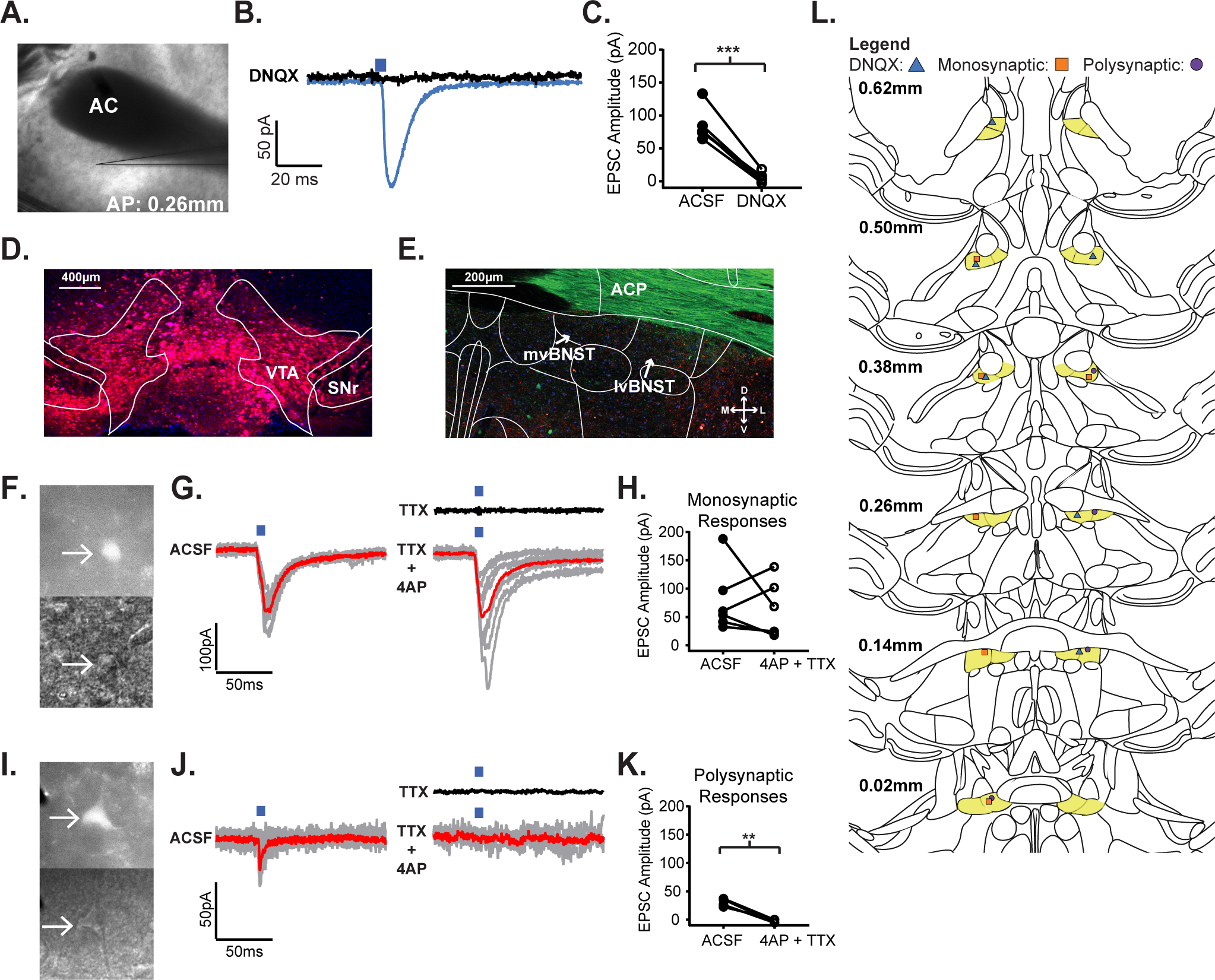
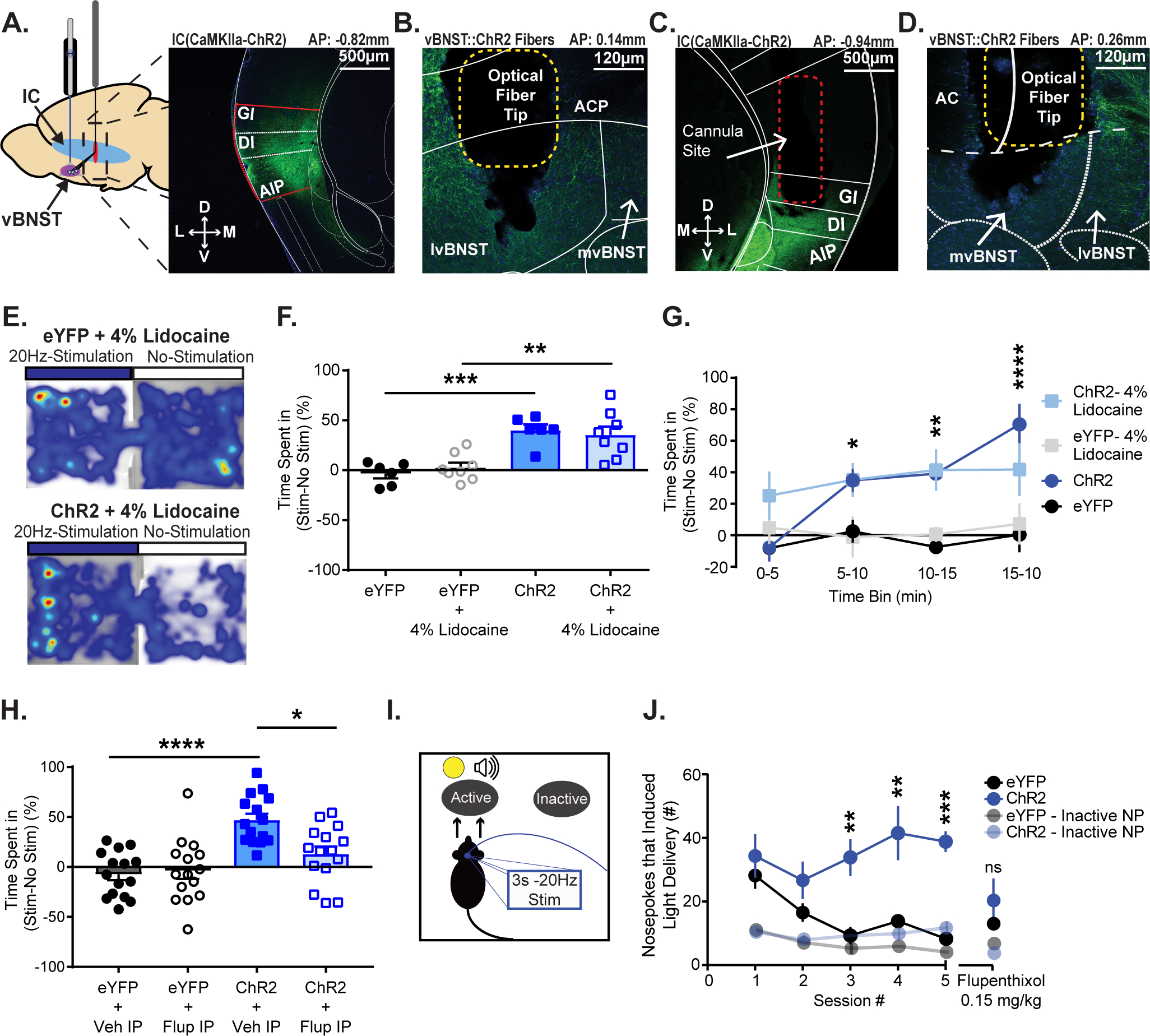
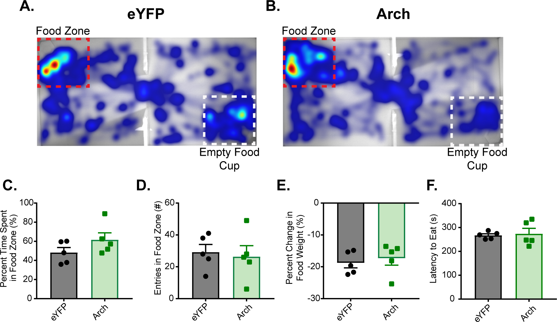
Individuals suffering from substance use disorder often experience relapse events that are attributed to drug craving. Insular cortex (IC) function is implicated in processing drug-predictive cues and is thought to be a critical substrate for drug craving, but the downstream neural circuit effectors of the IC that mediate reward processing are poorly described. Here, we uncover the functional connectivity of an IC projection to the ventral bed nucleus of the stria terminalis (vBNST), a portion of the extended amygdala that has been previously shown to modulate dopaminergic activity within the ventral tegmental area (VTA), and investigate the role of this pathway in reward-related behaviors. We utilized ex vivo slice electrophysiology and in vivo optogenetics to examine the functional connectivity of the IC-vBNST projection and bidirectionally control IC-vBNST terminals in various reward-related behavioral paradigms. We hypothesized that the IC recruits mesolimbic dopamine signaling by activating VTA-projecting, vBNST neurons. Using slice electrophysiology, we found that the IC sends a glutamatergic projection onto vBNST-VTA neurons. Photoactivation of IC-vBNST terminals was sufficient to reinforce behavior in a dopamine-dependent manner. Moreover, silencing the IC-vBNST projection was aversive and resulted in anxiety-like behavior without affecting food consumption. This work provides a potential mechanism by which the IC processes exteroceptive triggers that are predictive of reward.

Keywords: BNST; VTA; electrophysiology; insula; optogenetics; reward.


Conflict of interest statement

Declaration of Interests

The authors declare no competing interests.

Figures

Figure 1.
Figure 1.
The insular cortex sends glutamatergic afferents to VTA-projecting neurons in the vBNST. A) 4x magnification DIC image of a coronal vBNST slice from an IC(CaMKIIa::ChR2) expressing mouse where an optically evoked postsynaptic current was recorded. B) Optically-evoked postsynaptic current trace recorded in a vBNST neuron following IC(CaMKIIa::ChR2) stimulation, before and after application of the AMPA/KA receptor antagonist, DNQX. C) Peak amplitude current response to IC(CaMKIIa::ChR2) terminal stimulation in the vBNST before and after application of DNQX. D) 10x magnification of injected retrograde tracer, pAAV-CAG-TdTomato into the VTA for retrograde labeling of VTA-projecting neurons in the vBNST (VTA: ventral tegmental area; SNr: substantia nigra). E) 20x magnification of retrograde labeling from an IC(CaMKIIa::ChR2)//VTA(CAG::Retrograde-tdTomato) expressing mouse (mvBNST: medial ventral bed nucleus of the strial terminalis; lvBNST: lateral ventral bed nucleus of the stria terminalis). F) Two 40x magnification DIC images of the same monosynaptic neuron from an IC(CaMKIIa::ChR2)//VTA(CAG::Retrograde-tdTomato) expressing mouse under Top: wide-field fluorescent illumination to excite tdTomato in VTA expressing neurons; Bottom: Infrared illumination of the neuron. G) Optically-evoked postsynaptic current trace recorded in a VTA-projecting, vBNST neuron following IC(CaMKIIa::ChR2) terminal stimulation, Left: in ACSF, Black: in TTX, and Right: in 4AP + TTX; the red line represents the averaged trace. H) Peak amplitude current response to IC(CaMKIIa::ChR2) stimulation before and after application of 4AP + TTX. I) Two 40x magnification DIC images of the same polysynaptic neuron from an IC(CaMKIIa::ChR2)//VTA(CAG::Retrograde-tdTomato) expressing mouse under Top: widefield fluorescent illumination to excite mCherry in VTA expressing neurons; Bottom: Infrared illumination of the neuron. J) Optically-evoked postsynaptic current trace recorded in a VTA-projecting, vBNST neuron following IC(CaMKIIa::ChR2) terminal stimulation, Left: in ACSF, Black: in TTX, and Right: in 4AP + TTX; the red line represents the averaged trace. K) Peak amplitude response to IC(CaMKIIa::ChR2) terminal stimulation before and after application of 4AP + TTX. L) Patching cartography map showing light-evoked responses for: Blue Triangle: DNQX responses, Orange Square: Monosynaptic responses, and Purple Circle: Polysynaptic responses. (*p≤0.05, **p≤0.01, ***p≤0.001, ****p<0.0001)
Figure 2.
Figure 2.
Photostimulation of the IC-vBNST pathway is reinforcing. A) Left: Schematic depicting IC virus injection and vBNST cannula implantation. Right: Nissl stain of the ChR2 injection site in the IC of a C57/Bl6J mouse (GI: granular insula; DI: dysgranular insula; AIP: agranular insula). B) Nissl stain of ChR2 expression in IC terminals and optical fiber terminal site within the vBNST (ACP: anterior commissure posterior limb; mvBNST: medial ventral bed nucleus of the strial terminalis; lvBNST: lateral ventral bed nucleus of the stria terminalis). C) Nissl stain of cannula site for lidocaine infusions in the IC of an IC(CaMKIIa::ChR2) expressing mouse. D) Nissl stain of corresponding ChR2 expression in IC terminals and optical fiber terminal site within the vBNST. E) Heat maps from both an IC(CaMKIIa::eYFP+4%Lidocaine) (Top) and an IC(CaMKIIa::ChR2+4%Lidocaine) (Bottom) expressing subject during RTPP. F) Measured percent time spent in stimulus-paired chamber vs. non-stimulus-paired chamber during a RTPP task. We found that optical stimulation increased percent time spent in the stimulation chamber in both groups (saline, lidocaine) of ChR2 mice. G) Measured time spent in stimulus chamber vs. non-stimulus chamber to examine preference for the stimulation over time. Mice began showing preference for the stimulation chamber significantly more than controls by the second 5min bin. H) Measured percent time subjects spent in the stimulus-paired chamber vs. non-stimulus chamber during RTPP. Mice underwent the test once with IP administration of saline (Veh), then again after a two-day break with IP administration of 0.15mg/kg Flupenthixol. We found that dopamine receptor antagonism blunted preference for the stimulation chamber. I) Intracranial self-stimulation schematic. J) Mice learned to nosepoke for self-stimulation of the IC-vBNST pathway. Once stable responding occurred (5 sessions), all mice were given IP administration of flupenthixol 30 min prior to the session. Inactive nosepokes are shown at 50% opacity. Flupenthixol reduced active nosepoke responding in the ChR2 group so they were no longer significantly different compared to the controls. (*p≤0.05, **p≤0.01, ***p≤0.001, ****p<0.0001)
Figure 3.
Figure 3.
Silencing the IC-vBNST pathway is anxiogenic without affecting locomotor activity. A) C57/Bl6 mice were infected with the inhibitory opsin, Arch in the IC cell bodies and had an optical fiber implant in the vBNST for terminal silencing. Nissl stain of the Arch injection site (GI: granular insula; DI: dysgranular insula; AIP: agranular insula). B) 40x magnification of IC cell bodies expressing Arch. C) Nissl stain of Arch expression in IC terminals and optical fiber site within the vBNST (ACP: anterior commissure posterior limb; mvBNST: medial ventral bed nucleus of the strial terminalis; lvBNST: lateral ventral bed nucleus of the stria terminalis). D) 40x magnification of IC(CaMKIIa::Arch) terminals in the vBNST. E) Mice underwent an open field assay with continuous photoinhibition staggered off and on for 3 minute bins over 21 minutes (7 bins). Heat maps from both a IC(CaMKIIa::eYFP) (left) and an IC(CaMKIIa::Arch) (Right) expressing subject during the open field assay. The white box in the center represents the “center zone”. F) We then measured the total distance traveled in the open field assay when the laser was either OFF or ON. We found no effect of IC terminal inhibition on locomotor activity. G) We then examined changes in time spent in the center of the open field as a measure for anxiety. We found that Arch inhibition significantly reduced the time spent in the center in a within-groups comparison. H) Mice expressing either IC(CaMKIIa::eYFP) or IC(CaMKIIa::Arch) underwent a Real Time Place Preference assay. Heat maps from both a IC(CaMKIIa::eYFP) (Top) and a IC(CaMKIIa::Arch) (Bottom) expressing subject during RTPP. I) We found that optical inhibition of Arch-expressing IC terminals in the vBNST caused aversion for the stimulationpaired chamber. J) Measured time spent in stimulus chamber vs. non-stimulus chamber to examine aversion for the photoinhibition over time. Mice begin showing significant aversion for the stimulation chamber by the last 5min bin. (*p≤0.05, **p≤0.01, ***p≤0.001, ****p<0.0001)
Figure 4.
Figure 4.
IC-vBNST photoinhibition does not affect consummatory behavior. In a food approach task, percent time subject spent in the food zone was measured in a 15 minute free feeding task in a twochamber arena. Mice received continuous vBNST photoinhibition in the food zone that contained a cup filled with sucrose pellets. A) Example heat map of eYFP-expressing mouse during the food approach task. The red box outlines the food zone that also contained the cup filled with sucrose pellets, and the white box out lined the zone containing a clean, empty food cup. B) Example heat map of an Arch-expressing mouse during the same food approach task. C) Measured percent time spent in the food zone and found no difference between groups. D) Measured total number of entries in the food zone and found no difference between groups. E) Measured percent change in food before and after the food approach task and found no difference between groups. F) Measured latency to eat during the food approach task and found no difference between groups.

References

    1. Yang M, Mamy J, Gao P, Xiao S. From Abstinence to Relapse: A Preliminary Qualitative Study of Drug Users in a Compulsory Drug Rehabilitation Center in Changsha, China. PLoS ONE. 2015;10(6). doi:10.1371/journal.pone.0130711 - DOI - PMC - PubMed
    1. Vandaele Y, Janak PH. Defining the place of habit in substance use disorders. Prog Neuropsychopharmacol Biol Psychiatry. 2018;87(Pt A):22–32. doi:10.1016/j.pnpbp.2017.06.029 - DOI - PMC - PubMed
    1. Yang M, Mamy J, Gao P, Xiao S. From Abstinence to Relapse: A Preliminary Qualitative Study of Drug Users in a Compulsory Drug Rehabilitation Center in Changsha, China. PLoS ONE. 2015;10(6). doi:10.1371/journal.pone.0130711 - DOI - PMC - PubMed
    1. Naqvi NH, Rudrauf D, Damasio H, Bechara A. Damage to the Insula Disrupts Addiction to Cigarette Smoking. Science. 2007;315(5811):531–534. doi:10.1126/science.1135926 - DOI - PMC - PubMed
    1. Contreras M, Ceric F, Torrealba F. Inactivation of the Interoceptive Insula Disrupts Drug Craving and Malaise Induced by Lithium. Science. 2007;318(5850):655–658. doi:10.1126/science.1145590 - DOI - PubMed

Publication types