Review
. 2020 Jul 30:6:68.
doi: 10.1038/s41420-020-00305-y. eCollection 2020.

Extracellular vesicles in cardiovascular diseases

Affiliations
Review

Extracellular vesicles in cardiovascular diseases

Shihui Fu et al. Cell Death Discov. .

Abstract

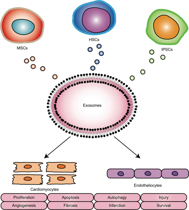
Due to the continued high incidence and mortality rate worldwide, there is still a need to develop new strategies for the prevention, diagnosis and treatment of cardiovascular diseases (CVDs). Proper cardiovascular function depends on the coordinated interplay and communication between cardiomyocytes and noncardiomyocytes. Extracellular vesicles (EVs) are enclosed in a lipid bilayer and represent a significant mechanism for intracellular communication. By containing and transporting various bioactive molecules, such as micro-ribonucleic acids (miRs) and proteins, to target cells, EVs impart favourable, neutral or detrimental effects on recipient cells, such as modulating gene expression, influencing cell phenotype, affecting molecular pathways and mediating biological behaviours. EVs can be released by cardiovascular system-related cells, such as cardiomyocytes, endotheliocytes, fibroblasts, platelets, smooth muscle cells, leucocytes, monocytes and macrophages. EVs containing miRs and proteins regulate a multitude of diverse functions in target cells, maintaining cardiovascular balance and health or inducing pathological changes in CVDs. On the one hand, miRs and proteins transferred by EVs play biological roles in maintaining normal cardiac structure and function under physiological conditions. On the other hand, EVs change the composition of their miR and protein cargoes under pathological conditions, which gives rise to the development of CVDs. Therefore, EVs hold tremendous potential to prevent, diagnose and treat CVDs. The current article reviews the specific functions of EVs in different CVDs.

Keywords: Cardiovascular diseases; Molecular biology.


Conflict of interest statement

Conflict of interestThe authors declare that they have no conflict of interest.

Figures

Fig. 1
Fig. 1. Origins, contents and roles of extracellular vesicles.
Extracellular vesicles (EVs) are enclosed in a lipid bilayer and represent a by-product released by cells. In addition to direct cell-cell contact or the transport of secreted molecules, EVs also participate in intercellular communication. By containing and transporting various bioactive molecules, such as proteins, lipids, messenger ribonucleic acids (mRNAs), micro-ribonucleic acids (microRNAs, miRs) and deoxyribonucleic acids (DNAs), to target cells, EVs impart favourable, neutral or detrimental effects on recipient cells, such as modulating gene expression, influencing cell phenotype, affecting molecular pathways and mediating biological behaviours.
Fig. 2
Fig. 2. Origins, contents and roles of extracellular vesicles in cardiovascular diseases.
Extracellular vesicles (EVs) can be released by cardiovascular system-related cells, such as cardiomyocytes, endotheliocytes, fibroblasts, platelets, smooth muscle cells (SMCs), leukocytes, monocytes and macrophages. On the one hand, EVs play biological roles in maintaining normal cardiac structure and function under physiological conditions. On the other hand, EVs change their composition under pathological conditions and contribute to the development of cardiovascular diseases (CVDs). Therefore, EVs hold tremendous potential to monitor and treat CVDs.
Fig. 3
Fig. 3. Origins and roles of stem cell-derived extracellular vesicles.
Extracellular vesicles (EVs) mimic the cardioprotective properties of various stem cells, including mesenchymal stromal cells (MSCs), haematopoietic stem cells (HSCs) and induced pluripotent stem cell (iPSCs). These EVs modulate cardiomyocytes and endotheliocytes by stimulating cell proliferation, decreasing cell apoptosis, inhibiting cell autophagy, promoting angiogenesis, reducing tissue fibrosis, preventing cardiovascular injury, treating myocardial infarction and improving cardiac survival.

References

    1. Benjamin EJ, et al. American Heart Association Statistics Committee and Stroke Statistics Subcommittee. Heart disease and stroke statistics-2017 update: a report from the American Heart Association. Circulation. 2017;135:e146–e603. doi: 10.1161/CIR.0000000000000485. - DOI - PMC - PubMed
    1. Gartz M, Strande JL. Examining the paracrine effects of exosomes in cardiovascular disease and repair. J. Am. Heart Assoc. 2018;7:pii: e007954. - PMC - PubMed
    1. Maia J, et al. Exosome-based cell-cell communication in the tumor microenvironment. Front. Cell Dev. Biol. 2018;6:18. - PMC - PubMed
    1. Sahoo S, Losordo DW. Exosomes and cardiac repair after myocardial infarction. Circ. Res. 2014;114:333–344. - PubMed
    1. Colombo M, Raposo G, Théry C. Biogenesis, secretion, and intercellular interactions of exosomes and other extracellular vesicles. Annu. Rev. Cell Dev. Biol. 2014;30:255–289. - PubMed