

MicroRNAs in the Vitreous Humor of Patients with Retinal Detachment and a Different Grading of Proliferative Vitreoretinopathy: A Pilot Study
- PMID: 32821520
- PMCID: PMC7409223
- DOI: 10.1167/tvst.9.6.23
MicroRNAs in the Vitreous Humor of Patients with Retinal Detachment and a Different Grading of Proliferative Vitreoretinopathy: A Pilot Study
Abstract
Purpose: Although the expression of microRNAs (miRNAs) in retinal pigment epithelial (RPE) cells undergoing epithelial-mesenchymal transition (EMT) is involved in the pathogenesis of proliferative vitreoretinopathy (PVR), its expression in the vitreous of patients with primary retinal detachment (RD) and different PVR grading has not yet been investigated. We assessed the expression of miRNAs in the vitreous humor (VH) of patients diagnosed with RD and different grading of PVR.
Methods: The VH was extracted from the core of the vitreous chamber in patients who had undergone standard vitrectomy for primary RD. RNA was extracted and TaqMan Low-Density Arrays (TLDAs) were used for miRNA profiling that was performed by single TaqMan assays. A gene ontology (GO) analysis was performed on the differentially expressed miRNAs.
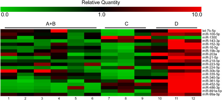
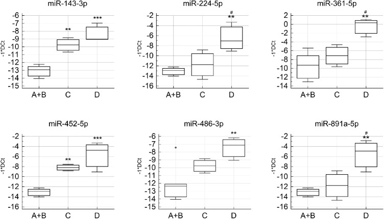
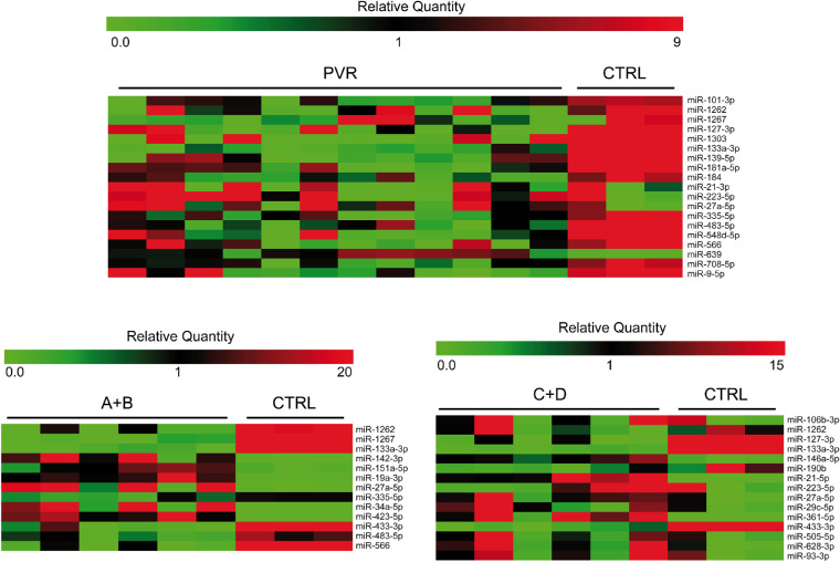
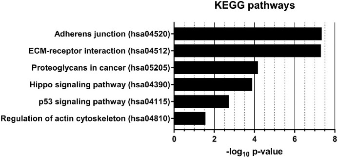
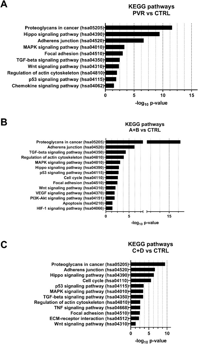
Results: A total of 15 eyes with RD, 3 eyes for each grade of PVR (A, B, C, and D) and 3 from unaffected individuals, were enrolled in this prospective comparative study. Twenty miRNAs were altered in the comparison among pathological groups. Interestingly, the expression of miR-143-3p, miR-224-5p, miR-361-5p, miR-452-5p, miR-486-3p, and miR-891a-5p increased with the worsening of PVR grading. We also identified 34 miRNAs showing differential expression in PVR compared to control vitreous samples. GO analysis showed that the deregulated miRNAs participate in processes previously associated with PVR pathogenesis.
Conclusions: The present pilot study suggested that dysregulated vitreal miRNAs may be considered as a biomarker of PVR and associated with the PVR-related complications in patients with RD.
Translational relevance: The correlation between vitreal miRNAs and the pathological phenotypes are essential to identify the novel miRNA-based mechanisms underlying the PVR disease that would improve the diagnosis and treatment of the condition.
Keywords: microRNA; profiling; proliferative vitreoretinopathy; retinal detachment.
Copyright 2020 The Authors.
Conflict of interest statement
Disclosure: M.D. Toro, None; M. Reibaldi, None; T. Avitabile, None; C. Bucolo, None; S. Salomone, None; R. Rejdak, None; K. Nowomiejska, None; S. Tripodi, None; C. Posarelli, None; M. Ragusa, None; C. Barbagallo, None
Figures
References
-
- Pastor JC, Rojas J, Pastor-Idoate S, Di Lauro S, Gonzalez-Buendia L, Delgado-Tirado S. Proliferative vitreoretinopathy: a new concept of disease pathogenesis and practical consequences, Prog Retin Eye Res. 2016; 51: 125–155. - PubMed
-
- Gagliano C, Toro MD, Avitabile T, Stella S, Uva MG. Intravitreal steroids for the prevention of PVR after surgery for retinal detachment. Curr Pharm Des. 2015; 21: 4698–4702. - PubMed
-
- Yao J, Hu LL, Li XM, et al. .. Comprehensive circular RNA profiling of proliferative vitreoretinopathy and its clinical significance. Biomed Pharmacother. 2019; 111: 548–554. - PubMed
-
- Cardillo JA, Stout JT, LaBree L. Post-traumatic proliferative vitreoretinopathy. The epidemiologic profile, onset, risk factors, and visual outcome. Ophthalmology. 1997; 104: 1166–1173. - PubMed
-
- Tseng W, Cortez RT, Ramirez G, Stinnett S, Jaffe GJ. Prevalence and risk factors for proliferative vitreoretinopathy in eyes with rhegmatogenous retinal detachment but no previous vitreoretinal surgery. Am J Ophthalmol. 2004; 137: 1105–1115. - PubMed
MeSH terms
Substances
LinkOut - more resources
Full Text Sources
Medical
Research Materials
Miscellaneous
