. 2021 Feb;216(2):362-368.
doi: 10.2214/AJR.20.23429. Epub 2020 Aug 21.

Virtual Imaging Trials for Coronavirus Disease (COVID-19)

Affiliations

Virtual Imaging Trials for Coronavirus Disease (COVID-19)

Ehsan Abadi et al. AJR Am J Roentgenol. 2021 Feb.

Abstract

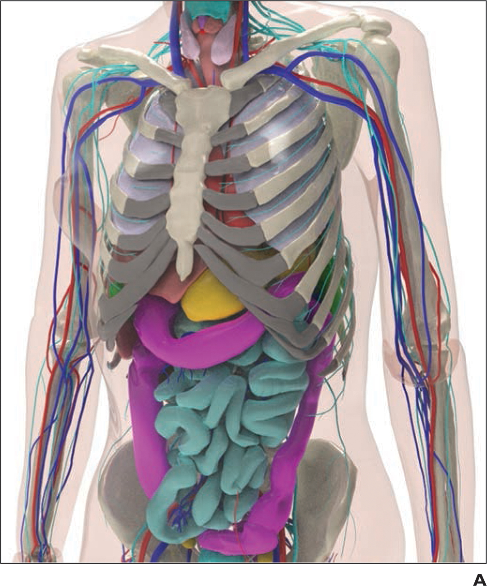
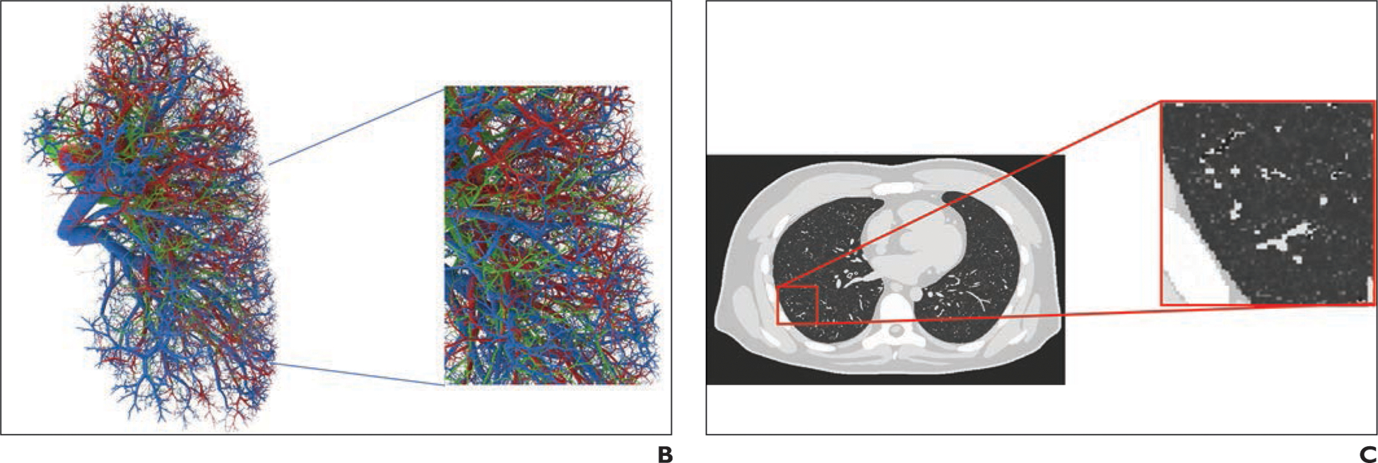
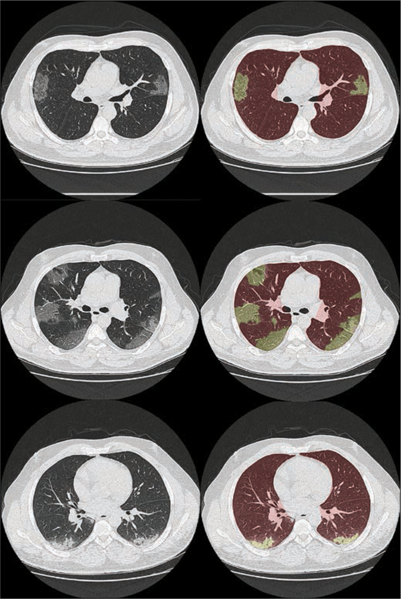
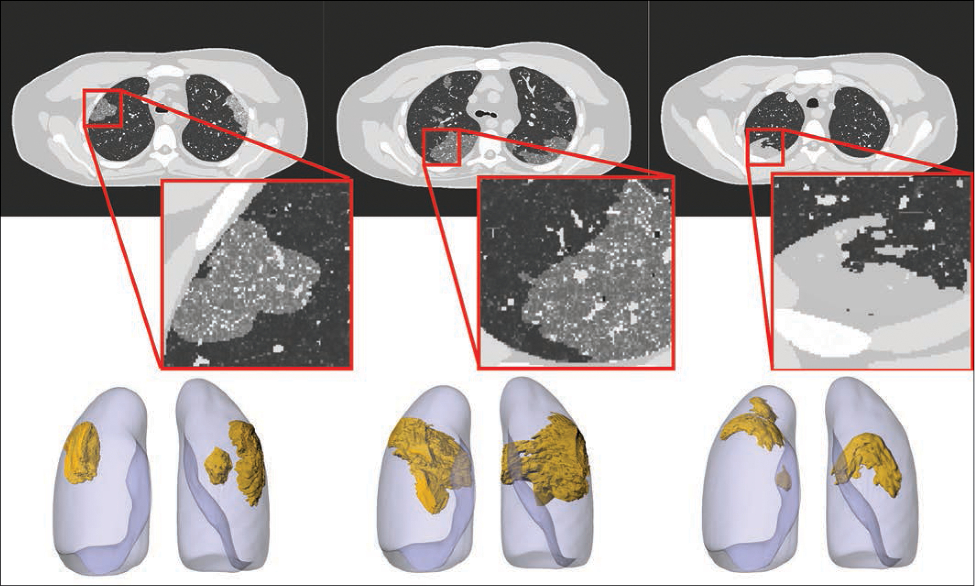
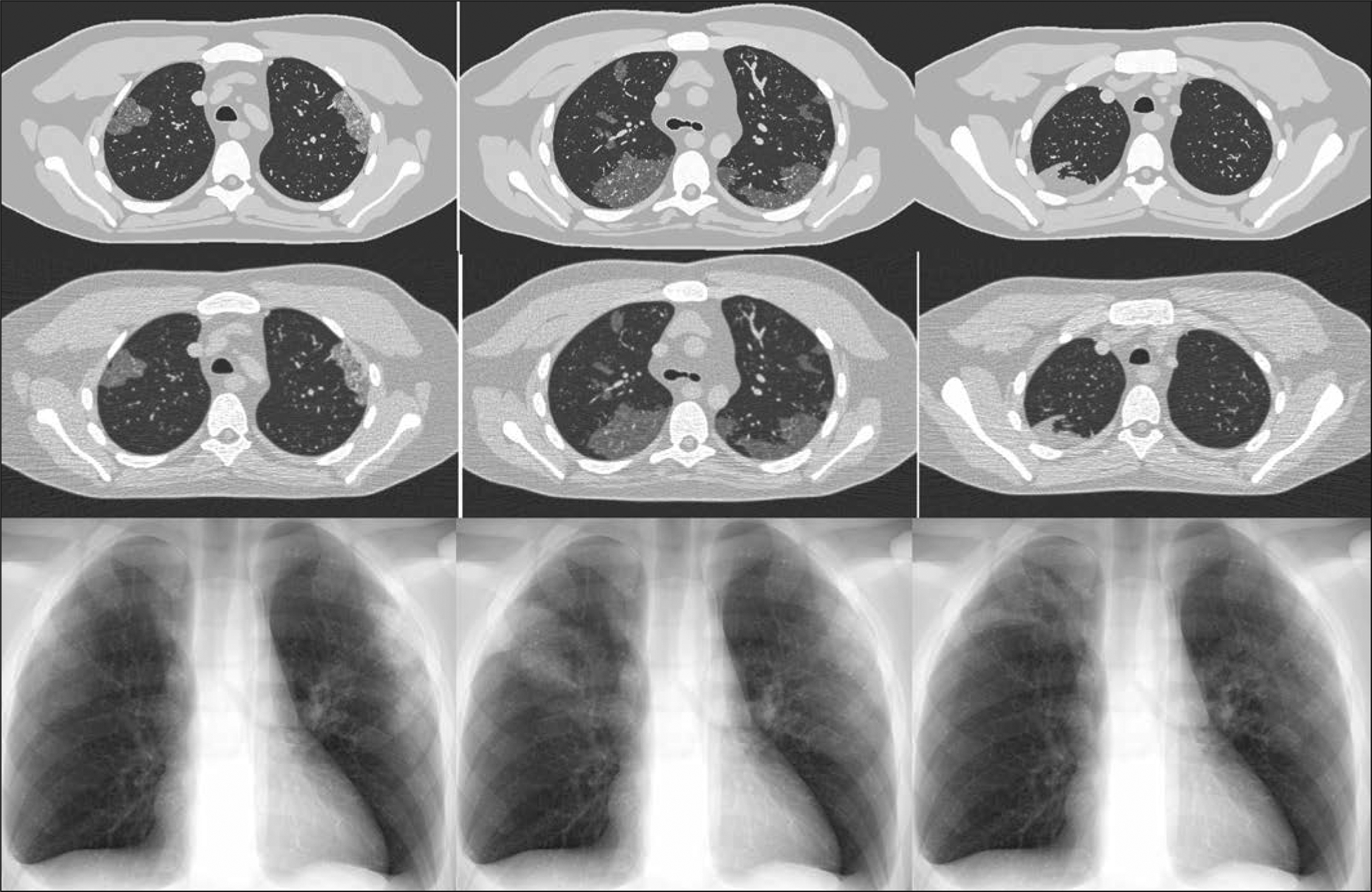
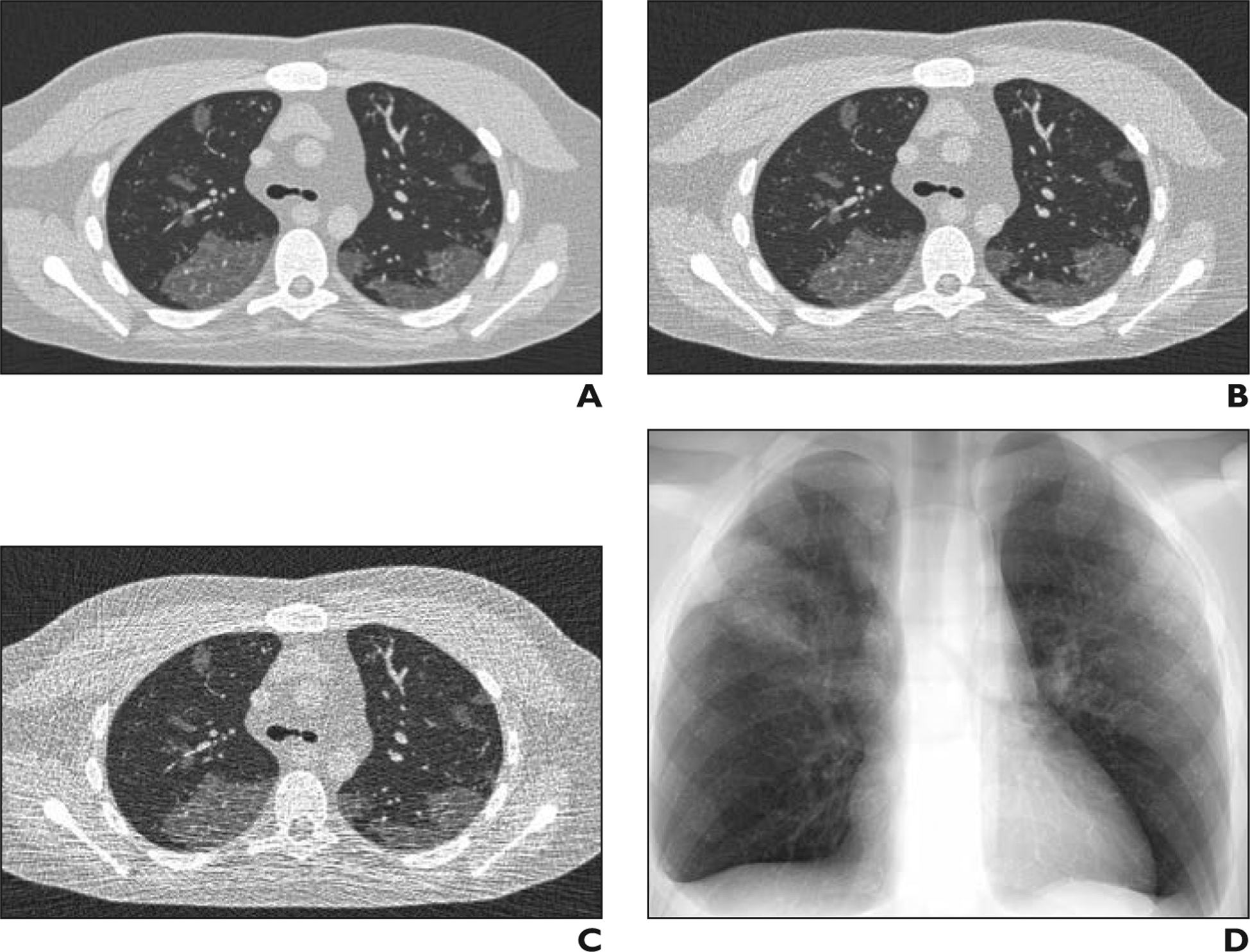
OBJECTIVE. The virtual imaging trial is a unique framework that can greatly facilitate the assessment and optimization of imaging methods by emulating the imaging experiment using representative computational models of patients and validated imaging simulators. The purpose of this study was to show how virtual imaging trials can be adapted for imaging studies of coronavirus disease (COVID-19), enabling effective assessment and optimization of CT and radiography acquisitions and analysis tools for reliable imaging and management of COVID-19. MATERIALS AND METHODS. We developed the first computational models of patients with COVID-19 and as a proof of principle showed how they can be combined with imaging simulators for COVID-19 imaging studies. For the body habitus of the models, we used the 4D extended cardiac-torso (XCAT) model that was developed at Duke University. The morphologic features of COVID-19 abnormalities were segmented from 20 CT images of patients who had been confirmed to have COVID-19 and incorporated into XCAT models. Within a given disease area, the texture and material of the lung parenchyma in the XCAT were modified to match the properties observed in the clinical images. To show the utility, three developed COVID-19 computational phantoms were virtually imaged using a scanner-specific CT and radiography simulator. RESULTS. Subjectively, the simulated abnormalities were realistic in terms of shape and texture. Results showed that the contrast-to-noise ratios in the abnormal regions were 1.6, 3.0, and 3.6 for 5-, 25-, and 50-mAs images, respectively. CONCLUSION. The developed toolsets in this study provide the foundation for use of virtual imaging trials in effective assessment and optimization of CT and radiography acquisitions and analysis tools to help manage the COVID-19 pandemic.

Keywords: COVID-19; CT; coronavirus disease; radiography; virtual imaging trials.


Figures

Fig. 1—
Fig. 1—
Example of 4D extended cardiac-torso (XCAT) phantom developed at Duke University. A, Representative computational model shows female XCAT phantom with anthropomorphic organs and structures. B, Representative computational model shows lung stroma intraorgan structure of XCAT phantom that was developed using anatomically informed mathematic model. Inset shows enlarged view for better visibility of details and small structures. C, Voxelized rendition (ground truth) of XCAT phantom highlights detailed model of lung parenchyma. Inset shows enlarged view for better visibility of details and small structures.
Fig. 1—
Fig. 1—
Example of 4D extended cardiac-torso (XCAT) phantom developed at Duke University. A, Representative computational model shows female XCAT phantom with anthropomorphic organs and structures. B, Representative computational model shows lung stroma intraorgan structure of XCAT phantom that was developed using anatomically informed mathematic model. Inset shows enlarged view for better visibility of details and small structures. C, Voxelized rendition (ground truth) of XCAT phantom highlights detailed model of lung parenchyma. Inset shows enlarged view for better visibility of details and small structures.
Fig. 2—
Fig. 2—
CT images (left) of patients who had tested positive for coronavirus disease (COVID-19) with ground-glass opacity (top and middle) and consolidation (bottom) abnormalities. Right images show overlay of lung and abnormality segmentation on corresponding CT images.
Fig. 3—
Fig. 3—
Four-dimensional extended cardiac-torso (XCAT) phantom developed at Duke University shows three coronavirus disease (COVID-19) abnormalities with different shapes. Left and center phantoms show ground-glass opacities and right phantom shows consolidation. Top images show voxelized renditions (ground truth) of phantoms, and insets show enlarged view for better visibility of details and small structures. Bottom images show surface-based rendition of same COVID-19 abnormalities incorporated into adult male XCAT phantom.
Fig. 4—
Fig. 4—
Ground-truth phantom images (top) of 4D extended cardiac-torso phantom developed at Duke University show different coronavirus disease (COVID-19) abnormalities. Simulated CT (middle) and radiographic (bottom) images were generated using DukeSim, also developed at Duke University. Ground-glass opacity (left and middle) and consolidation (right) abnormalities are visible on both CT and radiographic images.
Fig. 5—
Fig. 5—
Simulated CT images of coronavirus disease (COVID-19) on 4D extended cardiac-torso phantom developed at Duke University. A–D, Images of same phantom show simulated CT at 50 (A), 25 (B), and 5 (C) mAs as well as simulated chest radiograph (D).

References

    1. World Health Organization website. WHO director-general’s opening remarks at the media briefing on COVID-19: 11 March 2020. www.who.int/dg/speeches/detail/who-director-general-s-opening-remarks-at.... Accessed April 10, 2020
    1. Worldometer website. COVID-19 coronavirus pandemic. www.worldometers.info/coronavirus/. Accessed April 9, 2020
    1. Johns Hopkins University Center for Systems Science and Engineering website. Coronavirus COVID-19 global cases. gisanddata.maps.arcgis.com/apps/opsdashboard/index.html#/bda7594740fd402.... Accessed April 9, 2020
    1. Ai T, Yang Z, Hou H, et al. Correlation of chest CT and RT-PCR testing in coronavirus disease 2019 (COVID-19) in China: a report of 1014 cases. Radiology 2020. February 26 [Epub ahead of print] - PMC - PubMed
    1. Li L, Qin L, Xu Z, et al. Artificial intelligence distinguishes COVID-19 from community acquired pneumonia on chest CT. Radiology 2020. March 19 [Epub ahead of print] - PMC - PubMed