Review
. 2020 Aug 21;16(8):e1008762.
doi: 10.1371/journal.ppat.1008762. eCollection 2020 Aug.

COVID-19 pandemic: Insights into structure, function, and hACE2 receptor recognition by SARS-CoV-2

Affiliations
Review

COVID-19 pandemic: Insights into structure, function, and hACE2 receptor recognition by SARS-CoV-2

Anshumali Mittal et al. PLoS Pathog. .

Abstract

Severe Acute Respiratory Syndrome Coronavirus-2 (SARS-CoV-2) is a newly emerging, highly transmissible, and pathogenic coronavirus in humans that has caused global public health emergencies and economic crises. To date, millions of infections and thousands of deaths have been reported worldwide, and the numbers continue to rise. Currently, there is no specific drug or vaccine against this deadly virus; therefore, there is a pressing need to understand the mechanism(s) through which this virus enters the host cell. Viral entry into the host cell is a multistep process in which SARS-CoV-2 utilizes the receptor-binding domain (RBD) of the spike (S) glycoprotein to recognize angiotensin-converting enzyme 2 (ACE2) receptors on the human cells; this initiates host-cell entry by promoting viral-host cell membrane fusion through large-scale conformational changes in the S protein. Receptor recognition and fusion are critical and essential steps of viral infections and are key determinants of the viral host range and cross-species transmission. In this review, we summarize the current knowledge on the origin and evolution of SARS-CoV-2 and the roles of key viral factors. We discuss the structure of RNA-dependent RNA polymerase (RdRp) of SARS-CoV-2 and its significance in drug discovery and explain the receptor recognition mechanisms of coronaviruses. Further, we provide a comparative analysis of the SARS-CoV and SARS-CoV-2 S proteins and their receptor-binding specificity and discuss the differences in their antigenicity based on biophysical and structural characteristics.


Conflict of interest statement

"The authors have declared that no competing interests exist".

Figures

Fig 1
Fig 1. Origin and transmission of pathogenic HCoVs.
Yellow and red arrows indicate mild and severe infections in humans, respectively. The figure is inspired from Jie Cui and colleagues [46], and the illustrations of coronaviruses (left) are adapted from “Desiree Ho, Innovative Genomics Institute,” available at https://innovativegenomics.org/free-covid-19-illustrations/. HCoV, Human Coronavirus; MERS-CoV, Middle East Respiratory Syndrome Coronavirus; SARS-CoV, Severe Acute Respiratory Syndrome Coronavirus; SARS-CoV-2, SARS-CoV, Severe Acute Respiratory Syndrome Coronavirus-2.
Fig 2
Fig 2. Phylogenetic relationships in the Coronavirinae subfamily.
The subfamily is formed by 4 genera: Alphacoronavirus, Betacoronavirus (lineages A, B, C, and D), Gammacoronavirus, and Deltacoronavirus. We randomly picked 62 SARS-CoV-2 genome sequences, representing 15 different countries, together with other Coronavirinae subfamily members. The phylogenetic tree was created using NgPhylogeny.fr tool. The analysis indicates that SARS-CoV-2 has a close relationship with bat coronavirus RaTG13 and SARS-CoV; therefore, it is classified as a new member of the lineage B Betacoronavirus. SARS-CoV, Severe Acute Respiratory Syndrome Coronavirus; SARS-CoV-2, Severe Acute Respiratory Syndrome Coronavirus-2.
Fig 3
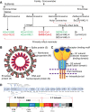
Fig 3. Classification and structure of coronavirus.
(A) Classification of coronaviruses: the 7 known HCoVs are shown in green and red. HCoVs in red bind the host receptor ACE2. (B) Schematic of the SARS-CoV-2 structure; the illustration of the virus is adapted from “Desiree Ho, Innovative Genomics Institute,” available at https://innovativegenomics.org/free-covid-19-illustrations/. (C) Cartoon depicts key features and the trimeric structure of the SARS-CoV-2 S protein. (D) Schematic of SARS-CoV-2 genome (top) and S protein (bottom); annotations are adapted from NCBI (NC_045512.2) and Expasy (https://covid-19.uniprot.org/uniprotkb/P0DTC2), respectively. ACE2, angiotensin-converting enzyme 2; CTD, C-terminal domain; E, envelope; HCoV, Human Coronavirus; HR1/2, heptad repeat 1/2; M, membrane; N, nucleocapsid; Nsp, nonstructural protein; NTD, N-terminal domain; orf, open reading frame; RBD, receptor-binding domain; RBM, receptor-binding motif; RdRp, RNA-dependent RNA polymerase; S protein, spike protein; SARS-CoV-2, Severe Acute Respiratory Syndrome Coronavirus-2; UTR, untranslated region.
Fig 4
Fig 4. Cryo-EM structure of RdRp of SARS-CoV-2.
(A) The domain architecture of RdRp or nsp12 of SARS-CoV-2 is subdivided into NiRAN, interface, fingers, palm, and thumb subdomains; A–G indicate conserved motifs. (B) The cryo-EM structure of apo-RdRp complex (shown as front view, PDB: 7BV1) consists of nsp12, nsp7 (brown), and 2 chains of nsp8 (nsp8.1 and nsp8.2, both in gray). The nsp8.1 interacts directly with nsp12, whereas the nsp8.2 binds to nsp7, which in turn interacts with nsp12. The RNA template is expected to enter the active site, which is formed by motifs A and C through a groove clamped by motifs F and G. Motif E and the thumb subdomain support the primer strand. The RdRp subdomain color scheme is according to Fig 4A. (C) The cryo-EM structure (in top view) of the RdRp complex bound to RNA (PDB: 6YYT) shows 2 chains of nsp8 stabilizing the extending RNA with their alpha helices. The apo-RdRp complex structure (PDB: 7BV1) is shown for comparison. The active site is expanded to show the RNA molecules coming out of the groove formed by the finger and the thumb subdomains. The figures were prepared using Pymol. cryo-EM, cryo-electron microscopy; NiRAN, nidovirus RdRp-associated nucleotidyltransferase; PDB, Protein Data Bank; RdRp, RNA-dependent RNA polymerase; SARS-CoV-2, Severe Acute Respiratory Syndrome Coronavirus-2.
Fig 5
Fig 5. Structure of the SARS-CoV-2 S protein alone and in complex with ACE2 receptor.
(A) Side view of the trimeric SARS-CoV-2 S ectodomain in the prefusion state (PDB: 6VSB). The protomer in green is in the “up” conformation, and the other 2 protomers in red and cyan are in “down” conformation. (B) Top view of the trimeric S protein showing RBDs in red, blue, and green on each protomer. (C) Structure of a single protomer showing the receptor-binding subunit S1 (blue) and the membrane-fusion subunit S2 (green). The furin-like protease site at the boundary of S1/S2 subunits is depicted. (D) The S1 subunit showing the RBM in the CTD region (blue) and the NTD region (brown). The S2 subunit showing the fusion peptide (red), second cleavage site S2′ (black), and HR1 (pink). (E) Structure of the RBD, core subdomain (green), and RBM (blue) (PDB: 6LZG). (F) SARS-CoV-2-RBD:ACE2 receptor polar interface shown by specific residues. (G) Structure of the SARS-CoV-2-RBD in complex with ACE2 receptor (PDB: 6LZG). (H) Structural similarity between the SARS-CoV-RBD:hACE2 (green) and SARS-CoV-2-S-CTD:hACE2 (yellow) complexes. (I) Crystal structure of the SARS-CoV-2-RBD (green) in complex with a monoclonal antibody CR3022 (orange). The RBM and CR3022 binding sites do not overlap and are distantly located on the RBD (PDB: 6W41). The figures were prepared using Pymol. ACE2, angiotensin-converting enzyme 2; CTD, C-terminal domain; hACE2, human ACE2; HR1, heptad repeat 1; NTD, N-terminal domain; PDB, Protein Data Bank; RBD, receptor-binding domain; RBM, receptor-binding motif; S protein, spike protein; SARS-CoV, Severe Acute Respiratory Syndrome Coronavirus; SARS-CoV-2, Severe Acute Respiratory Syndrome Coronavirus-2.

References

    1. Hamre D, Procknow JJ. A new virus isolated from the human respiratory tract. Proc Soc Exp Biol Med. 1966;121: 190–193. 10.3181/00379727-121-30734 - DOI - PubMed
    1. Kahn JS, McIntosh K. History and recent advances in coronavirus discovery. Pediatr Infect Dis J. 2005;24: S223–227, discussion S226. 10.1097/01.inf.0000188166.17324.60 - DOI - PubMed
    1. Tyrrell DA, Bynoe ML. Cultivation of viruses from a high proportion of patients with colds. Lancet. 1966;1: 76–77. 10.1016/s0140-6736(66)92364-6 - DOI - PubMed
    1. WHO. Coronavirus never before seen in humans is the cause of SARS [Internet]. 2003 [cited 2020 Jun 27]. Available from: https://www.who.int/mediacentre/news/releases/2003/pr31/en/
    1. Holmes KV. SARS-associated coronavirus. N Engl J Med. 2003;348: 1948–1951. 10.1056/NEJMp030078 - DOI - PubMed