Review
. 2020 Nov 1;93(1115):20200562.
doi: 10.1259/bjr.20200562. Epub 2020 Sep 9.

What's New for Clinical Whole-body MRI (WB-MRI) in the 21st Century

Affiliations
Review

What's New for Clinical Whole-body MRI (WB-MRI) in the 21st Century

Nina Tunariu et al. Br J Radiol. .

Abstract

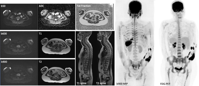
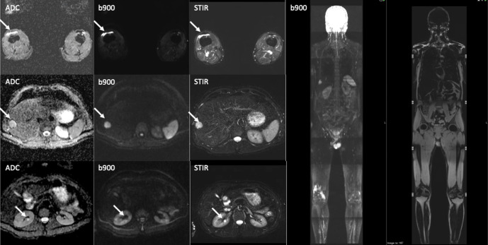
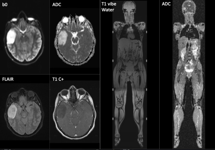
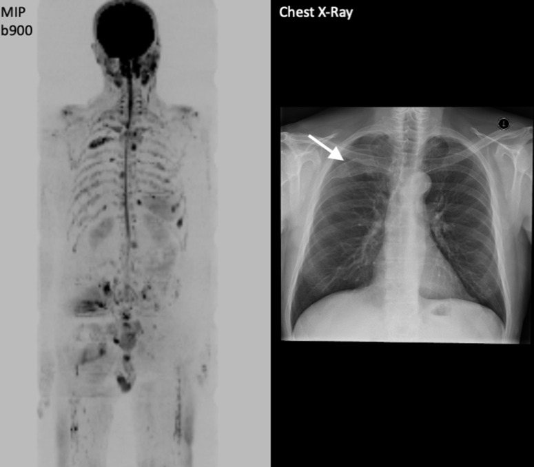
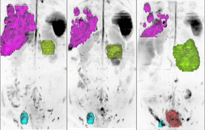
Whole-body MRI (WB-MRI) has evolved since its first introduction in the 1970s as an imaging technique to detect and survey disease across multiple sites and organ systems in the body. The development of diffusion-weighted MRI (DWI) has added a new dimension to the implementation of WB-MRI on modern scanners, offering excellent lesion-to-background contrast, while achieving acceptable spatial resolution to detect focal lesions 5 to 10 mm in size. MRI hardware and software advances have reduced acquisition times, with studies taking 40-50 min to complete.The rising awareness of medical radiation exposure coupled with the advantages of MRI has resulted in increased utilization of WB-MRI in oncology, paediatrics, rheumatological and musculoskeletal conditions and more recently in population screening. There is recognition that WB-MRI can be used to track disease evolution and monitor response heterogeneity in patients with cancer. There are also opportunities to combine WB-MRI with molecular imaging on PET-MRI systems to harness the strengths of hybrid imaging. The advent of artificial intelligence and machine learning will shorten image acquisition times and image analyses, making the technique more competitive against other imaging technologies.


Figures

Figure 1.
Figure 1.
Example of a typical WB-MRI protocol including axial DWI (3 b values and ADC map), axial T1W and T2W, CAIPIRINHA (Controlled Aliasing in Parallel Imaging Results in Higher Acceleration)-derived relative fat fraction and sagittal T1W and T2W spine images. Note the sagittal spine images were acquired without anterior saturation bands to visualise the sternum. The inverted coronal 3D b900 MIP and FDG-PET images show similar anatomical coverage between techniques.
Figure 2.
Figure 2.
A screening whole body-MRI in a healthy male volunteer showing incidental benign renal cysts, hepatic haemangioma and a right knee effusion (white arrows). The liver haemangioma was confirmed on ultrasound examination.
Figure 3.
Figure 3.
Screening whole-body MRI in a 33-year-old female with p53 cancer predisposition syndrome. MRI of the brain including T1W post-contrast examination shows an asymptomatic right temporal lobe low-grade glioma. which was treated by curative surgery.
Figure 4.
Figure 4.
A 51-year-old male with solitary infiltration of the posterior third rib on skeletal survey led to discussion of local radiotherapy as primary treatment. 3D b900 inverse grey scale MIP from WB-MRI confirmed multifocal disease, resulting in a change to system treatment.
Figure 5.
Figure 5.
99mTc-MDP Bone scan and WB-MRI images in a 60-year-old male with metastatic castrate resistant prostate carcinoma (mCRPC) illustrating the FLARE phenomenon. Bone scan performed in April 2018 after 12 weeks of chemotherapy showed several new bone lesions e.g. at T11 (arrows). Contemporaneous WB-MRI shows response with significant increase in ADC values (40%) of the lesion at T11 that appears “new” on bone scan. Note that the apparent new bone lesions on April 2018 in the lower thoracic spine bone scan were visible on the MRI from Jan 2018, but were occult/non-visible on the bone scan from Jan 2018.
Figure 6.
Figure 6.
Coronal 3D MIP and axial, DWI and FDG-PET images show bone marrow involvement in a 80-year-old male with large B-cell lymphoma. The initial PET staging was reported as Stage 1 AE with a dominant sternal mass (a) and no other lesions. WB-MRI highlighted other focal bone lesions within T10 vertebral body (b), right acetabulum (c) and left femur (d) that were classified as inflammatory on PET imaging in view of the lower SUV values and their location adjacent to osteophytes and degenerative change. Following, tumour board discussion, the disease stage was changed to IVAE and treatment altered.
Figure 7.
Figure 7.
A 65-year-old female with history of right invasive lobular breast carcinoma presenting with raised tumour marker CA15-345 U/ml and back pain. The tumour showed lymphovascular invasion; with ER8 PR0 Her-2 negative. WB-MRI show multiple vertebral and pelvic bone metastases that were occult on PET-CT without FDG tracer uptake.
Figure 8.
Figure 8.
A 60-year-old male with lung adenocarcinoma. FDG PET-CT and WB-MRI demonstrate liver, bone and brain metastases, which were not visible on the staging CT (not shown). White arrows show concordant lesions detected by both FDG PET-CT and WB-MRI. However, WB-MRI showed additional liver and bone lesions (grey arrows), while post-contrast T1W brain images show cerebral metastases not visible on FDG PET-CT (not shown).
Figure 9.
Figure 9.
3D MIP inverted b900 images in a 47-year-old female with advanced ovarian cancer. The different tumour sites are colour-coded according to the organ involved liver metastases (pink), peritoneal disease (green), pelvic lymphadenopathy (blue) and local relapse (orange). Pre- and post-chemotherapy images show response heterogeneity with decrease in the liver and nodal disease, but increase in peritoneal and local disease.

References

    1. Lam DL, Larson DB, Eisenberg JD, Forman HP, Lee CI. Communicating potential radiation-induced cancer risks from medical imaging directly to patients. AJR Am J Roentgenol 2015; 205: 962–70. doi: 10.2214/AJR.15.15057 - DOI - PubMed
    1. Hendee WR, O'Connor MK. Radiation risks of medical imaging: separating fact from fantasy. Radiology 2012; 264: 312–21. doi: 10.1148/radiol.12112678 - DOI - PubMed
    1. Ballinger ML, Best A, Mai PL, Khincha PP, Loud JT, Peters JA, et al. Baseline surveillance in Li-Fraumeni syndrome using whole-body magnetic resonance imaging: a meta-analysis. JAMA Oncol 2017; 3: 1634–9. doi: 10.1001/jamaoncol.2017.1968 - DOI - PMC - PubMed
    1. Taylor SA, Mallett S, Beare S, Bhatnagar G, Blunt D, Boavida P, et al. Diagnostic accuracy of whole-body MRI versus standard imaging pathways for metastatic disease in newly diagnosed colorectal cancer: the prospective streamline C trial. Lancet Gastroenterol Hepatol 2019; 4: 529–37. doi: 10.1016/S2468-1253(19)30056-1 - DOI - PMC - PubMed
    1. Taylor SA, Mallett S, Ball S, Beare S, Bhatnagar G, Bhowmik A, et al. Diagnostic accuracy of whole-body MRI versus standard imaging pathways for metastatic disease in newly diagnosed non-small-cell lung cancer: the prospective streamline L trial. Lancet Respir Med 2019; 7: 523–32. doi: 10.1016/S2213-2600(19)30090-6 - DOI - PMC - PubMed