Review
. 2020 Oct 12;38(4):454-472.
doi: 10.1016/j.ccell.2020.07.013. Epub 2020 Aug 20.

Identifying and Targeting Human Tumor Antigens for T Cell-Based Immunotherapy of Solid Tumors

Affiliations
Review

Identifying and Targeting Human Tumor Antigens for T Cell-Based Immunotherapy of Solid Tumors

Vid Leko et al. Cancer Cell. .

Abstract

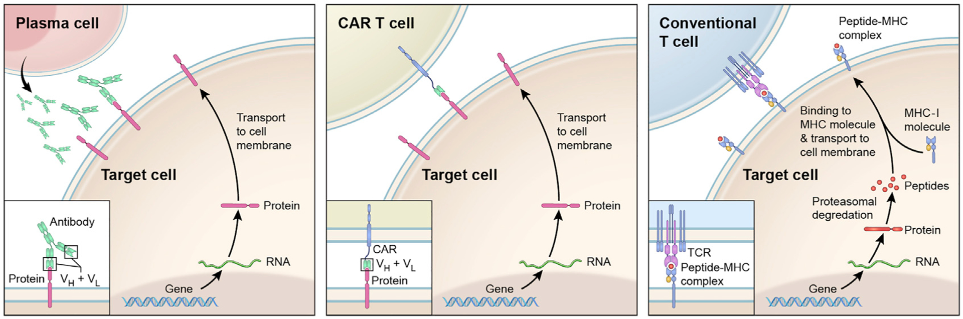
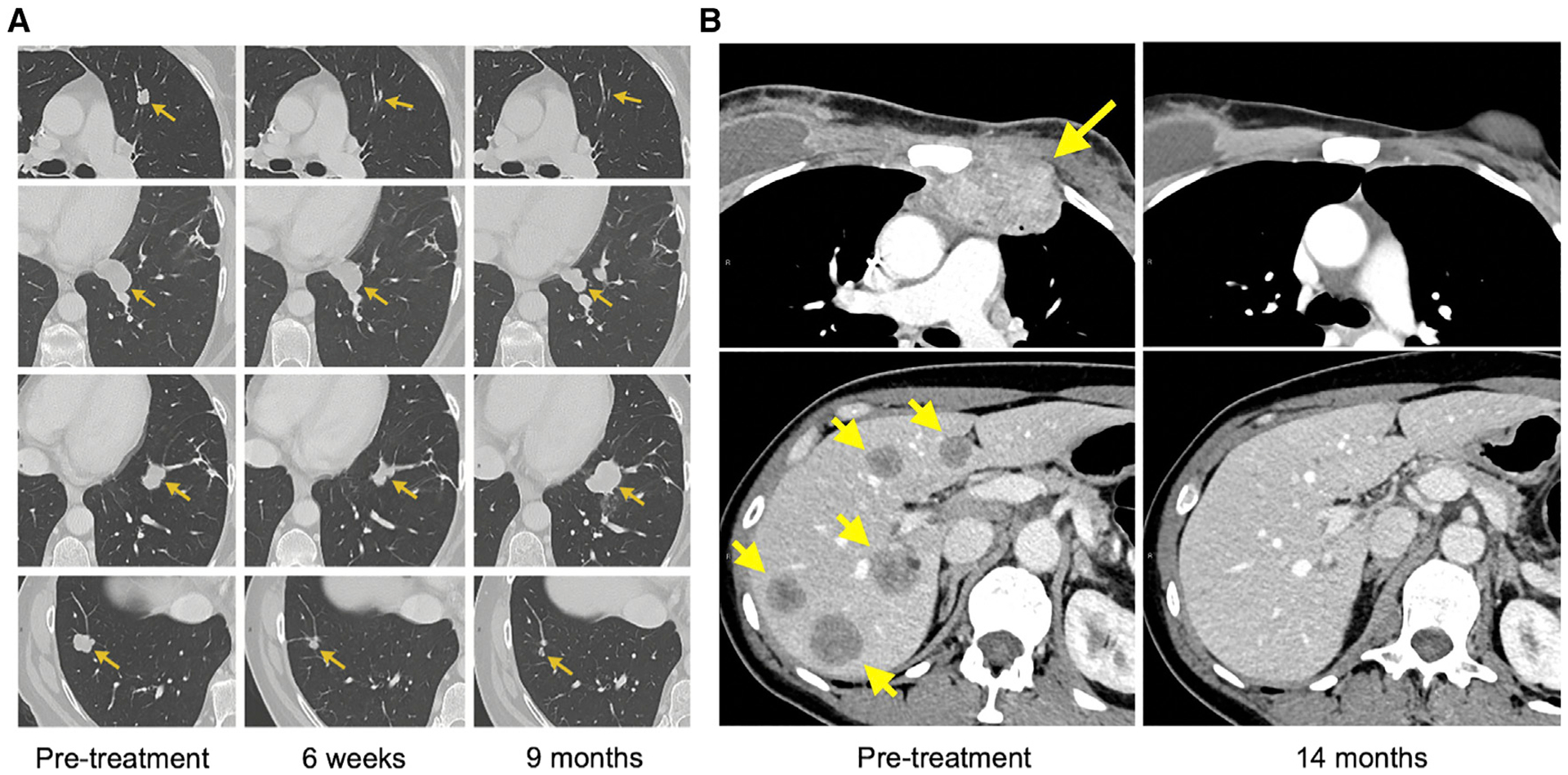
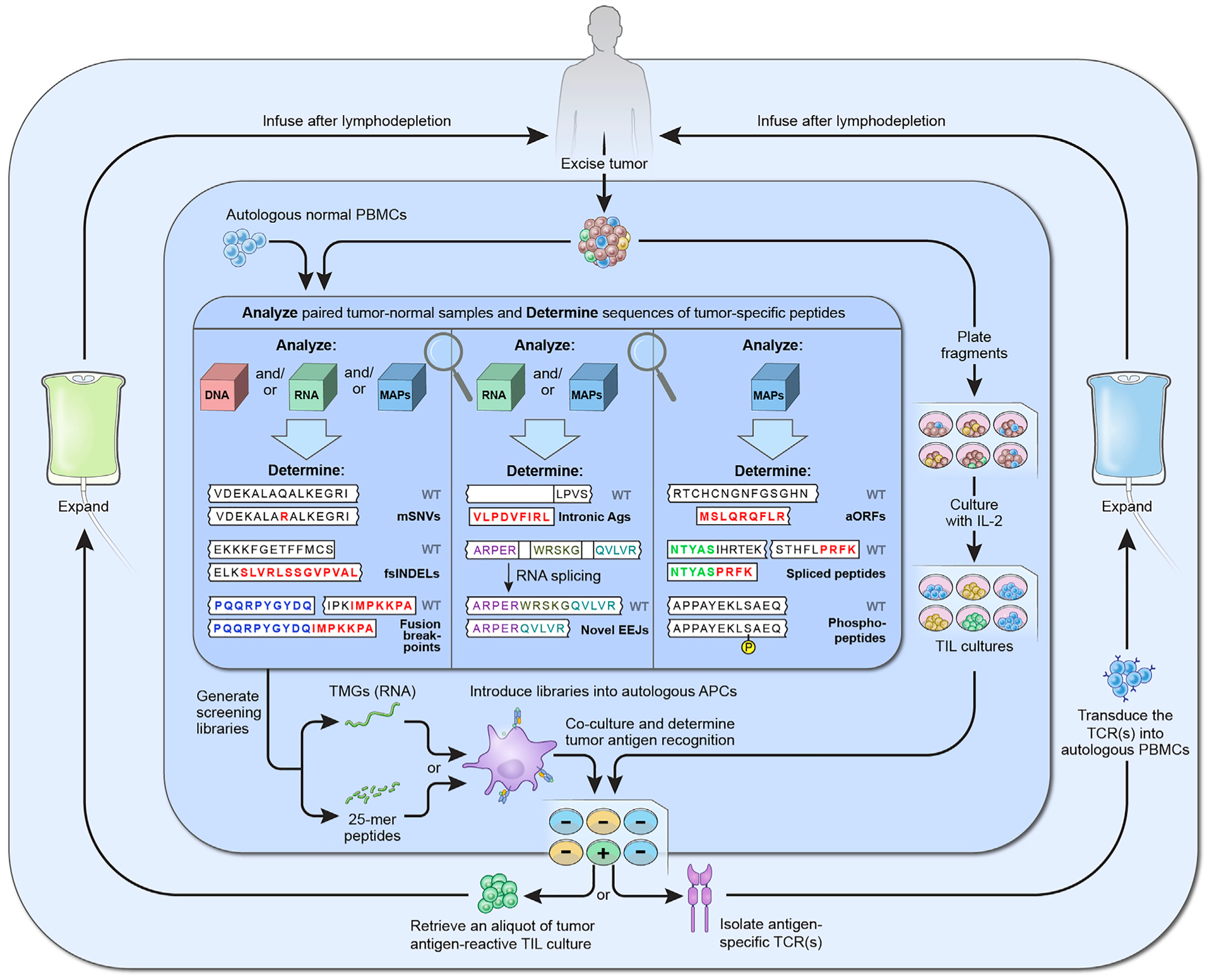
Cancer elimination in humans can be achieved with immunotherapy that relies on T lymphocyte-mediated recognition of tumor antigens. Several types of these antigens have been recognized based on their cellular origins and expression patterns, while their detection has been greatly facilitated by recent achievements in next-generation sequencing and immunopeptidomics. Some of them have been targeted in clinical trials with various immunotherapy approaches, while many others remain untested. Here, we discuss molecular identification of different tumor antigen types, and the clinical safety and efficacy of targeting them with immunotherapy. Additionally, we suggest strategies to increase the efficacy and availability of antigen-directed immunotherapies for treatment of patients with metastatic cancer.

Keywords: adoptive T cell transfer; cancer antigens; cancer immunology; cancer immunotherapy; tumor antigens; tumor immunology.


Figures

Figure 1.
Figure 1.. General Principles of Antigen Recognition by the Adaptive Immune System
Upon stimulation, B lymphocytes differentiate into plasma cells (left panel) to produce antibodies (immunoglobulins). These can bind to a variety of intact proteins (but also other molecules) that are exposed on the cell surface. The antigen-binding portions of an antibody are composed of variable light (VL) and variable heavy chain (VH) domains (insert). CAR T cells (middle panel) recognize the same types of molecules as do the antibodies. These cells have been modified to express CARs, which are constructed by fusing antibody-derived antigen-binding domains (VH + VL) with intracellular T cell signaling domains (insert). In contrast, conventional T cells (right panel) recognize peptides that are derived from cellular proteins and are displayed on cell surface in complex with MHC molecules. For clarity, only the presentation on MHC class I molecules is depicted, in a simplified manner.
Figure 2.
Figure 2.. Examples of Durable Tumor Regressions in Patients Who Were Treated with TILs That Recognized Tumor Neoantigens
(A) Regression of lung tumors in a patient with metastatic colorectal carcinoma who received KRASG12D-reactive TILs. The tumor in the second row exhibited a partial response and was surgically resected; the pathology showed no viable tumor cells. The tumor in the third row progressed 9 months after the therapy and was then surgically resected. The pathology demonstrated loss of the MHC molecule necessary for the presentation of the KRASG12D. (B) Complete regression of a chest wall mass (upper panels) and multiple liver tumors (lower panels) in a patient with metastatic breast cancer who received TILs directed against four different tumor mutations.
Figure 3.
Figure 3.. A Suggested Approach for Comprehensive Identification of Tumor Neoantigen-Reactive T Cells and Their Use for Personalized Immunotherapy
Three shaded areas in the figure represent suggested strategies for paired tumor-normal analysis to identify candidate tumor neoantigens (inner), TIL screening to identify neoantigen-reactive T cells (middle), and the use of reactive cells for immunotherapy (outer). Firstly, following surgical excision of one or more metastases, one tumor segment is further divided into 12–24 fragments, which are cultured with IL-2 in order to expand TILs (middle area, right). The other segment is analyzed together with a sample of normal cells, typically PBMCs, to identify sequences of candidate tumor antigens (inner area). This analysis can be performed on the cellular genome (DNA), transcriptome (RNA), and/or immunopeptidome (MAPs). Although most current analyses are limited to mSNVs and non-frameshift and fsINDELs, they could be further expanded to allow identification of other potential antigen types, such as fusion breakpoints, intronic antigens, novel EEJs, antigens translated from aORFs, and posttranslationally spliced or enzymatically modified peptides, such as phosphopeptides. Next, sequences of candidate antigens are used as templates to synthesize peptide and TMG screening libraries, which are then introduced to the autologous APCs (middle area, bottom). After the addition of TILs and the overnight co-cultures, such methods as measuring interferon-γ production or upregulation of T cell surface activation molecules (not depicted) are used to identify the TILs that recognize tumor neoantigens. Finally, reactive TILs are expanded to very large numbers ex vivo and reinfused back into the patient (outer area, left). Alternatively, TCRs that conferred tumor antigen recognition are retrovirally transduced into autologous PBMCs, which are then expanded and reinfused into the patient (outer area, right). Both approaches entail pretreating the patient with lymphodepleting chemotherapy, which enables engraftment of transferred lymphocytes, and are followed by intravenous IL-2 administration to stimulate their survival and proliferation.

References

    1. Aarnoudse CA, van den Doel PB, Heemskerk B, and Schrier PI (1999). Interleukin-2-induced, melanoma-specific T cells recognize CAMEL, an unexpected translation product of LAGE-1. Int. J. Cancer 82, 442–448. - PubMed
    1. Aleksic M, Liddy N, Molloy PE, Pumphrey N, Vuidepot A, Chang KM, and Jakobsen BK (2012). Different affinity windows for virus and cancer-specific T-cell receptors: implications for therapeutic strategies. Eur. J. Immunol 42, 3174–3179. - PMC - PubMed
    1. Almeida LG, Sakabe NJ, deOliveira AR, Silva MC, Mundstein AS, Cohen T, Chen YT, Chua R, Gurung S, Gnjatic S, et al. (2009). CTdata-base: a knowledge-base of high-throughput and curated data on cancer-testis antigens. Nucleic Acids Res. 37, D816–D819. - PMC - PubMed
    1. Bassani-Sternberg M, Braunlein E, Klar R, Engleitner T, Sinitcyn P, Audehm S, Straub M, Weber J, Slotta-Huspenina J, Specht K, et al. (2016). Direct identification of clinically relevant neoepitopes presented on native human melanoma tissue by mass spectrometry. Nat. Commun 7, 13404. - PMC - PubMed
    1. Bregni M, Badoglio M, Pedrazzoli P, and Lanza F (2016). Is allogeneic transplant for solid tumors still alive? Bone Marrow Transpl. 51, 751–752. - PubMed

Publication types

MeSH terms