

Viral anterior uveitis
- PMID: 32823392
- PMCID: PMC7690545
- DOI: 10.4103/ijo.IJO_928_20
Viral anterior uveitis
Abstract
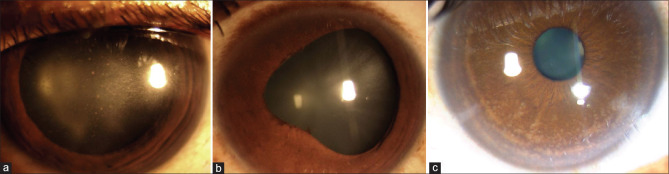
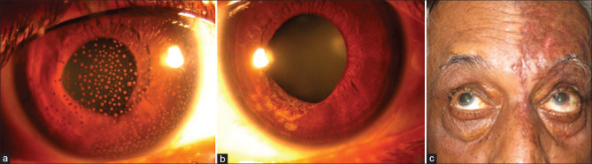
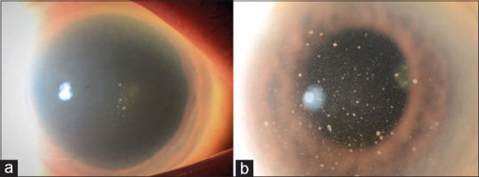
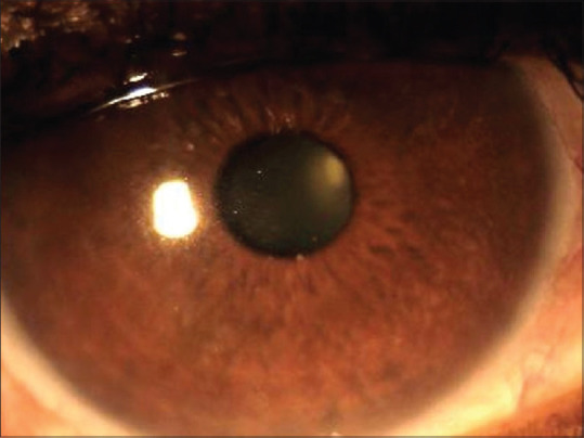
Viral anterior uveitis (VAU) needs to be suspected in anterior uveitis (AU) associated with elevated intraocular pressure, corneal involvement, and iris atrophic changes. Common etiologies of VAU include herpes simplex, varicella-zoster, cytomegalovirus, and rubella virus. Clinical presentations can vary from granulomatous AU with corneal involvement, Posner-Schlossman syndrome, Fuchs uveitis syndrome, and endothelitis. Due to overlapping clinical manifestations between the different viruses, diagnostic tests like polymerase chain reaction and Goldmann-Witmer coefficient analysis on the aqueous humor may help in identifying etiology to plan and monitor treatment.
Keywords: Cytomegalovirus; herpes simplex virus; ocular hypertension; rubella virus; varicella-zoster virus; viral anterior uveitis.
Conflict of interest statement
None
Figures
Comment in
-
Commentary: Clinical pearls and pitfalls in diagnosing viral anterior uveitis.Indian J Ophthalmol. 2020 Sep;68(9):1774. doi: 10.4103/ijo.IJO_1397_20. Indian J Ophthalmol. 2020. PMID: 32823393 Free PMC article. No abstract available.
References
-
- Groen-Hakan F, Babu K, Tugal-Tutkun I, Pathanapithoon K, de Boer JH, Smith JR, et al. Challenges of diagnosing viral anterior uveitis. Ocul Immunol Inflamm. 2017;25:710–20. - PubMed
-
- Cunningham ET., Jr The expanding spectrum of viral anterior uveitis. Ophthalmology. 2011;118:1903–4. - PubMed
-
- Jap A, Chee S-P. Viral anterior uveitis. Curr Opin Ophthalmol. 2011;22:483–8. - PubMed
-
- Chan NS, Chee S-P. Demystifying viral anterior uveitis: A review. Clin Exp Ophthalmol. 2019;47:320–33. - PubMed
-
- Relvas LJ, Caspers L, Chee S-P, Zierhut M, Willermain F. Differential diagnosis of viral-induced anterior uveitis. Ocul Immunol Inflamm. 2018;26:726–31. - PubMed
Publication types
MeSH terms
Substances
LinkOut - more resources
Full Text Sources
Medical
