Review
. 2020 Sep;68(9):1775-1786.
doi: 10.4103/ijo.IJO_1352_20.

Post-fever retinitis - Newer concepts

Affiliations
Review

Post-fever retinitis - Newer concepts

Padmamalini Mahendradas et al. Indian J Ophthalmol. 2020 Sep.

Abstract

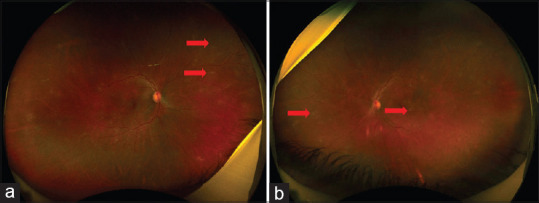
Post-fever retinitis (PFR) is an infectious or para-infectious uveitic entity caused by bacterial or viral agents and seen mainly in tropical countries. Systemic symptoms such as joint pain, skin rash are common during the febrile stage. On the basis of only clinical presentation, it is difficult to pin-point the exact etiology for PFR. Serological investigations, polymerase chain reaction, and knowledge of concurrent epidemics in the community may help to identify the etiological organism. Bacterial causes of PFR such as rickettsia and typhoid are treated with systemic antibiotics, with or without systemic steroid therapy, whereas PFR of viral causes such as chikungunya, dengue, West Nile virus, and Zika virus have no specific treatment and are managed with steroids. Nevertheless, many authors have advocated mere observation and the uveitis resolved with its natural course of the disease. In this article, we have discussed the clinical features, pathogenesis, investigations, and management of PFR.

Keywords: Chikungunya; Rickettsia; West Nile virus; Zika virus; dengue; post-fever retinitis; systemic steroids; typhoid.


Conflict of interest statement

None

Figures

Figure 1
Figure 1
A 6-year-old girl child presented with decreased vision in both the eyes for 12 days, after an episode of fever with skin rash 25 days ago. Child had a black eschar and OX K on Weil Felix test was positive suggesting of Scrub typhus. Fundus examination revealed disc edema, hemorrhages and a thick epiretinal membrane (ERM) in OD (a), similar findings with posterior vitreous detachmentin LE. (b) OCT scan confirmed vitreomacular traction with subretinal fluid (SRF) in OD (c) and showed indistinct retinal layer with thinning at the macula, minimal SRF with choroidal thinning in OS (d)
Figure 2
Figure 2
A 24-year-old female presented with complaints of blur vision in OD for 10 days preceding a viral fever as diagnosed by her physician. Investigations revealed positive WFT (OX2 and OX19) suggestive of ITT. Ocular examination showed normal anterior segments, but fundus exam revealed 1 + vitritis, cotton-wool spot such as retinitis lesions inferior to the disc with macular fan suggestive of resolving macular edema. (a) OCT scan passing over the retinitis lesion showed vitreous cells, thickening of inner retina with after shadowing and minimal subretinal fluid. (b)
Figure 3
Figure 3
(a) Wide field fundus photography of a 35-year-old Indian Male of the right eye showing supero-nasal areas of sheathing (block red arrow) in a patient of post typhoid fever. (b) left eye showing retinitis patches (block red arrow)
Figure 4
Figure 4
Fundus fluorescein angiography of the same patient in the late phases showing staining of the disc with staining and leakage from the retinal veins
Figure 5
Figure 5
Optical coherence tomography of the left eye of the same patient showing posterior vitreous cells (red hollow arrow) and inner retinal involvement (solid red arrow)
Figure 6
Figure 6
A 58-year-old male presented with decreased vision in both eyes since 15 days, after an episode of high fever and testing positive for NS1 antigen. Fundus examination of both eyes revealed retinal hemorrhages and cotton wool spots (a). Structural OCT scan showed cystoid macular edema (b). OCT Angiography (c) showed decreased vessel density in deep retinal plexus and flow void areas (arrowhead outline). After treatment, Fundus examination revealed almost resolved retinal hemorrhages (a). OCT scan showed resolved cystoid macular edema (b). OCTA showed reduction in flow void areas (c)
Figure 7
Figure 7
A 50-year-old female presented with decreased vision in both eyes, H/o Fever and NS1 antigen was positive. Orange lesion at fovea termed foveolitis (a). Structural OCT scan (b) right eye showed cystoid macular edema, left eye showed inner retinal thickening (arrow outline), and both eyes showed subfoveal outer retinal thickening known as foveolitis (arrowhead outline). OCT A showed decreased vessel density in deep retinal plexus and flow void areas (arrow heads). After treatment reduced cotton wool spots (a). OCT showed resolving CME (b). OCTA showed reduction in flow void areas (c)
Figure 8
Figure 8
(a and b) Fundus photography in a 64-year-old diabetic patient with serologically proven WNV infection shows multiple deep chorioretinal lesions, diffuse periarterial sheating, and moderate diabetic retinopathy with macular edema. (c and d) Mid-phase fluorescein angiograms shows inactive chorioretinal lesions, with a target-like appearance (central hypofluorescence and peripheral hyperfluorescence) more evident in the left eye. Several chorioretinal lesions extend in a linear pattern from the optic disc. Note the presence of retinal capillary leakage resulting from diabetic maculopathy
Figure 9
Figure 9
(a) Fundus photography in a 62-year-old diabetic patient with serologically confirmed WNV infection shows extensive deep retinal hemorrhages, retinal whitening, and diffuse periarterial sheathing. (b) Fluorescein angiography shows extensive areas of retinal capillary non-perfusion consistent with occlusive retinal vasculitis, with associated peripheral inactive chorioretinal lesions
Figure 10
Figure 10
A 49-year-old male, presented with complaints of decreased vision in both the eyes for 14 days, after an episode of fever with skin rash 6 weeks back. On investigation, Chikungunya Ig M antibody was positive. Ocular examination revealed vitritis +, hyperemic disc, retinitis, retina hemorrhages in both eyes with incomplete macular star in the right eye secondary to chikungunya retinitis
Figure 11
Figure 11
Fundus autofluorescence shows hyper and hypo autofluorescence lesions suggestive of resolving Zika virus retino-choroiditis
Figure 12
Figure 12
Fundus fluorescein angiography of the right eye showing early staining of the optic disc head with multiple areas of hyperfluorescence superonasal to the macula

References

    1. Vishwanath S, Badami K, Sriprakash KS, Sujatha BL, Shashidhar SD, Shilpa YD. Post-fever retinitis: A single center experience from South India. Int Ophthalmol. 2014;34:851–7. - PubMed
    1. Kawali A, Mahendradas P, Mohan A, Mallavarapu M, Shetty B. Epidemic retinitis. Ocul Immunol Inflamm. 2019;27:571–7. - PubMed
    1. Rahi M, Gupte MD, Bhargava A, Varghese GM, Arora R. DHR-ICMR Guidelines for diagnosis & management of Rickettsial diseases in India. Indian J Med Res. 2015;141:417–22. - PMC - PubMed
    1. Kato T, Watanabe K, Katori M, Terada Y, Hayasaka S. Conjunctival injection, episcleral vessel dilation, and subconjunctival hemorrhage in patients with new Tsutsugamushi disease. Jpn J Ophthalmol. 1997;41:196–9. - PubMed
    1. Khairallah M, Ladjimi A, Chakroun M, Messaoud R, Yahia SB, Zaouali S, et al. Posterior segment manifestations of rickettsia conorii infection. Ophthalmology. 2004;111:529–34. - PubMed