

An update on autoimmune retinopathy
- PMID: 32823399
- PMCID: PMC7690499
- DOI: 10.4103/ijo.IJO_786_20
An update on autoimmune retinopathy
Abstract
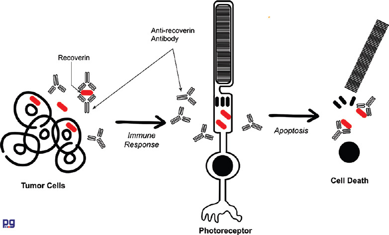
Autoimmune retinopathy (AIR) refers to a group of rare autoimmune retinal degenerative diseases presumably caused by cross-reactivity of serum autoantibodies against retinal antigens. The pathogenesis of AIR remains largely presumptive and there are a significant number of antiretinal antibodies that have been detected in association with AIR. The diagnosis of AIR is largely based on the demonstration of antiretinal antibodies in the serum along with suggestive clinical features and ancillary investigations. A high index of suspicion along with early diagnosis and treatment may play a critical role to lower the risk of irreversible immunological damage to the retinal cells in these patients. A multi-disciplinary approach for complete management and evaluation is helpful in such conditions. Various therapeutic options have been described for the treatment of AIR, though there is no consensus on standard treatment protocol.
Keywords: Autoimmune retinopathy; cancer-associated retinopathy; enolase; melanoma-associated retinopathy; recoverin.
Conflict of interest statement
None
Figures
References
-
- Sawyer RA, Selhorst JB, Zimmerman LE, Hoyt WF. Blindness caused by photoreceptor degeneration as a remote effect of cancer. Am J Ophthalmol. 1976;81:606–13. - PubMed
-
- Klingele TG, Burde RM, Rappazzo JA, Isserman MJ, Burgess D, Kantor O. Paraneoplastic retinopathy. J Clin Neuroophthalmol. 1984;4:239–45. - PubMed
-
- Gass J. Acute Vogt-Koyonagi-Harada-like Syndrome Occuring in a Patient with Metastatic Cutaneous. Melanoma Amsterdam, Netherland. 1984
-
- Thirkill CE, Roth AM, Keltner JL. Cancer-associated retinopathy. Arch Ophthalmol Chic Ill. 1960;1987(105):372–5. - PubMed
-
- Mizener JB, Kimura AE, Adamus G, Thirkill CE, Goeken JA, Kardon RH. Autoimmune retinopathy in the absence of cancer. Am J Ophthalmol. 1997;123:607–18. - PubMed
Publication types
MeSH terms
Substances
LinkOut - more resources
Full Text Sources
Medical
