

Expression Patterns of Coagulation Factor XIII Subunit A on Leukemic Lymphoblasts Correlate with Clinical Outcome and Genetic Subtypes in Childhood B-cell Progenitor Acute Lymphoblastic Leukemia
- PMID: 32823516
- PMCID: PMC7463512
- DOI: 10.3390/cancers12082264
Expression Patterns of Coagulation Factor XIII Subunit A on Leukemic Lymphoblasts Correlate with Clinical Outcome and Genetic Subtypes in Childhood B-cell Progenitor Acute Lymphoblastic Leukemia
Abstract
Background: Based on previous retrospective results, we investigated the association of coagulation FXIII subunit A (FXIII-A) expression pattern on survival and correlations with known prognostic factors of B-cell progenitor (BCP) childhood acute lymphoblastic leukemia (ALL) as a pilot study of the prospective multi-center BFM ALL-IC 2009 clinical trial.
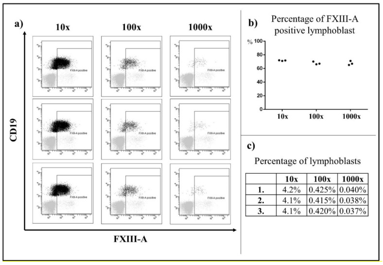
Methods: The study included four national centers (n = 408). Immunophenotyping by flow cytometry and cytogenetic analysis were performed by standard methods. Copy number alteration was studied in a subset of patients (n = 59). Survival rates were estimated by Kaplan-Meier analysis. Correlations between FXIII-A expression patterns and risk factors were investigated with Cox and logistic regression models.
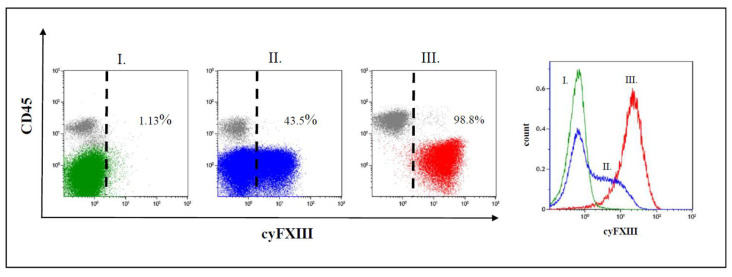
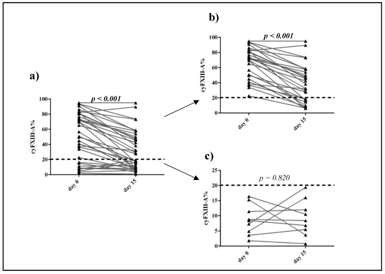
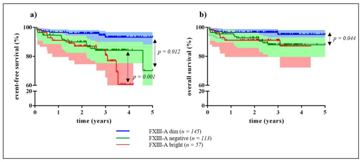
Results: Three different patterns of FXIII-A expression were observed: negative (<20%), dim (20-79%), and bright (≥80%). The FXIII-A dim expression group had significantly higher 5-year event-free survival (EFS) (93%) than the FXIII-A negative (70%) and FXIII-A bright (61%) groups. Distribution of intermediate genetic risk categories and the "B-other" genetic subgroup differed significantly between the FXIII-A positive and negative groups. Multivariate logistic regression confirmed independent association between the FXIII-A negative expression characteristics and the prevalence of intermediate genetic risk group.
Conclusions: FXIII-A negativity is associated with dismal survival in children with BCP-ALL and is an indicator for the presence of unfavorable genetic alterations.
Keywords: B-cell progenitor; Factor XIII subunit A; acute lymphoblastic leukemia; children; genetic risk categories; middle-income countries; survival.
Conflict of interest statement
The funding organization(s) played no role in the study design; in the collection, analysis, and interpretation of data; in the writing of the report; or in the decision to submit the report for publication.
Figures
References
-
- Acute Lymphoblastic Leukaemia Committee (All)—International Bfm Study Group. [(accessed on 18 June 2020)]; Available online: https://bfminternational.wordpress.com/structure/committees/acute-lympho....
-
- Howard S.C., Davidson A., Luna-Fineman S., Israels T., Chantada G., Lam C.G., Hunger S.P., Bailey S., Ribeiro R.C., Arora R.S., et al. A framework to develop adapted treatment regimens to manage pediatric cancer in low- and middle-income countries: The pediatric oncology in developing countries (podc) committee of the international pediatric oncology society (siop) Pediatr. Blood Cancer. 2017;64:e26879. doi: 10.1002/pbc.26879. - DOI - PubMed
-
- Stary J., Zimmermann M., Campbell M., Castillo L., Dibar E., Donska S., Gonzalez A., Izraeli S., Janic D., Jazbec J., et al. Intensive chemotherapy for childhood acute lymphoblastic leukemia: Results of the randomized intercontinental trial all ic-bfm 2002. J. Clin. Oncol. Off. J. Am. Soc. Clin. Oncol. 2014;32:174–184. doi: 10.1200/JCO.2013.48.6522. - DOI - PubMed
-
- Arber D.A., Orazi A., Hasserjian R., Thiele J., Borowitz M.J., Le Beau M.M., Bloomfield C.D., Cazzola M., Vardiman J.W. The 2016 revision to the world health organization classification of myeloid neoplasms and acute leukemia. Blood. 2016;127:2391–2405. doi: 10.1182/blood-2016-03-643544. - DOI - PubMed
Grants and funding
LinkOut - more resources
Full Text Sources
