Review
. 2020 Aug 13;25(16):3686.
doi: 10.3390/molecules25163686.

G-Quadruplexes at Telomeres: Friend or Foe?

Affiliations
Review

G-Quadruplexes at Telomeres: Friend or Foe?

Tracy M Bryan. Molecules. .

Abstract

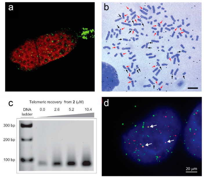
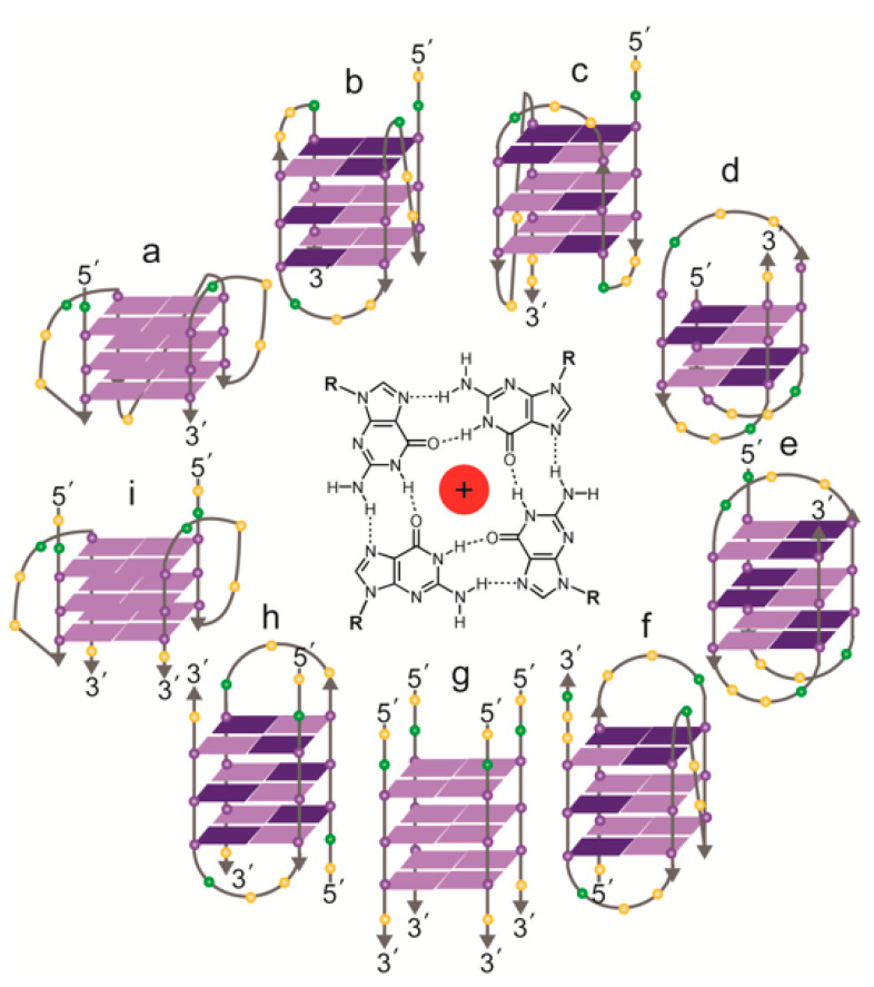
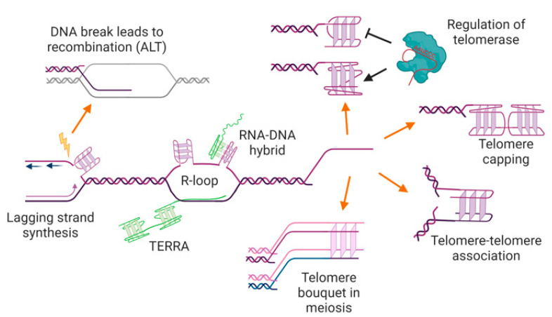
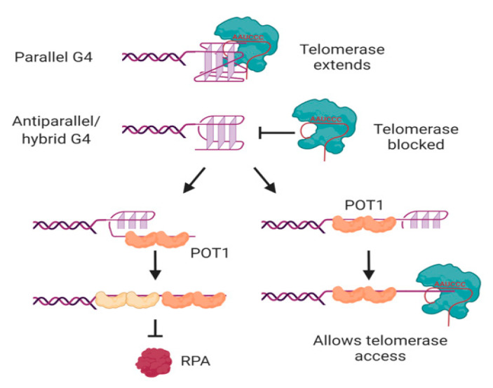
Telomeres are DNA-protein complexes that cap and protect the ends of linear chromosomes. In almost all species, telomeric DNA has a G/C strand bias, and the short tandem repeats of the G-rich strand have the capacity to form into secondary structures in vitro, such as four-stranded G-quadruplexes. This has long prompted speculation that G-quadruplexes play a positive role in telomere biology, resulting in selection for G-rich tandem telomere repeats during evolution. There is some evidence that G-quadruplexes at telomeres may play a protective capping role, at least in yeast, and that they may positively affect telomere maintenance by either the enzyme telomerase or by recombination-based mechanisms. On the other hand, G-quadruplex formation in telomeric DNA, as elsewhere in the genome, can form an impediment to DNA replication and a source of genome instability. This review summarizes recent evidence for the in vivo existence of G-quadruplexes at telomeres, with a focus on human telomeres, and highlights some of the many unanswered questions regarding the location, form, and functions of these structures.

Keywords: G-quadruplex; telomerase; telomere.


Conflict of interest statement

The author declares no conflict of interest.

Figures

Figure 1
Figure 1
Examples of direct evidence for formation of G-quadruplexes at telomeres. (a) Immunofluorescence of a Stylonychia lemnae cell using an antibody raised against telomeric G-quadruplexes (green). DNA is counterstained in red; the replication band is the unstained region extending across the cell. Image from [35]. (b) Autoradiograph of metaphase spread of human T98G cells cultured with labeled G4 ligand 3H-360A for 48 h. Black arrows indicate silver grains on the terminal regions and red arrows indicate silver grains on the interstitial regions. Bar = 10 µm. Image from [36]. (c) Pull-down of telomeric DNA from human HT1080 cells using the indicated concentrations of a derivative of G4 ligand pyridostatin attached to an affinity tag (2). Genomic DNA was sheared into 100–300 bp pieces prior to pulldown, and telomeric sequences detected by PCR amplification. Reprinted by permission from Springer Nature [37]. (d) Immunofluorescence of a human 293T cell using the BG4 antibody against G-quadruplexes (green) together with fluorescence in situ hybridization against telomeric DNA (red). Arrows indicate G4-telomere colocalizations. Image by A.L. Moye and T.M. Bryan.
Figure 2
Figure 2
Possible relationships between t-loops and G-quadruplex structures. Top left: schematic of a t-loop formed by intercalation of a telomeric 3′ overhang into the duplex portion of a telomere; it is also possible for the other strand to participate in stabilizing the junction [81]. It is possible that G-quadruplexes could form in the displaced G-strand (the “D-loop”), or involving the 3′ overhang at the junction (not shown). It has been shown that RNA transcribed from telomeres (TERRA; green) localizes to the t-loop junction, possibly through DNA-RNA G-quadruplex formation [81]. Bottom left: electron microscopy image of a t-loop in genomic DNA isolated from human HeLa cells; image by Jack D. Griffith. Top right: it is possible that G-quadruplexes form at the telomeric overhang at times in the cell cycle when t-loops are resolved, although there is no direct evidence for this at present. Bottom right: Electron micrograph showing G-quadruplex formation in a long single-stranded telomeric fragment; image by Jack D. Griffith. Arrows indicate bead-like structures that represent higher-order interactions between multiple G-quadruplexes [82]. Figure created with BioRender.com.
Figure 3
Figure 3
Topologies of solved structures of human telomeric G-quadruplexes, either intramolecular (af) or intermolecular (gi). Centre: a G-quartet, comprising four guanines, stabilized by a central cation. (a) Crystal structure of AG3(T2AG3)3 in K+ (parallel monomer) [84]; (b) NMR structure of TAG3(T2AG3)3 in K+ (hybrid form 1) [85]; (c) NMR structure of TAG3(T2AG3)3TT or TTAG3(T2AG3)3TT in K+ (hybrid form 2) [85,87]; (d) NMR structure of G3T2A(BrG)G2T(TAG3T)2 in K+ (antiparallel form 3) [86]; (e) NMR structure of AG3(T2AG3)3 in Na+ (antiparallel) [89]; (f) NMR structure of (T2AG3)3TTA(BrG)G2T2A in Na+ (antiparallel) [90]; (g) NMR structure of T2AG3T in K+ (parallel tetramer) [92]; (h) NMR structure of UAG3T(BrU)AG3T in K+ (antiparallel dimer) [93]; (i) crystal structure of (TAG3T)2 in K+ (parallel dimer) [84]; the same topology was seen in equilibrium with (h) by NMR with TAG3UTAG3T in K+ [93]. Guanines: purple spheres; thymines: yellow spheres; adenines: green spheres. Syn guanines shown in dark purple, anti guanines in light purple.
Figure 4
Figure 4
Potential locations, functions, and consequences of G-quadruplexes at telomeres. G-quadruplexes may form in the single-stranded telomere overhang, where they may positively or negatively regulate telomerase and/or have a capping function, preventing access to the DNA repair machinery. G-quadruplexes at overhangs may also mediate interactions between two telomeres (e.g., during sister chromatid cohesion, or in the macronucleus of ciliated protozoa), or be involved in telomere clustering in meiosis. G-quadruplexes could also form in the double-stranded region of the telomere during DNA replication or transcription, where they may trigger genome instability and/or recombination-mediated telomere maintenance. The RNA transcribed from telomeres, TERRA, can also form into G-quadruplexes, either unimolecular or as an RNA-DNA hybrid. See text for details and references. Figure created with BioRender.com.
Figure 5
Figure 5
Interactions of telomerase, POT1 and RPA with human telomeric G-quadruplexes. Telomerase can bind and extend parallel, but not antiparallel or hybrid, G-quadruplexes. POT1 binds to antiparallel or hybrid G-quadruplexes through a mechanism in which G4 unfolding precedes “trapping” of the unfolded DNA by POT1. The two OB folds of each POT1 molecule bind to consensus binding site TTAGGGTTAG; sequential binding of two POT1 molecules therefore coats the 4-repeat telomeric DNA (left). Although RPA also has the ability to unwind G-quadruplexes, POT1 competes with this activity. If binding of POT1 occurs at the 5′ region of the DNA, the 3′ tail can form a substrate for telomerase (right). Not shown is POT1’s binding partner TPP1, which also influences G4 unwinding dynamics and telomerase activity. See text for details and references. Figure created with BioRender.com.

References

    1. Wellinger R.J., Sen D. The DNA structures at the ends of eukaryotic chromosomes. Eur. J. Cancer. 1997;33:735–749. doi: 10.1016/S0959-8049(97)00067-1. - DOI - PubMed
    1. McClintock B. The stability of broken ends of chromosomes in Zea mays. Genetics. 1941;26:234–282. - PMC - PubMed
    1. Muller H.J. The re-making of chromosomes. Collect. Net. 1938;13:181–198.
    1. Olovnikov A.M. Principle of marginotomy in template synthesis of polynucleotides. Dokl. Akad. Nauk Sss. 1971;201:1496–1499. - PubMed
    1. Watson J.D. Origin of concatemeric T7 DNA. Nat. New Biol. 1972;239:197–201. doi: 10.1038/newbio239197a0. - DOI - PubMed

Substances

LinkOut - more resources