. 2020 Aug 13;12(8):885.
doi: 10.3390/v12080885.

Low-Level Ionizing Radiation Induces Selective Killing of HIV-1-Infected Cells with Reversal of Cytokine Induction Using mTOR Inhibitors

Affiliations

Low-Level Ionizing Radiation Induces Selective Killing of HIV-1-Infected Cells with Reversal of Cytokine Induction Using mTOR Inhibitors

Daniel O Pinto et al. Viruses. .

Abstract

HIV-1 infects 39.5 million people worldwide, and cART is effective in preventing viral spread by reducing HIV-1 plasma viral loads to undetectable levels. However, viral reservoirs persist by mechanisms, including the inhibition of autophagy by HIV-1 proteins (i.e., Nef and Tat). HIV-1 reservoirs can be targeted by the "shock and kill" strategy, which utilizes latency-reversing agents (LRAs) to activate latent proviruses and immunotarget the virus-producing cells. Yet, limitations include reduced LRA permeability across anatomical barriers and immune hyper-activation. Ionizing radiation (IR) induces effective viral activation across anatomical barriers. Like other LRAs, IR may cause inflammation and modulate the secretion of extracellular vesicles (EVs). We and others have shown that cells may secrete cytokines and viral proteins in EVs and, therefore, LRAs may contribute to inflammatory EVs. In the present study, we mitigated the effects of IR-induced inflammatory EVs (i.e., TNF-α), through the use of mTOR inhibitors (mTORi; Rapamycin and INK128). Further, mTORi were found to enhance the selective killing of HIV-1-infected myeloid and T-cell reservoirs at the exclusion of uninfected cells, potentially via inhibition of viral transcription/translation and induction of autophagy. Collectively, the proposed regimen using cART, IR, and mTORi presents a novel approach allowing for the targeting of viral reservoirs, prevention of immune hyper-activation, and selectively killing latently infected HIV-1 cells.

Keywords: HIV-1; HIV-1 therapy; Ionizing radiation; autophagy; cell death; extracellular vesicles; inflammation; latency reversal; mTOR inhibition; shock and kill.


Conflict of interest statement

The authors declare no conflict of interest. The sponsors had no role in the design, execution, interpretation, or writing of the study.

Figures

Figure 1
Figure 1
Catalase and superoxide dismutase dictate the survival of irradiated HIV-1-infected cells. (A) Chronically HIV-1-infected T-cell line ACH2 and pro-monocytes U1, and corresponding uninfected CEM and U937, treated with 10 Gy of ionizing radiation (IR), lysed (5 days post-IR) and probed for catalase (CAT) and superoxide dismutase (SOD) via Western blot. (B) Densitometry analysis of the CAT and SOD bands was performed. (C) Two dosages of exogenous CAT (3.08 U) and SOD (16.82 U) were added to U1 and ACH2 cells in two dosages to over the time period of 5 days (Day 1 and 3) to test for cell viability recovery after IR treatment.
Figure 2
Figure 2
IR and autophagy inducing (AI) drugs affect the cell viability of HIV-1-infected cells. (A) Cell viability of HIV-1-infected monocytes cells (U1) was performed 48 h post-treatment with IR (5 Gy), Rapa (50 nM; AI), and IR/Rapa. (B) The same treatment was performed on HIV-1-infected T-cells (ACH2). (C) The therapeutic window of Rapamycin (Rapa) at 0, 15, 150, and 300 nM was assessed for U1 cells. (D) Additionally, the same titration was performed in ACH2 cells.
Figure 3
Figure 3
A combination of IR, AI, and serum starvation induces cell death of HIV-1-infected cells. Cell viability was assessed at two-time points (72 h and 120 h). All cells were pretreated for 3 days with cART (10 µM cocktail). Additionally, cells were treated with IR-/+(10 Gy), 0.1% DMSO (control), Rapa (300 nM; AI via mTOR inhibition) or SP600125 (300 nM; JNK pathway inhibitor). (A) Time titration for U1 and (B) ACH2 cells.
Figure 3
Figure 3
A combination of IR, AI, and serum starvation induces cell death of HIV-1-infected cells. Cell viability was assessed at two-time points (72 h and 120 h). All cells were pretreated for 3 days with cART (10 µM cocktail). Additionally, cells were treated with IR-/+(10 Gy), 0.1% DMSO (control), Rapa (300 nM; AI via mTOR inhibition) or SP600125 (300 nM; JNK pathway inhibitor). (A) Time titration for U1 and (B) ACH2 cells.
Figure 4
Figure 4
AI drugs mediate viral RNA levels in infected myeloid and T-cells. AI drugs (mTOR inhibitors; Rapa and INK128 at 300 nM) were administered as described in Figure 3, and cells were allowed to incubate for 5 days. Rapa is an mTOR complex-1 inhibitor, while INK128 is a more potent mTOR complex-1/2 inhibitor. Intracellular short RNA transcript TAR and env RNA was measured via RT-qPCR for U1 (A) and ACH2 (B) cells.
Figure 5
Figure 5
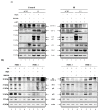
IR and AI treatment decrease Pr55/p24, CD63, and TNF-α protein expression in HIV-1-infected cells. EVs from supernatant material from cells harvested in Figure 4 were isolated with NT80/82. ACH2 and U1 cells (A) and PBMC patient samples (n = 4) (B) were Western blotted for TNF-α, Pr55/p24, CD63 and actin. Non-specific bands were denoted by “ns”.
Figure 6
Figure 6
mTORi drugs mitigate the effects of EV-TNF-α and viral proteins on recipient cells. (A) Nanotraped EVs (100 μL; NT80/82) from four PBMCs treated with control (untreated), IR (10 Gy), IR/Rapa (10 Gy/300 nM), and IR/INK128 (10 Gy/300 nM) were cultured with HLM-1 recipient cells which contain a triple mutation in Tat and transcription can be induced by IR and TNF-α. Western Blot analysis was used to detect the presence of viral proteins (i.e., p24, gp120, gp41), EVs marker (i.e., CD63), cytokine (i.e., mTNF-α and TNF-α), and actin. (B) Cell pellets from PBMCs 9–12 infected with HIV-1 89.6 were treated with IR (0.5 Gy), Rapa (50 nM) or INK128 (50 nM), and 3% FBS media for 5 days prior to Western Blot analysis for markers of cell death (i.e., PARP-1 and Bax) and virus (i.e., Nef).
Figure 6
Figure 6
mTORi drugs mitigate the effects of EV-TNF-α and viral proteins on recipient cells. (A) Nanotraped EVs (100 μL; NT80/82) from four PBMCs treated with control (untreated), IR (10 Gy), IR/Rapa (10 Gy/300 nM), and IR/INK128 (10 Gy/300 nM) were cultured with HLM-1 recipient cells which contain a triple mutation in Tat and transcription can be induced by IR and TNF-α. Western Blot analysis was used to detect the presence of viral proteins (i.e., p24, gp120, gp41), EVs marker (i.e., CD63), cytokine (i.e., mTNF-α and TNF-α), and actin. (B) Cell pellets from PBMCs 9–12 infected with HIV-1 89.6 were treated with IR (0.5 Gy), Rapa (50 nM) or INK128 (50 nM), and 3% FBS media for 5 days prior to Western Blot analysis for markers of cell death (i.e., PARP-1 and Bax) and virus (i.e., Nef).
Figure 7
Figure 7
Combination of cART, Starvation, IR, and mTORi drugs suppress the effects of TNF-α and viral proteins on recipient cells. The HIV-1-infected myeloid and T-cells, located in latent reservoirs, persist despite cART pretreatment. Ionizing radiation (IR) activates HIV-1 and induces cellular stress, which leads to cell death, with increased resilience in myeloid cells. The infected cells also secrete EVs associated with pro-inflammatory cytokines (i.e., mTNF-α and TNF-α) and viral molecules (i.e., p24, Pr55, gp120, gp41, and TAR RNA) with adverse effects on neighboring recipient cells. Drugs that inhibit mTOR (i.e., Rapa and INK128), along with serum starvation, suppress the release of cytokines and viral molecules, preventing LRA related side effects. Figure created with BioRender.com.

References

    1. Global AIDS Update 2019. UNAIDS; Geneva, Switzerland: 2019. p. 316.
    1. Hatano H., Jain V., Hunt P.W., Lee T.H., Sinclair E., Do T.D., Hoh R., Martin J.N., McCune J.M., Hecht F., et al. Cell-based measures of viral persistence are Associated with immune activation and programmed cell death protein 1 (PD-1)–expressing CD4+ T cells. J. Infect. Dis. 2013;208:50–56. doi: 10.1093/infdis/jis630. - DOI - PMC - PubMed
    1. Imamichi H., Dewar R.L., Adelsberger J.W., Rehm C.A., O’Doherty U., Paxinos E.E., Fauci A.S., Lane H.C. Defective HIV-1 proviruses produce novel protein-coding RNA species in HIV-infected patients on combination antiretroviral therapy. PNAS. 2016;113:8783–8788. doi: 10.1073/pnas.1609057113. - DOI - PMC - PubMed
    1. Iordanskiy S., Van Duyne R., Sampey G.C., Woodson C.M., Fry K., Saifuddin M., Guo J., Wu Y., Romerio F., Kashanchi F., et al. Therapeutic doses of irradiation activate viral transcription and induce apoptosis in HIV-1 infected cells. Virology. 2015;485:1–15. doi: 10.1016/j.virol.2015.06.021. - DOI - PMC - PubMed
    1. Fields J.A., Spencer B., Swinton M., Qvale E.M., Marquine M.J., Alexeeva A., Gough S., Soontornniyomkij B., Valera E., Masliah E., et al. Alterations in brain TREM2 and Amyloid-β levels are associated with neurocognitive impairment in HIV-infected persons on antiretroviral therapy. J. Neurochem. 2018 doi: 10.1111/jnc.14582. - DOI - PMC - PubMed

Publication types

MeSH terms

LinkOut - more resources