. 2020 Aug 13;21(16):5811.
doi: 10.3390/ijms21165811.

EphrinA1-Fc Attenuates Ventricular Remodeling and Dysfunction in Chronically Nonreperfused WT but not EphA2-R-M mice

Affiliations

EphrinA1-Fc Attenuates Ventricular Remodeling and Dysfunction in Chronically Nonreperfused WT but not EphA2-R-M mice

K'Shylah S Whitehurst et al. Int J Mol Sci. .

Abstract

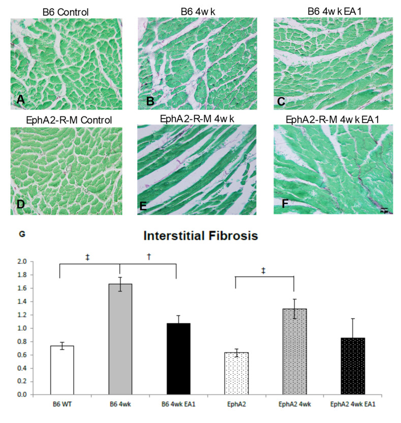
Background: EphrinA1-Fc abolishes acute I/R injury and attenuates nonreperfused cardiac injury 4 days after permanent occlusion in mice. The goal of this study was to assess the capacity of a single intramyocardial administration of ephrinA1-Fc at the time of coronary artery ligation, to determine the degree to which early salvage effects translate to reduced adverse remodeling after 4 weeks of nonreperfused myocardial infarction (MI) in wild-type B6 and EphA2-R-M (EphA2 receptor null) mice.

Methods: At 4 weeks post-MI, echocardiography, histologic and immunohistochemical analyses of B6 mouse hearts were performed. Primary mouse cardiac fibroblasts (FBs) isolated from B6 mice cultured in the presence of low and high dose ephrinA1-Fc, both with and without pro-fibrotic TGF-β stimulation and Western blots, were probed for relative expression of remodeling proteins MMP-2, MMP-9 and TIMP-1, in addition to DDR2 and (p)SMAD2/3/totalSMAD2/3.

Results: EphrinA1-Fc preserved a significant degree of contractile function, decreased adverse left ventricular remodeling, attenuated excessive compensatory hypertrophy, and decreased interstitial fibrosis in wild-type (WT) B6 mouse hearts. In contrast, most of these parameters were poorer in ephrinA1-Fc-treated EphA2-R-M mice. Of note, fibrosis was proportionately decreased, implying that other EphA receptor(s) are more important in regulating the pro-fibrotic response. Primary FBs showed disparate alteration of MMP-2, MMP-9 and TIMP-1, as well as DDR2 and p-SMAD2/3/totalSMAD2/3, which indicates that matrix remodeling and cardiac fibrosis in the injured heart are influenced by ephrinA1-Fc.

Conclusion: This study demonstrates the capacity of a single administration of ephrinA1-Fc at the onset of injury to attenuate long-term nonreperfused post-MI ventricular remodeling that results in progressive heart failure, and the important role of EphA2 in mitigating the deleterious effects.

Keywords: EphA2; ephrinA1; fibrosis; myocardial infarction; remodeling.


Conflict of interest statement

All authors declare that there are no conflict of interest.

Figures

Figure 1
Figure 1
Kaplan–Meier Estimate Analysis Demonstrating Survival Rates in WT and EphA2-R-M Mouse Hearts. Kaplan–Meier graph showing survival rates of IgG-Fc (IgG; n = 8)- and ephrinA1-Fc (EA1; n = 11)-treated WT B6 and EphA2-R-M mice 1 week (same as 4 weeks) post-MI. These data show the poorer survival of EphA2-R mice treated with EA1 (n = 4) compared to IgG (n = 10) and both WT groups.
Figure 2
Figure 2
Hematoxylin and Eosin (H&E) Stains for Morphometric Analyses and Calculated Expansion Index in WT and EphA2-R-M Mouse Hearts. Representative H&E stains (20×) of WT (AC) and EphA2-R-M (DF) hearts used for morphometric determination of left ventricular remodeling indices (left ventricular area, scar area and thickness, chamber area) demonstrating that EA1 had a nonsignificant tendency to reduce adverse remodeling in WT B6 mouse hearts, but actually worsened infarct expansion (G) in EphA2-R-M mouse hearts. B6 WT control (n = 10), EphA2-R-M control (n = 10), WT B6 (IgG-Fc 4w, n = 8), EphA2-R-M (IgG-Fc, n = 10), EA1-treated WT B6 (n = 11) and ephrinA1-Fc-treated EphA2-R-M (n = 4). Statistical significance * p < 0.05 and p < 0.01).
Figure 3
Figure 3
Myocyte Cross-Sectional Area in WT and EphA2-R-M Mouse Hearts. Myocyte cross-sectional area in WT and EphA2-R-M mice in uninjured control, untreated (IgG-Fc) vs. ephrinA1-Fc- treated mice at 4 weeks post-MI (stain: hematoxylin and eosin; 400×). Representative images in WT B6 (AC) but not in EphA2-R-M (DF) mouse hearts show that EA1 significantly attenuated compensatory hypertrophy (G), indicating that progression to the heart failure phenotype may be accelerated in mice deficient in EphA2-R. B6 WT control (n = 10), EphA2-R-M control (n = 10), WT B6 (IgG-Fc 4w, n = 8), EphA2-R-M (IgG-Fc, n = 10), EA1-treated WT B6 (n = 11) and ephrinA1-Fc-treated EphA2-R-M (n = 4).
Figure 4
Figure 4
Interstitial Fibrosis in WT and EphA2-R-M Mouse Hearts. WT (AC) and EphA2-R-M (DF) images depicting interstitial fibrosis 4 weeks after infarction (stain: picrosirius red/fast green; 400x). The fibrotic response was attenuated by EA1 in WT B6 mouse hearts but not EphA2-R-M mouse hearts (G). B6 WT control (n = 10), EphA2-R-M control (n = 10), WT B6 (IgG-Fc 4w, n = 8), EphA2-R-M (IgG-Fc, n = 10), EA1-treated WT B6 (n= 11) and ephrinA1-Fc-treated EphA2-R-M (n = 4); p < 0.05 and p < 0.001.
Figure 5
Figure 5
Cardiac Function in WT B6 and EphA2-R-M Mouse Hearts. EF (A) improved 10.7% from untreated to ephrinA1-Fc- treated B6 mice, but there was no statistically significant difference between treated and untreated EphA2 mice (* p < 0.05, p < 0.01, p < 0.001). B6 WT control (n = 11), EphA2-R-M control (n = 22), WT B6 (IgG-Fc 4w, n = 12), EphA2-R-M (IgG-Fc, n = 13), EA1-treated WT B6 (n = 11) and ephrinA1-Fc-treated EphA2-R-M (n = 4). (B) Representative traces for all WT and EphA2-R-M groups.
Figure 6
Figure 6
Active Myofibroblasts in Infarcted WT B6 (AC) and EphA2-R-M (DF) Mouse Hearts. Smooth muscle α-actin positive cells in uninjured B6 and EphA2 control heart tissue demonstrate expected staining of VSMCs in large vessels (200×). At 4 weeks post-MI, SMα-A positive myofibroblasts are visible in the infarcted myocardium in both B6 and EphA2, but to a much lesser extent in ephrinA1-Fc-treated B6 and EphA2-R-M mice (n = 3/group).
Figure 7
Figure 7
Expression Changes in MMP-2, MMP-9, TIMP1, DDR2 and (p)SMAD2/3/total SMAD2/3 in B6 and EphA2-R-M Mouse Hearts. MMP-9, MMP-2 and TIMP-1 expression in ephrinA1-Fc-treated +/-TGF-β primary cardiac fibroblasts are modulated in a dose-dependent manner (n = 3/group; significance: * vs. control, vs. 0.1 µM EA1, vs. TGF-β). Western blots of DDR2 and (p)SMAD2/3/totalSMAD2/3 show dose-dependent changes in response to ephrinA1-Fc-treatment +/-TGF-β (NS).

References

    1. Pasquale E.B. Eph-ephrin bidirectional signaling in physiology and disease. Cell. 2008;133:38–52. doi: 10.1016/j.cell.2008.03.011. - DOI - PubMed
    1. Kullander K., Klein R. Mechanisms and functions of Eph and ephrin signalling. Nat. Rev. Mol. Cell Biol. 2002;3:475–486. doi: 10.1038/nrm856. - DOI - PubMed
    1. Brantley D.M., Cheng N., Thompson E.J., Lin Q., Brekken R.A., Thorpe P.E., Muraoka R.S., Cerretti D.P., Pozzi A., Jackson D., et al. Soluble Eph A receptors inhibit tumor angiogenesis and progression in vivo. Oncogene. 2002;21:7011–7026. doi: 10.1038/sj.onc.1205679. - DOI - PubMed
    1. McCarron J.K., Stringer B.W., Day B.W., Boyd A.W. Ephrin expression and function in cancer. Future Oncol. 2010;6:165–176. doi: 10.2217/fon.09.146. - DOI - PubMed
    1. Lisabeth E.M., Falivelli G., Pasquale E.B. Eph receptor signaling and ephrins. Cold Spring Harb. Perspect. Biol. 2013;5:a009159. doi: 10.1101/cshperspect.a009159. - DOI - PMC - PubMed

MeSH terms