. 2020 Aug 13;9(8):1895.
doi: 10.3390/cells9081895.

The Hippo Pathway Effector YAP1 Regulates Intestinal Epithelial Cell Differentiation

Affiliations

The Hippo Pathway Effector YAP1 Regulates Intestinal Epithelial Cell Differentiation

Sepideh Fallah et al. Cells. .

Abstract

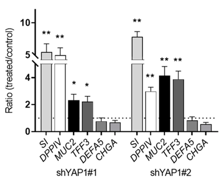
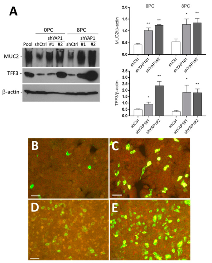
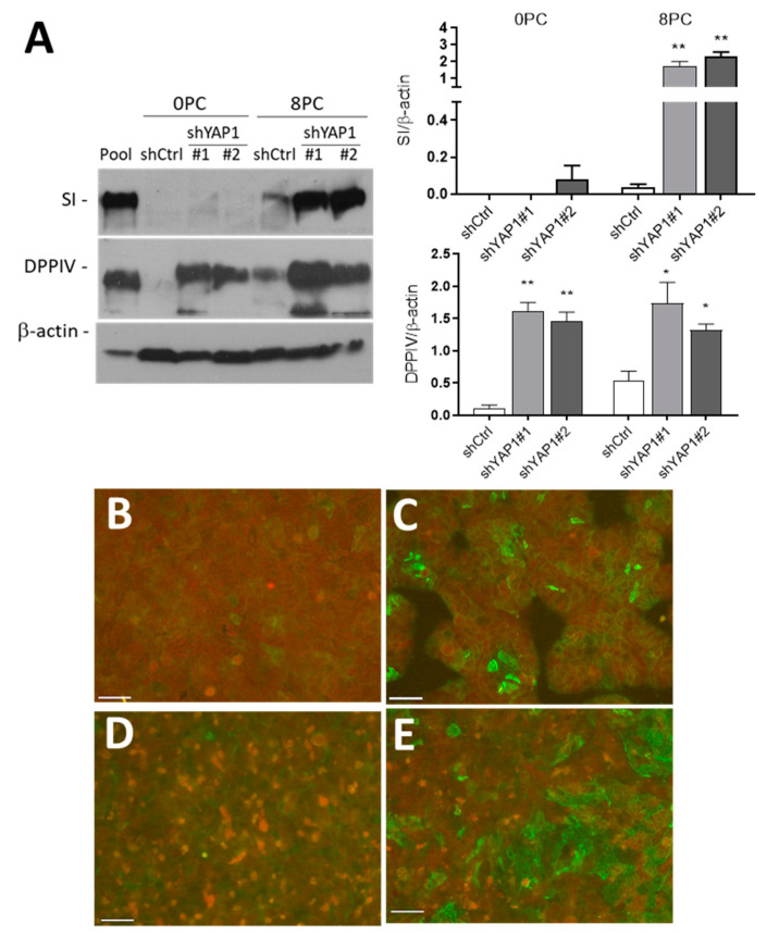
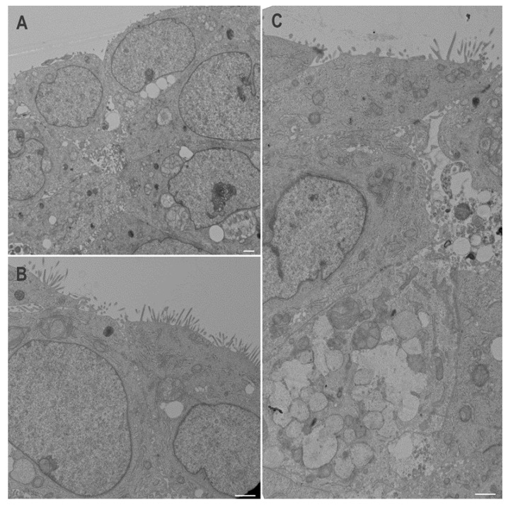
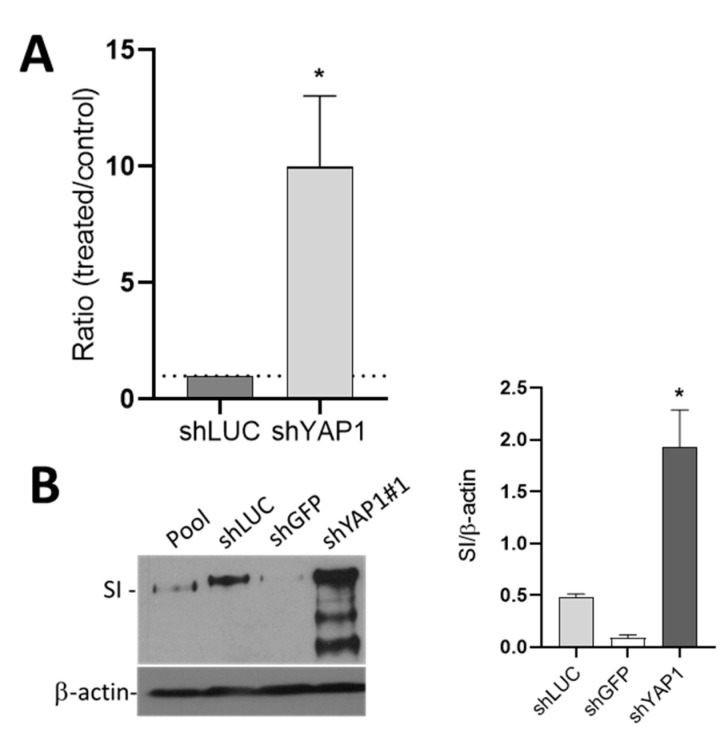
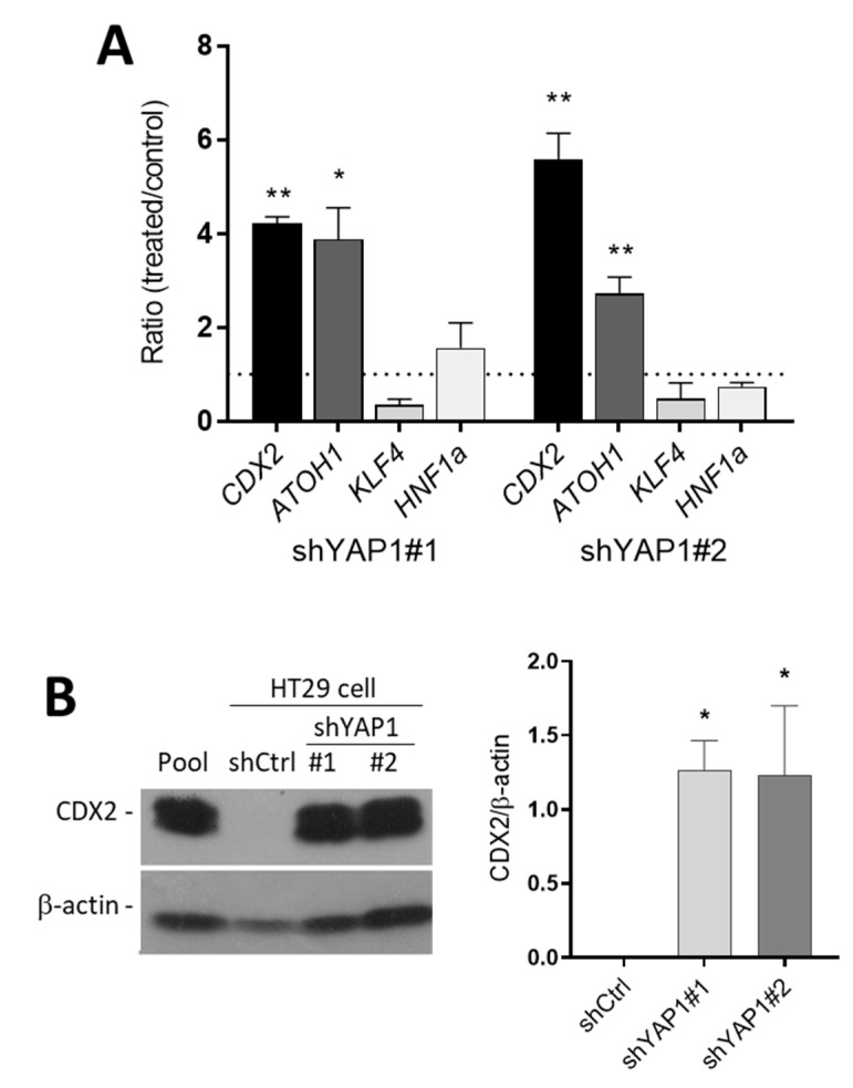
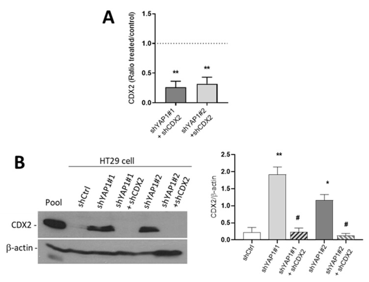
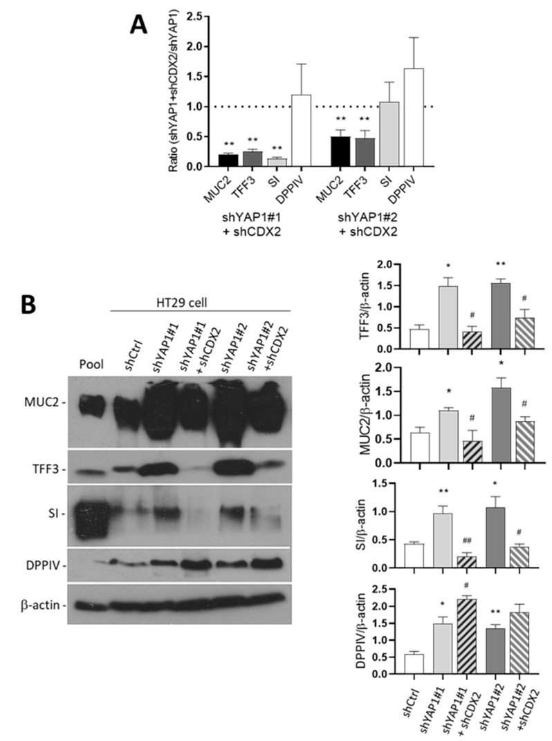
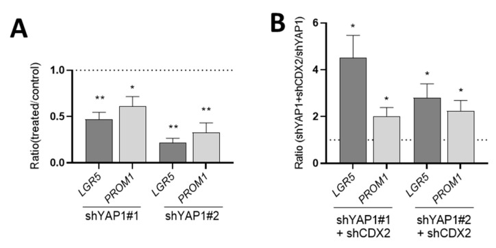
The human intestine is covered by epithelium, which is continuously replaced by new cells provided by stem cells located at the bottom of the glands. The maintenance of intestinal stem cells is supported by a niche which is composed of several signaling proteins including the Hippo pathway effectors YAP1/TAZ. The role of YAP1/TAZ in cell proliferation and regeneration is well documented but their involvement on the differentiation of intestinal epithelial cells is unclear. In the present study, the role of YAP1/TAZ on the differentiation of intestinal epithelial cells was investigated using the HT29 cell line, the only multipotent intestinal cell line available, with a combination of knockdown approaches. The expression of intestinal differentiation cell markers was tested by qPCR, Western blot, indirect immunofluorescence and electron microscopy analyses. The results show that TAZ is not expressed while the abolition of YAP1 expression led to a sharp increase in goblet and absorptive cell differentiation and reduction of some stem cell markers. Further studies using double knockdown experiments revealed that most of these effects resulting from YAP1 abolition are mediated by CDX2, a key intestinal cell transcription factor. In conclusion, our results indicate that YAP1/TAZ negatively regulate the differentiation of intestinal epithelial cells through the inhibition of CDX2 expression.

Keywords: CDX2; Hippo pathway; TAZ; YAP1; absorptive cells; differentiation; goblet cell; intestinal cell; stem cell.


Conflict of interest statement

The authors declare no conflict of interest. The funders had no role in the design of the study; in the collection, analyses, or interpretation of data; in the writing of the manuscript, nor in the decision to publish the results.

Figures

Figure 1
Figure 1
Nuclear expression of YAP1/TAZ in human intestinal crypt cells. Representative confocal imaging for the detection of YAP1/TAZ (green) (A,C), DEFA5 (red) (A,D) and DAPI (blue) (A,B) in the adult small intestine. Nuclear expression of YAP1/TAZ in some of the cells located at the bottom of the crypts was observed (arrowheads) except in Paneth cells (stars). Scale bar is equal to 10 μm.
Figure 2
Figure 2
Expression of stem cell markers, Hippo effectors, goblet and absorptive cells markers in HT29 cells. (A) Expression of LGR5, CD44, PROM1, EPCAM, YAP1, TAZ, MUC2 and SI transcripts in HT29 cells relative to a pool of cancer cells. * p < 0.05, ** p < 0.01. (B) Western blot analysis showing expression of YAP protein in HT29 cells in which the TAZ protein was consistently below detectable levels. Both YAP and TAZ proteins were found to be expressed by Caco-2 cells while only TAZ was detectable in HIEC. β-actin was used as a loading control. (C) The expression of YAP1 and TAZ was also investigated at the transcript levels in the adult small intestine (A Int) and the intestinal cell lines relative to the pool. Statistical significance for YAP1 vs. TAZ (paired T test): * p < 0.05, ** p < 0.005, n ≥ 3. (D) Indirect immunofluorescence of HT29 cells confirmed the presence of the YAP protein in a large proportion of the cells while a few LGR5 and MUC2 positive cells were detected in the normal HT29 cells. Scale bar = 50 μm.
Figure 3
Figure 3
Efficiency of YAP1 knockdown in HT29 cells. (A) qPCR analyses were performed to detect the expression of YAP1 and two of its target genes CYR61 and CTGF in shYAP1 stably expressing cells using two specific sequences (identified as shYAP1 #1 and #2) relative to shCtrl. (B) Western blot analysis showing the expression of YAP1 protein in shYAP1#1, shYAP1#2 and shCtrl expressing cells. A cancer cell pool was used as reference for YAP1 and TAZ detection. β-actin was used as a loading control. ** p < 0.005.
Figure 4
Figure 4
Effect of YAP1 knockdown on stem cell marker expression in subconfluent HT29 cells. (A) Transcript expression of the stem cell markers LGR5, CD44, PROM1, EPCAM, ASCL2 and OCT4 in YAP1 knockdown cells relative to shCtrl (dotted line). (B) Reduction in LGR5 protein in both shYAP1 cell lines compared with shCtrl. β-actin was used as a loading control. * p < 0.05, ** p < 0.005.
Figure 5
Figure 5
Expression of intestinal cell lineage markers in response to YAP1 knockdown. qPCR analyses of absorptive cell markers SI and DPPIV, goblet cell markers MUC2 and TFF3, Paneth cell marker DEFA5 and enteroendocrine cell marker CHGA were analyzed in shYAP1#1 and shYAP1#2 expressing cells relative to shCtrl cells (dotted line). * p < 0.05, ** p < 0.005.
Figure 6
Figure 6
YAP1 knockdown stimulates goblet cell differentiation. (A) Representative Western blot analysis and data compilation from three separate experiments showing higher expression of MUC2 and TFF3 in shYAP1#1 and shYAP1#2 expressing cells relative to shCtrl cells at both Day 0 (0PC) and Day 8 (8PC) post-confluence. β-actin was used as a loading control. * p < 0.05, ** p < 0.005. (BE) Indirect immunofluorescence analysis for the detection of MUC2 positive cells in shCtrl (B,D) and shYAP1#2 (C,E) at 0PC (B,C) and 8PC (C,E). Bar = 50 µm.
Figure 7
Figure 7
YAP1 knockdown stimulates absorptive cell differentiation. (A) Representative Western blot analysis and data compilation from three separate experiments showing higher expression of SI and DPPIV in shYAP1#1 and shYAP1#2 expressing cells relative to shCtrl cells at both Day 0 (0PC) and Day 8 (8PC) post-confluence. β-actin was used as a loading control. * p < 0.05, ** p < 0.005. (BE) Indirect immunofluorescence analysis for the detection of DPPIV positive cells in shCtrl (B,D) and shYAP1#2 (C,E) at 0PC (B,C) and 8 PC (C,E). Bar = 50 µm.
Figure 8
Figure 8
Transmission electron microscopy of HT29 stably expressing shCtrl or shYAP1 at 8PC. (A) Control cells expressing shTAZ are small and multilayered and express limited differentiation characteristics. (B,C) Cells expressing shYAP1 are larger and exhibit some absorptive-like features such as regular microvilli (B) and goblet cell-like features such as mucinous granule forming goblet-like aggregates (lower part of (C)). Scale bars = 1 μm.
Figure 9
Figure 9
The effect of shYAP1 expression on Caco-2/15 cell differentiation. (A) Transcript expression of the absorptive cell marker SI in Caco-2/15 cells expressing shYAP1 relative to shCtrl (shLUC). (B) Western blot analysis and data compilation showing higher expression of SI in shYAP1 Caco-2/15 cells relative to shCtrl at five days post-confluence. β–actin was used as a loading control. Statistical comparison between shYAP1 vs. shLUC: * p < 0.05.
Figure 10
Figure 10
Expression of intestinal differentiation-regulating transcription factors in response to YAP1 knockdown. (A) qPCR analysis of the expression of various transcription factors involved in intestinal epithelial cell differentiation in shYAP1#1 and shYAP1#2 expressing cells relative to shCtrl (dotted line) showing the significant increase in expression of CDX2 and ATOH1 in YAP1 knockdown cells. (B) Representative Western blot analysis and data compilation from three separate experiments showing higher expression of CDX2 protein in YAP1 knockdown HT29 cells. β-actin was used as a loading control. * p < 0.05, ** p < 0.005.
Figure 11
Figure 11
Efficiency of CDX2 knockdown in shYAP1 expressing HT29 cells. (A) qPCR analyses were performed to evaluate the expression of CDX2 in shCDX2 stably expressing shYAP1#1 and -#2 cells relative to normal shYAP1#1 and -#2 cells. (B) Representative Western blot analysis showing the expression of CDX2 protein in shCtrl vs. shYAP1#1 and shYAP1#2 ± shCDX2 expressing cells and data compilation of three separate experiments. A cancer cell pool was used as reference for CDX2 detection. β-actin was used as a loading control. * significant vs. shCtrl; # significant vs. shYAP1; */# p < 0.05, ** p < 0.005.
Figure 12
Figure 12
CDX2 mediates the upregulation of most of the intestinal differentiation markers in shYAP1 cells. (A) qPCR analysis showing that the abolition of CDX2 in shYAP1 cells resulted in a significant reduction of MUC2, TFF3 and SI expression while it remained without effect on DPPIV. (B) Representative Western blot analysis showing the expression of MUC2, TFF3, SI and DPPIV in shCtrl vs. shYAP1#1 and shYAP1#2 ± shCDX2 expressing cells and data compilation of three separate experiments. At the transcript level, the upregulation of these markers in shYAP1 cells appears to depend upon CDX2 since its abolition results in the restoration of HT29 basal levels for MUC2, TFF3 and SI while DPPIV appears to remain unaffected. A pool was used as a positive control. β-actin was used as a loading control. * significant vs. shCtrl; # significant vs. shYAP1; */# p < 0.05, **/## p < 0.005.
Figure 13
Figure 13
Abolition of CDX2 expression restores control LGR5 and PROM1 levels in five-days post-confluent shYAP1 cells. (A) qPCR analysis showing that the abolition of YAP1 resulted in a significant reduction of LGR5 and PROM1 expression. (B) qPCR analysis showing that the abolition of CDX2 in shYAP1 cells resulted in a significant increase in LGR5 and PROM1 expression relative to shYAP1 control cells. A: * significant vs. shCtrl, B: * significant vs. shYAP1; * p < 0.05, ** p < 0.005.
Figure 14
Figure 14
Integration of the molecular mechanisms regulating intestinal differentiation. All steps of intestinal epithelial cell differentiation occur in the crypt so that only fully mature cells reach the villus [1,8]. The crypt is divided into three distinct compartments: the stem cell (SC), the transit amplifying (TA) and terminal differentiation (TD) zones. Intestinal cell differentiation relies on pro-differentiation factors such as CDX2, HNF1α and GATA4 which are expressed in the TA and TD zones. Goblet cells differentiate in the TA zone, but absorptive cell differentiation is restrained by epigenetic mechanisms involving polycomb repressive complex 2 (PRC2-Suz12) and histone deacetylases (HDACs), which allow proliferation in the TA zone, ensuring a larger proportion of absorptive cells in the TD zone. YAP1/TAZ appear to act at an earlier phase, which involves stemness maintenance and inhibition of both absorptive and secretory lineages through a repression of pro-differentiation transcription factors such as CDX2. Adapted from [1,8].

Similar articles

Cited by

References

    1. Fallah S., Sénicourt B., Beaulieu J.-F. Proliferation in the Gastrointestinal Epithelium. In: Kuipers E.J., editor. Encyclopedia of Gastroenterology. 2nd ed. Academic Press; Oxford, UK: 2020. pp. 304–310.
    1. Van der Heijden M., Vermeulen L. Stem cells in homeostasis and cancer of the gut. Mol. Cancer. 2019;18:66. doi: 10.1186/s12943-019-0962-x. - DOI - PMC - PubMed
    1. Spit M., Koo B.K., Maurice M.M. Tales from the crypt: Intestinal niche signals in tissue renewal, plasticity and cancer. Open Biol. 2018;8:180120. doi: 10.1098/rsob.180120. - DOI - PMC - PubMed
    1. Andersson-Rolf A., Zilbauer M., Koo B.K., Clevers H. Stem Cells in Repair of Gastrointestinal Epithelia. Physiology. 2017;32:278–289. doi: 10.1152/physiol.00005.2017. - DOI - PMC - PubMed
    1. Sei Y., Feng J., Chow C.C., Wank S.A. Asymmetric cell division-dominant neutral drift model for normal intestinal stem cell homeostasis. Am. J. Physiol. Gastrointest. Liver Physiol. 2019;316:G64–G74. doi: 10.1152/ajpgi.00242.2018. - DOI - PMC - PubMed

Publication types

MeSH terms

LinkOut - more resources