

Bactericidal and In Vitro Cytotoxicity of Moringa oleifera Seed Extract and Its Elemental Analysis Using Laser-Induced Breakdown Spectroscopy
- PMID: 32823699
- PMCID: PMC7464216
- DOI: 10.3390/ph13080193
Bactericidal and In Vitro Cytotoxicity of Moringa oleifera Seed Extract and Its Elemental Analysis Using Laser-Induced Breakdown Spectroscopy
Abstract
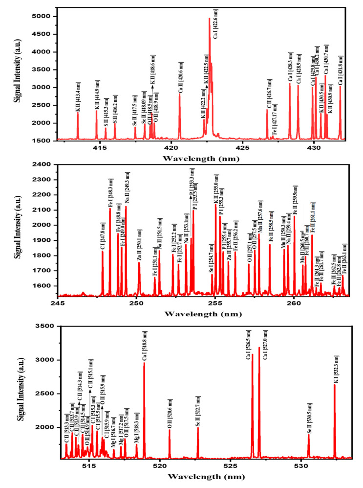
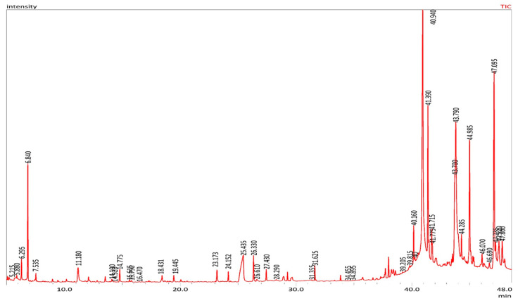
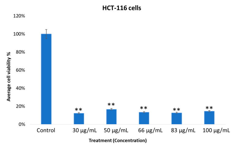
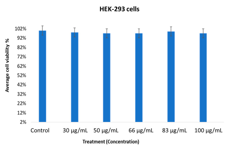
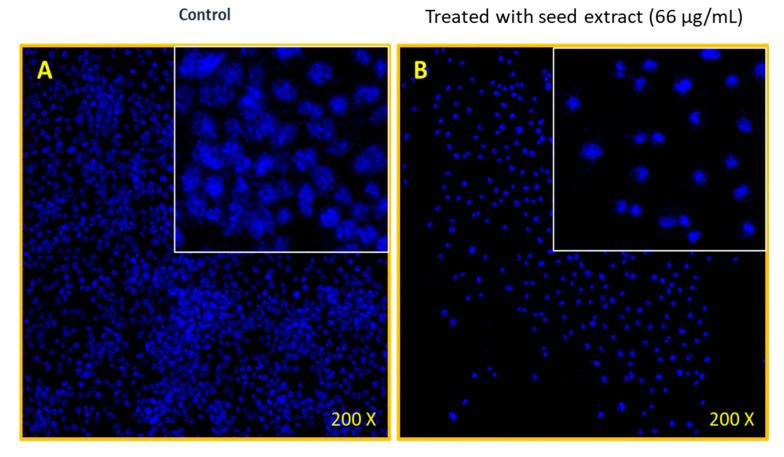
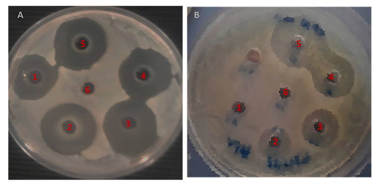
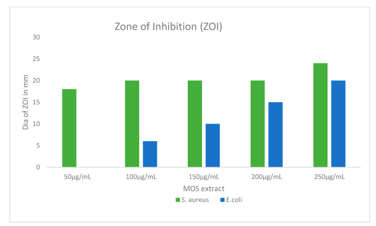
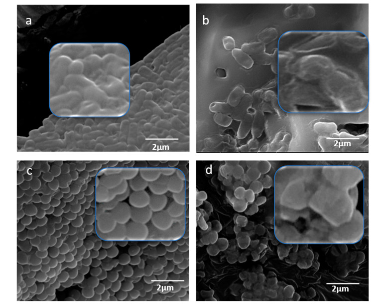
In the current study, we present the correlation between the capability of laser-induced breakdown spectroscopy (LIBS) to monitor the elemental compositions of plants and their biological effects. The selected plant, Moringa oleifera, is known to harbor various minerals and vitamins useful for human health and is a potential source for pharmaceutical interventions. From this standpoint, we assessed the antibacterial and in vitro cytotoxicity of the bioactive components present in Moringa oleifera seed (MOS) extract. Detailed elemental analyses of pellets of MOSs were performed via LIBS. Furthermore, the LIBS outcome was validated using gas chromatography-mass spectrometry (GC-MS). The LIBS signal was recorded, and the presence of the essential elements (Na, Ca, Se, K, Mg, Zn, P, S, Fe and Mn) in the MOSs were examined. The bactericidal efficacy of the alcoholic MOS extract was examined against Escherichia coli (E. coli) and Staphylococcus aureus(S. aureus) by agar well diffusion (AWD) assays and scanning electron microscopy (SEM), which depicted greater inhibition against Gram-positive bacteria. The validity and DNA nuclear morphology of human colorectal carcinoma cells (HCT-116) cells were evaluated via an MTT assay and DAPI staining. The MTT assay results manifested a profoundly inhibitory action of MOS extract on HCT116 cell growth. Additionally, MOS extracts produced inhibitory action in colon cancer cells (HCT-116), whereas no inhibitory action was seen using the same concentrations of MOS extract on HEK-293 cells (non-cancerous cells), suggesting that MOS extracts could be non-cytotoxic to normal cells. The antibacterial and anticancer potency of these MOS extracts could be due to the presence of various bioactive chemical complexes, such as ethyl ester and D-allose and hexadecenoic, oleic and palmitic acids, making them an ideal candidate for pharmaceutical research and applications.
Keywords: GC-MS; LIBS; Moringa oleifera; antibacterial; anticancer; seed extract.
Conflict of interest statement
The authors declare no conflict of interest.
Figures
Similar articles
-
Moringa oleifera as an Anti-Cancer Agent against Breast and Colorectal Cancer Cell Lines.PLoS One. 2015 Aug 19;10(8):e0135814. doi: 10.1371/journal.pone.0135814. eCollection 2015. PLoS One. 2015. PMID: 26288313 Free PMC article.
-
Antibacterial and Cytotoxic Effects of Moringa oleifera (Moringa) and Azadirachta indica (Neem) Methanolic Extracts against Strains of Enterococcus faecalis.Int J Dent. 2018 Sep 25;2018:1071676. doi: 10.1155/2018/1071676. eCollection 2018. Int J Dent. 2018. PMID: 30356384 Free PMC article.
-
GC-MS Analysis, Antibacterial, and Anticancer Activities of Hibiscus sabdariffa L. Methanolic Extract: In Vitro and In Silico Studies.Microorganisms. 2023 Jun 16;11(6):1601. doi: 10.3390/microorganisms11061601. Microorganisms. 2023. PMID: 37375103 Free PMC article.
-
Identification and Purification of Potential Bioactive Peptide of Moringa oleifera Seed Extracts.Plants (Basel). 2020 Oct 27;9(11):1445. doi: 10.3390/plants9111445. Plants (Basel). 2020. PMID: 33120901 Free PMC article.
-
Bioactive Components in Moringa Oleifera Leaves Protect against Chronic Disease.Antioxidants (Basel). 2017 Nov 16;6(4):91. doi: 10.3390/antiox6040091. Antioxidants (Basel). 2017. PMID: 29144438 Free PMC article. Review.
Cited by
-
Antioxidant, anticancer, and anti-inflammatory potential of Moringa seed and Moringa seed oil: A comprehensive approach.Food Sci Nutr. 2024 Jul 9;12(9):6157-6173. doi: 10.1002/fsn3.4312. eCollection 2024 Sep. Food Sci Nutr. 2024. PMID: 39554357 Free PMC article. Review.
-
Inhibition of Kinase Activity and In Vitro Downregulation of the Protein Kinases in Lung Cancer and Cervical Cancer Cell Lines and the Identified Known Anticancer Compounds of Ziziphus mucronata.Plants (Basel). 2025 Jan 28;14(3):395. doi: 10.3390/plants14030395. Plants (Basel). 2025. PMID: 39942957 Free PMC article.
-
Special Issue "Novel Antibacterial Agents".Pharmaceuticals (Basel). 2021 Apr 19;14(4):382. doi: 10.3390/ph14040382. Pharmaceuticals (Basel). 2021. PMID: 33921864 Free PMC article.
-
An insight into the neuroprotective and anti-neuroinflammatory effects and mechanisms of Moringa oleifera.Front Pharmacol. 2023 Jan 5;13:1035220. doi: 10.3389/fphar.2022.1035220. eCollection 2022. Front Pharmacol. 2023. PMID: 36686668 Free PMC article. Review.
-
Maximizing the extraction yield of plant gum exudate using response surface methodology and artificial neural networking and pharmacological characterization.Sci Rep. 2023 Jul 6;13(1):10954. doi: 10.1038/s41598-023-37847-x. Sci Rep. 2023. PMID: 37414773 Free PMC article.
References
-
- Oduro I., Ellis W.O., Owusu D. Nutritional potential of two leafy vegetables: Moringa oleifera and Ipomoea batatas leaves. Sci. Res. Essays. 2008;3:57–60. doi: 10.5897/SRE.9000686. - DOI
LinkOut - more resources
Full Text Sources
Other Literature Sources
Research Materials
Miscellaneous
