Review
. 2020 Aug 13;9(8):1897.
doi: 10.3390/cells9081897.

Involvement of JNK1 in Neuronal Polarization During Brain Development

Affiliations
Review

Involvement of JNK1 in Neuronal Polarization During Brain Development

Rubén Darío Castro-Torres et al. Cells. .

Abstract

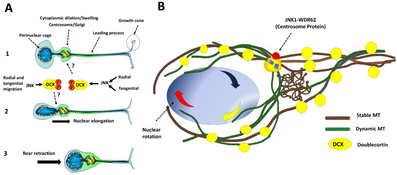
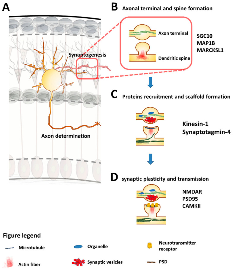
The c-Jun N-terminal Kinases (JNKs) are a group of regulatory elements responsible for the control of a wide array of functions within the cell. In the central nervous system (CNS), JNKs are involved in neuronal polarization, starting from the cell division of neural stem cells and ending with their final positioning when migrating and maturing. This review will focus mostly on isoform JNK1, the foremost contributor of total JNK activity in the CNS. Throughout the text, research from multiple groups will be summarized and discussed in order to describe the involvement of the JNKs in the different steps of neuronal polarization. The data presented support the idea that isoform JNK1 is highly relevant to the regulation of many of the processes that occur in neuronal development in the CNS.

Keywords: DCX; JNK1; MAP1B; NMDAR; SCG10; WDR62; asymmetric division; migration; synaptogenesis.


Conflict of interest statement

The authors declare no conflict of interest.

Figures

Figure 1
Figure 1
JNK1 controls polarization of dividing cells. Radial-glial-like cells (cortical NSCs) are located in the SVZ and show apicobasal polarization. The orientation of the mitotic spindle controls symmetric and asymmetric divisions: (A) Symmetric division preserves self-renewal and expands radial glial-like progeny, (B) Asymmetric division promotes the loss of apicobasal contact of daughter cells, promoting their differentiation. The activity of the Par3/Par6/aPKC and Gαi-LGN-NuMa complexes determines the orientation of the mitotic spindle. (C) The pericentriolar material of centrosome contains both JNK1 and WDR62. Genetic disabling of this elements induces abnormal spindle formation and aberrant centrosome biogenesis. Adapted from [50].
Figure 2
Figure 2
Role of JNKs in neuronal polarization. AE: Phenotypic changes along neuronal polarization in vitro. (A) (Stage 1) Embryonic hippocampal neurons extend the lamellipodia. Twelve hours later, the breakage of neuron symmetry occurs. (B) (Stage 2) Neurons become multipolar. One neurite is selected and experiences a positive feedback signal on its tip, which induces axon determination. (C) (Stage 3, axonogenesis) Signal propagates to the soma (red), inducing the stabilization and elongation of microtubules. The remaining neurites receive inhibitory signals (blue) (D) (Stage 4, Dendritogenesis) After axon elongation, the remaining neurites are extended. (E) (Stage 5, Spinogenesis) Finally, spines and axonal branching occur. (a–e) Molecular events that occur during neuronal polarization. (a) Axon determination is induced by growth factors. Centrosome and Golgi apparatuses are recruited. (b) Rho GTPases (Cdc42, Rac1) and the Par3/Par6/aPKC complex form a positive feedback circuit required for axon determination. (c) The persistent activation of Rac1 and Cdc42 leads to JNK activation, promoting neurite outgrowth through growth cones (red), while the activation of RhoA in minor neurites leads to JNK deactivation (blue). (d) Dendritogenesis is stimulated by BMP7 through Bone Morphogenic Protein Receptor (BMPR), Wnt7b through Frizzled receptor and Sema3A through Nrp1. Dendrite homeostasis is controlled through SCG10, MAP and MARCKSL1 substrates that are activated by JNK1 (red) (e) Spine density is regulated by substrates such as DCX and MARCKSL1, and axonal branching is promoted by the deactivation of JNK1 through BDNF.
Figure 3
Figure 3
Microtubule (MT) dynamics are regulated by JNK1. (A) MT stability and elongation. JIP scaffolding proteins and kinesin-1 facilitate the diffusion of JNK1 and the transport of relevant cargoes needed for axonal growth such as Tiam/Rac, SCG10, BDNF and TrkB receptors. Unphosphorylated MAP1B and phosphorylated Sthatmin family members by JNK1 promote microtubule stabilization. Moreover, JNK1 phosphorylates SCG10, resulting on its degradation. (B) MT shrinkage. The phosphorylation of MAP1B by JNK1 and unphosphorylated SCG10 promote microtubule depolymerization. In addition, unphosphorylated members of SCG10 proteins sequestrate tubulin dimers and favor microtubule catastrophe. Adapted from [96].
Figure 4
Figure 4
Effects of genetic Jnk1 disabling in the neuronal migration process during cortical embryonic development in mice. (A) Cortical glutamatergic neurons born in the lateral ventricles (blue). Cortical interneurons born in the GE (Red) (B) Interneurons from MGE (green) reach the corticostriatal junction at E12 and invade the subpallium. Interneurons enter to the developing cortex lateromedially following two migratory tangential streams (i, j). The (i) stream populates the superior layer, while the (j) stream populates deep layers and hippocampus. Jnk1-/- mice experience severe alterations due to a delay of entrance and erratic migration of cortical interneurons (k). (C) RG-like cells (red) self-renew and proliferate by symmetric division: (a-b) RG cells undergo asymmetric division and become migratory multipolar neurons. (c) Multipolar cells accumulate in IZ and form the MAZ. (d) Multipolar cells migrate towards the SP and seize radial processes to climb through them toward the pia. They acquire bipolar morphology. (e) Radial migration ceases and (f) neurons populate the upper (2/3 and 4) and deep (5 and 6) layers. This radial migration process is altered in Jnk1-/- mice at E18 of development. The multipolar-to-bipolar phase is faster than in wild-type (WT) (g to h versus a to d). The ventricular zone is smaller (*) and the cortical layers are disordered (**).
Figure 5
Figure 5
JNK1 regulates the phases of neuronal migration. (A) (1) Initially, the leading process is extended; (2) Later, the nucleus is pulled forward by the reorganization of microtubules that round the perinuclear cage; (3) Finally, rear retraction occurs. (B) Magnification of the perinuclear cage formed by MT. MTs (red and brown) are located around the nucleus (blue). The Centrosome/Golgi apparatus irradiates MTs toward the nucleus. Several targets of JNK1 are localized in the perinuclear cage (DCX, WDR62).
Figure 6
Figure 6
Synaptogenesis is controlled by JNK1. (A) Synaptogenesis occurs during neuronal differentiation. (B) The presynaptic (axon terminal) and postsynaptic domains (spine formation) are formed through the regulation of SGC10, MAP1B and MARCKSL1 substrates by JNK1. These events regulate actin (red) and microtubule dynamics (green). (C) Kinesin 1, together with JNK and JIP, control vesicle and organelle transport (blue), e.g., the formation of postsynaptic density scaffold proteins (black). (D) During synaptic transmission, the morphology of structures changes under the control of JNK1 (synaptic plasticity).

Similar articles

Cited by

References

    1. Sabapathy K. Role of the JNK Pathway in Human Diseases. Elsevier; Amsterdam, The Netherlands: 2012. pp. 145–169. - PubMed
    1. Sun Y., Liu W.-Z., Liu T., Feng X., Yang N., Zhou H.-F. Signaling pathway of MAPK/ERK in cell proliferation, differentiation, migration, senescence and apoptosis. J. Recept. Signal Transduct. 2015;35:600–604. doi: 10.3109/10799893.2015.1030412. - DOI - PubMed
    1. Coffey E.T. Nuclear and cytosolic JNK signalling in neurons. Nat. Rev. Neurosci. 2014;15:285–299. doi: 10.1038/nrn3729. - DOI - PubMed
    1. Cui J., Zhang M., Zhang Y.-Q., Xu Z. JNK pathway: diseases and therapeutic potential. Acta Pharmacol. Sin. 2007;28:601–608. doi: 10.1111/j.1745-7254.2007.00579.x. - DOI - PubMed
    1. Zeke A., Misheva M., Reményi A., Bogoyevitch M.A. JNK Signaling: Regulation and Functions Based on Complex Protein-Protein Partnerships. Microbiol. Mol. Boil. Rev. 2016;80:793–835. doi: 10.1128/MMBR.00043-14. - DOI - PMC - PubMed

Publication types