Review
. 2020 Aug 14;21(16):5845.
doi: 10.3390/ijms21165845.

Lipid Metabolism and Cancer Immunotherapy: Immunosuppressive Myeloid Cells at the Crossroad

Affiliations
Review

Lipid Metabolism and Cancer Immunotherapy: Immunosuppressive Myeloid Cells at the Crossroad

Augusto Bleve et al. Int J Mol Sci. .

Abstract

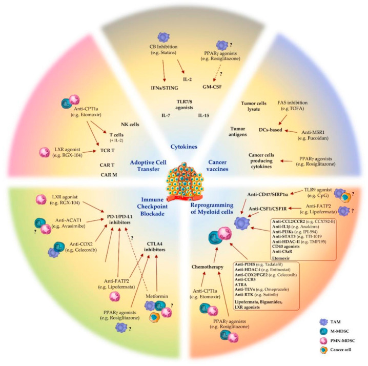
Cancer progression generates a chronic inflammatory state that dramatically influences hematopoiesis, originating different subsets of immune cells that can exert pro- or anti-tumor roles. Commitment towards one of these opposing phenotypes is driven by inflammatory and metabolic stimuli derived from the tumor-microenvironment (TME). Current immunotherapy protocols are based on the reprogramming of both specific and innate immune responses, in order to boost the intrinsic anti-tumoral activity of both compartments. Growing pre-clinical and clinical evidence highlights the key role of metabolism as a major influence on both immune and clinical responses of cancer patients. Indeed, nutrient competition (i.e., amino acids, glucose, fatty acids) between proliferating cancer cells and immune cells, together with inflammatory mediators, drastically affect the functionality of innate and adaptive immune cells, as well as their functional cross-talk. This review discusses new advances on the complex interplay between cancer-related inflammation, myeloid cell differentiation and lipid metabolism, highlighting the therapeutic potential of metabolic interventions as modulators of anticancer immune responses and catalysts of anticancer immunotherapy.

Keywords: cancer immunotherapy; cholesterol; fatty acids; lipid metabolism; myeloid-derived suppressor cells (MDSCs); obesity; tumor-associated macrophages (TAMs).


Conflict of interest statement

The authors declare no conflict of interest.

Figures

Figure 1
Figure 1
Targeting lipid metabolism of myeloid cells in cancer immunotherapy. Pharmacological modulation of molecular regulators of lipid metabolism in tumor-associated macrophages (TAM), monocytic (M-) and granulocytic (PMN-) myeloid-derived suppressor cells (MDSC) showed efficacy in dampening tumor immunosuppression and improving cancer immunotherapies: immune checkpoint blockade; adoptive cell transfer; cytokines administration or modulation; cancer vaccines; reprogramming of polarized inflammation in myeloid cells. Abbreviations: LXR, liver X receptor; ACAT1, acetyl-CoA acetyltransferase 1; COX2, cyclooxygenase 2; FATP2, fatty acid transport protein 1; PPARγ, peroxisome proliferator-activated receptor γ; PD-1, programmed-death protein 1; PD-L1, PD-1 ligand; CTLA4, cytotoxic T-lymphocyte antigen 4; CPT1a, carnitine palmitoyltransferase 1a; CAR T/M, chimeric antigen receptor T cell/Macrophage; TCR T, T cell receptor-engineered T cell; NK, natural killer; CB, cholesterol biosynthesis; IFN, interferon; STING, stimulator of interferon genes; TLR, Toll-like receptor; GM-CSF, granulocyte-macrophage colony-stimulator factor; FAS, fatty acid synthesis; MSR1, macrophage scavenger receptor 1; DC, dendritic cell; SIRP1α, signal regulatory protein 1α; CSF1, macrophage colony-stimulating factor; CSF1R, CSF1 receptor; PI3K, phosphoinositide 3-kinase; HDAC, histone deacetylases; C5aR, complement component 5 receptor; PDE5, phosphodiesterase 5; COX2, cyclooxygenase 2; PGE2, prostaglandin E2; CCR5, chemokine receptor 5; ATRA, all-trans retinoic acid; TEV, tumor-derived extracellular vesicles; RTK, receptor tyrosine kinase. Text in brackets represents examples of drugs targeting the molecule or pathway of reference. Dashed arrows and question marks underline controversial or not fully clarified evidence.
Figure 2
Figure 2
Influence of obesity on cancer-associated inflammation, suppressor myeloid cells and cancer progression. Obesity represents a risk factor for many cancers. Weight gain generates an excess of adipose tissue depots associated with dyslipidemias (i.e., hyper-cholesterolemia and -triglyceridemia) and metabolic alterations (e.g., hyperinsulinemia), which can promote tumor progression. In addition, obesity produces a chronic inflammatory state, which primes myelopoiesis and the immunosuppressive phenotype of tumor-associated myeloid cells. Reprogramming of intracellular lipid metabolism also occurs in myeloid cells from normal-weight cancer bearers in response to TME-derived factors. Several molecular players are associated with either pro- or anti-inflammatory (M1/M2) states of macrophage activation, as exemplified by tumor-associated macrophages (TAM) and myeloid-derived suppressor cells (MDSC). M1 macrophage polarization activates fatty acid synthesis (FAS), whereas immunosuppressive M2/TAMs and MDSCs shift toward fatty acid oxidation (FAO). Abbreviations: BMI, body mass index; IGF-1, insulin-like growth factor 1; HSC, hematopoietic stem cell; GMP, granulocyte-macrophage progenitor; ACLY, ATP-citrate lyase; UCP2, uncoupling protein 2; SREBP, sterol regulatory element-binding protein; E-FABP, epidermal fatty acids binding protein; CH25H, cholesterol 25-hydroxylase; ApoA1, apolipoprotein A1; HDL, high-density lipoprotein; FA, fatty acid; oxPAPC, oxidized 1-palmitoyl-2-arachidonyl-sn-glycero-3-phosphorylcholine; LpL, lipoprotein lipase; MSR1, macrophage scavenger receptor 1; PPARγ, peroxisome proliferator-activated receptor γ; PGC1β, PPARγ coactivator 1β; CPT1a/2, carnitine palmitoyltransferase 1a/2; 15-LOX2, 15-lipoxygenase 2; 5-LOX, 5-lipoxygenase; ABCA1/G1, ATP-binding cassette A1/G1; 22-/24-/25-/27-OHC, 22-/24-/25-/27-hydroxycholesterol; FATP2, fatty acid transport protein 1; LAL, lysosomal acid lipase; LXR, liver X receptor; LOX-1, oxidized low-density lipoprotein receptor 1; PUFA, polyunsaturated fatty acid. Question marks underline controversial or not fully clarified evidence. Words in italics represent lipidic metabolites.

References

    1. Velten L., Haas S.F., Raffel S., Blaszkiewicz S., Islam S., Hennig B.P., Hirche C., Lutz C., Buss E.C., Nowak D., et al. Human haematopoietic stem cell lineage commitment is a continuous process. Nat. Cell Biol. 2017;19:271–281. doi: 10.1038/ncb3493. - DOI - PMC - PubMed
    1. Ueha S., Shand F.H.W., Matsushima K. Myeloid cell population dynamics in healthy and tumor-bearing mice. Int. Immunopharmacol. 2011;11:783–788. doi: 10.1016/j.intimp.2011.03.003. - DOI - PubMed
    1. Wu W.-C., Sun H.-W., Chen H.-T., Liang J., Yu X.-J., Wu C., Wang Z., Zheng L. Circulating hematopoietic stem and progenitor cells are myeloid-biased in cancer patients. Proc. Natl. Acad. Sci. USA. 2014;111:4221–4226. doi: 10.1073/pnas.1320753111. - DOI - PMC - PubMed
    1. Mantovani A., Allavena P., Sica A., Balkwill F.R. Cancer-related inflammation. Nature. 2008;454:436–444. doi: 10.1038/nature07205. - DOI - PubMed
    1. Gabrilovich D.I., Ostrand-Rosenberg S., Bronte V. Coordinated regulation of myeloid cells by tumours. Nat. Rev. Immunol. 2012;12:253–268. doi: 10.1038/nri3175. - DOI - PMC - PubMed

Substances