Review
. 2020 Aug 15;9(8):1903.
doi: 10.3390/cells9081903.

Myosin XVI in the Nervous System

Affiliations
Review

Myosin XVI in the Nervous System

Elek Telek et al. Cells. .

Abstract

The myosin family is a large inventory of actin-associated motor proteins that participate in a diverse array of cellular functions. Several myosin classes are expressed in neural cells and play important roles in neural functioning. A recently discovered member of the myosin superfamily, the vertebrate-specific myosin XVI (Myo16) class is expressed predominantly in neural tissues and appears to be involved in the development and proper functioning of the nervous system. Accordingly, the alterations of MYO16 has been linked to neurological disorders. Although the role of Myo16 as a generic actin-associated motor is still enigmatic, the N-, and C-terminal extensions that flank the motor domain seem to confer unique structural features and versatile interactions to the protein. Recent biochemical and physiological examinations portray Myo16 as a signal transduction element that integrates cell signaling pathways to actin cytoskeleton reorganization. This review discusses the current knowledge of the structure-function relation of Myo16. In light of its prevalent localization, the emphasis is laid on the neural aspects.

Keywords: development; mammalian; myosin; myosin XVI; neural; neurodegenerative; unconventional.


Conflict of interest statement

The authors declare no conflict of interest. The funders had no role in the design of the study; in the collection, analyses, or interpretation of data; in the writing of the manuscript, or in the decision to publish the results.

Figures

Figure 1
Figure 1
Domain structure of unconventional Myo16 isoforms. The number of amino acids corresponds to the rat Myo16 protein (accession number: Q9ERC1). Myo16a (1322 aa, 148.75 kDa) is composed of an N-terminal ankyrin domain (Myo16Ank) possessing a conserved myosin phosphatase N-terminal element (MyPhoNE), a KVxF sequence motif (protein phosphatase type 1 catalytic subunit (PP1c) binding motif) and eight ankyrin repeats (Ank). Myo16Ank is followed by the motor domain, a single IQ motif and a short C terminal region. The domain organization of the N-terminus of Myo16b (1912 aa, 210.56 kDa) is identical to that of Myo16a. Myo16b, following the IQ motif, has a longer C-terminal tail extension (Myo16Tail) including a WAVE1 interacting region (WIR), a neuronal tyrosine-phosphorylated adaptor for phosphoinositide 3-kinase (NYAP) homology motif (NHM) and proline-rich regions (PRR). The phosphotyrosines (Y1416 and Y1441) in the NHM motif are indicated (pY).
Figure 2
Figure 2
The C-terminal Myo16Tail in the reorganization of the actin cytoskeleton through the PI3K-WAVE1 pathway. Myo16Tail phosphorylation is triggered by Contactin5 through an unknown neuronal receptor (indicated with an X mark). Fyn binding to Myo16 is indirectly regulated by Contactin5 through a phosphatase (presumably via protein tyrosine phosphatase α (PTPα)). Tyrosine-phosphorylated Myo16 interacts directly with the p85 subunit of the phosphoinositide 3-kinase (PI3K) that is mediated by the NHM motif. The binding eventuates the activation of PI3K and its downstream effectors Akt and Rac1. Myo16 also interacts with the Sra1 and Nap1 subunits of WAVE1 through its WAVE1 interacting region. By simultaneous binding, Myo16 can bridge PI3K to WAVE1 and can contribute to the activation of WAVE1, thereby triggering the Arp2/3 complex-mediated remodeling of the actin cytoskeleton.
Figure 3
Figure 3
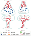
Schematic representation of the effects of Myo16 at parallel fiber-Purkinje cell synapses. Myo16 plays a role in the organization of the postsynaptic terminals of Purkinje cells by influencing the PI3K-WRC-Arp2/3 pathway that eventuates the downregulation of actin cytoskeleton dynamics. At the presynaptic site, Myo16 is implicated in the regulation of the number of synaptic vesicles, as well as of the morphological features of the parallel fiber terminal of granule cells. The X mark in the upper panel indicates the lack of interaction between Myo16, PI3K and WAVE1.
Figure 4
Figure 4
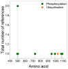
Post-translational prediction of Myo16 motor domain. Sequence analysis was performed using PhosphoSitePlus [71]. The total number of references shows the reliability of the prediction. Green squares indicate the phosphorylation sites while orange circle shows the ubiquitination site.

References

    1. Frénal K., Foth B.J., Soldati D. Myosin Class XIV and Other Myosins in Protists. In: Coluccio L.M., editor. Myosins, A Superfamily of Molecular Motors. Springer Netherlands; Heidelberg, Germany: 2008. pp. 421–440. (Series: Proteins and Cell Regulation).
    1. Coluccio L.M., Myosins A. Superfamily of Molecular Motors. 2nd ed. Springer International Publishing; Cham, Switzerland: 2020. (Series: Advances in Experimental Medicine and Biology).
    1. Brown M.E., Bridgman P.C. Myosin Function in Nervous and Sensory Systems. J. Neurobiol. 2004;58:118–130. doi: 10.1002/neu.10285. - DOI - PubMed
    1. Suter D.M. Functions of Myosin Motor Proteins in the Nervous System. In: Gallo G., Lanier L., editors. Neurobiology of Actin, Advances in Neurobiology 5. Springer Science and Business Media; Berlin, Germany: 2010. pp. 45–72.
    1. Wu X., Jung G., Hammer J.A., 3rd Functions of unconventional myosins. Curr. Opin. Cell Biol. 2000;12:42–51. doi: 10.1016/S0955-0674(99)00055-1. - DOI - PubMed

Publication types

LinkOut - more resources