Review
. 2020 Aug 16;21(16):5879.
doi: 10.3390/ijms21165879.

Zooming into the Dark Side of Human Annexin-S100 Complexes: Dynamic Alliance of Flexible Partners

Affiliations
Review

Zooming into the Dark Side of Human Annexin-S100 Complexes: Dynamic Alliance of Flexible Partners

Judith Weisz et al. Int J Mol Sci. .

Abstract

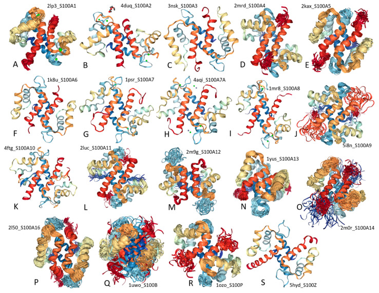
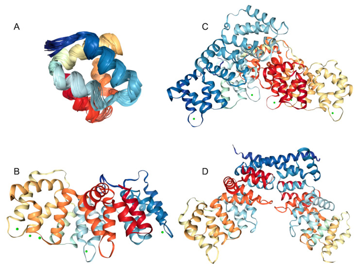
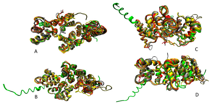
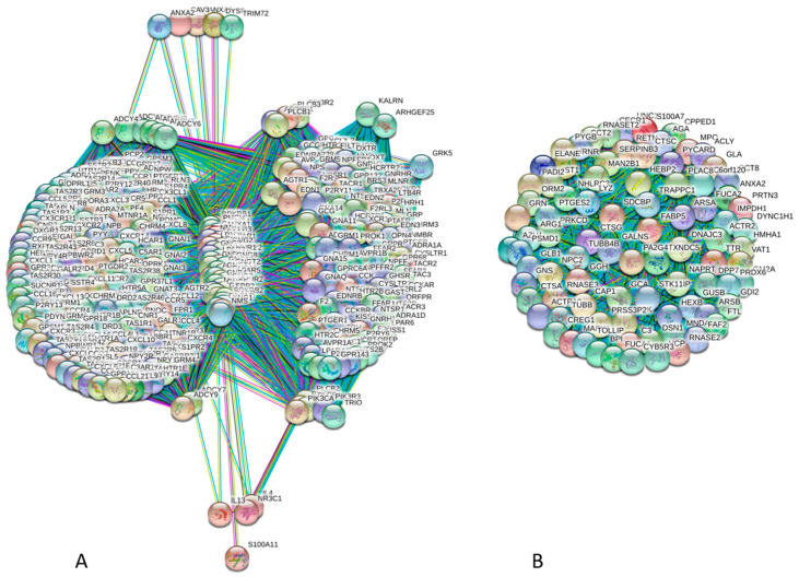
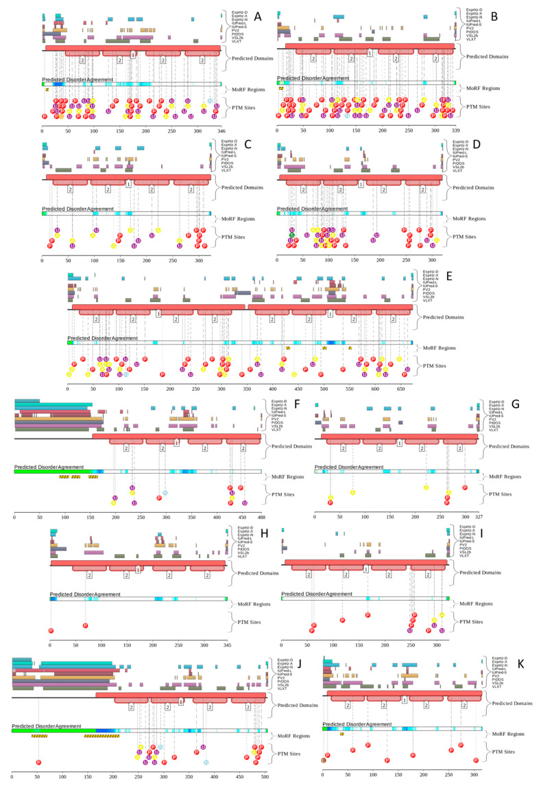
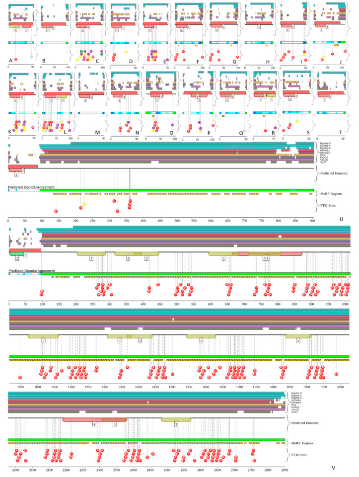
Annexins and S100 proteins form two large families of Ca2+-binding proteins. They are quite different both structurally and functionally, with S100 proteins being small (10-12 kDa) acidic regulatory proteins from the EF-hand superfamily of Ca2+-binding proteins, and with annexins being at least three-fold larger (329 ± 12 versus 98 ± 7 residues) and using non-EF-hand-based mechanism for calcium binding. Members of both families have multiple biological roles, being able to bind to a large cohort of partners and possessing a multitude of functions. Furthermore, annexins and S100 proteins can interact with each other in either a Ca2+-dependent or Ca2+-independent manner, forming functional annexin-S100 complexes. Such functional polymorphism and binding indiscrimination are rather unexpected, since structural information is available for many annexins and S100 proteins, which therefore are considered as ordered proteins that should follow the classical "one protein-one structure-one function" model. On the other hand, the ability to be engaged in a wide range of interactions with multiple, often unrelated, binding partners and possess multiple functions represent characteristic features of intrinsically disordered proteins (IDPs) and intrinsically disordered protein regions (IDPRs); i.e., functional proteins or protein regions lacking unique tertiary structures. The aim of this paper is to provide an overview of the functional roles of human annexins and S100 proteins, and to use the protein intrinsic disorder perspective to explain their exceptional multifunctionality and binding promiscuity.

Keywords: Ca2+-binding protein; S100 protein; annexin; intrinsically disordered protein; multifunctionality; protein–protein interactions.


Conflict of interest statement

The authors declare no conflict of interest.

Figures

Figure 1
Figure 1
Structural characterization of human S100 proteins. solution NMR structures (plots A, D, E, J, L, M, N, O, P, Q, and R) or X-ray crystallographic structures (plots B, C, F, G, H, I, K, and S) are shown for S100-A1 (A, PDB ID: 2LP3), S100-A2 (B, PDB ID: 4DUQ), S100-A3 (C, PDB ID: 3NSK), S100-A4 (D, PDB ID: 2MRD), S100-A5 (E, PDB ID: 2KAX), S100-A6 (F, PDB ID: 1K8U), S100-A7 (G, PDB ID: 1PSR), S100-A7A (H, PDB ID: 4AQI), S100-A8 (I, PDB ID: 1MR8), S100-A9 (J, PDB ID: 5I8N), S100-A10 (K, PDB ID: 4FTG), S100-A11 (L, PDB ID: 2LUC), S100-A12 (M, PDB ID: 2M9G), S100-A13 (N, PDB ID: 1YUS), S100-A14 (O, PDB ID: 2M0R), S100-A16 (P, PDB ID: 2L50), S100-B (Q, PDB ID: 1UWO), S100-P (R, PDB ID: 1OZO), and S100-Z (S, PDB ID: 5HYD). Note that no structural information is currently available for S100-A7B, S100-G, basalin (S100-A17), and hornerin (S100-A18).
Figure 2
Figure 2
Structural characterization of human annexins. A. NMR solution structure of a single annexin repeat from ANXA1 (PDB ID: 1BO9). B. An X-ray crystal structure of the full-length ANXA3 containing four annexin repeats (PDB ID: 1AXN). C. An X-ray crystal structure of the monomeric ANXA6 containing eight annexin repeats (PDB ID: 1M9I). D. An X-ray crystal structure of the dimeric ANXA13 (PDB ID: 6B3I).
Figure 3
Figure 3
Multiple structural alignment of human annexins (ANXA2, PDB ID: 1W7B, red structure; ANXA3, PDB ID: 1AXN, gray structure; ANXA4, PDB ID: 2ZOC, orange structure; ANXA5, PDB ID: 1AVR, yellow structure; C-terminal half of ANXA6, PDB ID: 1M9I, tan structure; ANXA8, PDB ID: 1W3W, silver structure; and ANXA13, PDB ID: 6B3I, green structure) conducted by MultiProt server [69]. Structures were plotted using the VMD software [70].
Figure 4
Figure 4
Illustrative examples of highly connected members of annexin and S100 proteins. These PPI networks were generated for ANXA1 (A, UniProt ID: P04083) and S1007 (B, UniProt ID: P31151) by computational platform STRING using the highest confidence of 0.9. STRING integrates all the information on protein-protein interactions (PPIs), complements it with computational predictions, and returns PPI network showing all possible PPIs of a query protein(s) [114]. The ANXA1-centered PPI network contains 354 nodes connected by 37,162 edges, whereas there are 86 nodes and 3655 edges in the S1007-centered PPI network.
Figure 5
Figure 5
Analysis of the intra-family and inter-family interactivity of human annexins and S100 protein. (A) The inter-family interactivity is illustrated by the PPI network between 12 human annexins and 23 human S100 proteins generated by STRING with the medium confidence of 0.4. In this network, 35 human calcium-binding proteins are connected by 173 edges. (B) The distributions of intra- and inter-family interactions for human annexins and S100 proteins. For each protein three bars are shown corresponding to the total number of interactions with other annexins and S100 proteins (black bar), the number of its interactions with annexins (red bar) and the number of its interactions with S100 proteins (green bar).
Figure 6
Figure 6
Evaluation of the global interactivity of human annexins and S100 proteins by STRING platform. (A) Global annexin-S100-centered interactome that includes 535 nodes connected by 44,201 edges. In this analysis, the medium confidence level of 0.4 was used. (B) Comparison of the involvement of each member of the human annexin and S100 families in annexin-S100 interfamily network and in a global annexin-S100-centered interactome.
Figure 7
Figure 7
Evaluation of the per-residue intrinsic disorder predispositions of human annexins (A,B) and S100 proteins (C). This analysis was conducted using PONDR® VSL2-based. Plot A contains PONDR® VSL2-generated profiles of the aligned annexins. Plot B represents disorder profiles of aligned core domains of human annexins. Plot C shows disorder profiles of 904-residue long basalin (S100-A17) and 2850-residue-long hornerin (S100-A18). N-terminal regions of both proteins contain short (~100 residues) partially ordered domains, with the remaining 800 and 2750 residues of S100-A17 and S100-A18 being highly disordered. Inset to this plot represents PONDR® VSL2 profiles of the N-terminal sections of these two proteins and all other human S100 proteins.
Figure 8
Figure 8
2D representation of the results of evaluation of disorder levels of human annexin and S100 proteins. Here, the percentages of residues in these proteins predicted to be disordered by PONDR® VSL2 are compared with their percentages of disordered residues predicted by PONDR® VLXT. The goal of this plot is to show the overall agreement between the outputs of different disorder predictors used in this study and to show high level of disorder in analyzed proteins.
Figure 9
Figure 9
Functional disorder profiles of human annexins generated by the D2P2 platform (http://d2p2.pro) that generates an interactive display that comments on the structural components, disordered segments, post-translational modifications (PTMs), and the presence of disorder-based binding sites in a protein of interest [139]. A, ANXA1; B, ANXA2; C, ANXA3; D, ANXA5; E, ANXA6; F, ANXA7; G, ANXA8; H, ANXA9; I, ANXA10; J, ANXA11; K, ANXA13. Note that no D2P2 profile is currently available for human ANXA4. At the top of each figure, there is a side by side comparison of seven separate disorder predictors (Espritz-D, Espritz-X, Espritz-N, IUPred-L, IUPred-S, PV2, PrDOS, VSL2b, and VLXT), bars indicate positive hits for disorder prediction. Below these colored bars there are two bars showing the position of predicted SCOP domains and conserved Pfam domains. The middle of each plot contains a bar labeled “Predicted Disorder Agreement” presenting the level of agreement between all of the disorder predictors, which is shown as color intensity in an aligned gradient bar below the stack of predictions. The green segments represent disorder that is not found within a predicted SCOP domain. The blue segments are where the disorder predictions intersect the SCOP domain prediction. Below the disorder agreement line, disorder-based binding region (i.e., disordered regions that fold upon interaction with binding partners and known as molecular recognition features, MoRFs) predicted by ANCHOR are displayed as yellow blocks with zigzag infill. Finally, the bottom of the plot shows positions of various PTMs within the query proteins. These are shown by differently colored circles containing letter A (acetylation), L (lipidation), N (nitrosylation), P (phosphorylation), S (sumoylation), and U (ubiquitylation).
Figure 10
Figure 10
D2P2-generated functional disorder profiles of human S100 proteins. A. S100A1; B. S100A3; C. S100A4; D. S100A5; E. S100A6; F. S100A7; G. S100A7A; H. S100A7L2; I. S100A8; J. S100A9; K. S100A10; L. S100A11; M. S100A12; N. S100A13; O. S100A14; P. S100A16; Q. S100B; R. S100G; S. S100P; T. S100Z; U. S100A17; V. S100A18. All the keys are described in the legend to Figure 9.

Similar articles

Cited by

References

    1. Miwa N., Uebi T., Kawamura S. S100-annexin complexes—biology of conditional association. FEBS J. 2008;275:4945–4955. doi: 10.1111/j.1742-4658.2008.06653.x. - DOI - PubMed
    1. Nockolds C.E., Kretsinger R.H., Coffee C.J., Bradshaw R.A. Structure of a calcium-binding carp myogen. Proc. Natl. Acad. Sci. USA. 1972;69:581–584. doi: 10.1073/pnas.69.3.581. - DOI - PMC - PubMed
    1. Raynal P., Pollard H.B. Annexins: The problem of assessing the biological role for a gene family of multifunctional calcium- and phospholipid-binding proteins. Biochim. Biophys. Acta. 1994;1197:63–93. doi: 10.1016/0304-4157(94)90019-1. - DOI - PubMed
    1. Gerke V., Moss S.E. Annexins: From structure to function. Physiol. Rev. 2002;82:331–371. doi: 10.1152/physrev.00030.2001. - DOI - PubMed
    1. Dunker A.K., Lawson J.D., Brown C.J., Williams R.M., Romero P., Oh J.S., Oldfield C.J., Campen A.M., Ratliff C.M., Hipps K.W., et al. Intrinsically disordered protein. J. Mol. Graph. Model. 2001;19:26–59. doi: 10.1016/S1093-3263(00)00138-8. - DOI - PubMed

MeSH terms

LinkOut - more resources