. 2020 Aug 17;10(8):1437.
doi: 10.3390/ani10081437.

Tumor Infiltrating Lymphocytes in Pet Rabbit Mammary Carcinomas: A Study with Relevance to Comparative Pathology

Affiliations

Tumor Infiltrating Lymphocytes in Pet Rabbit Mammary Carcinomas: A Study with Relevance to Comparative Pathology

Sandra Schöniger et al. Animals (Basel). .

Abstract

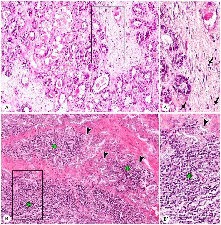
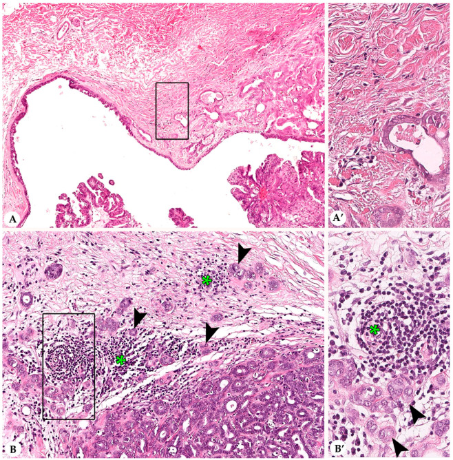
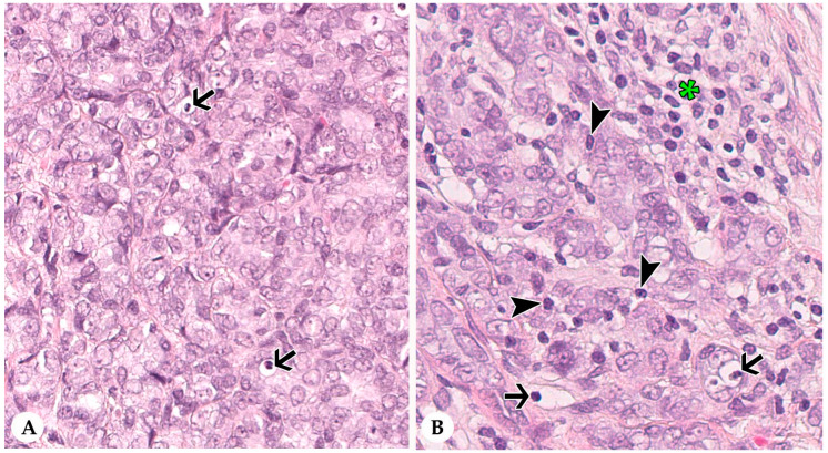
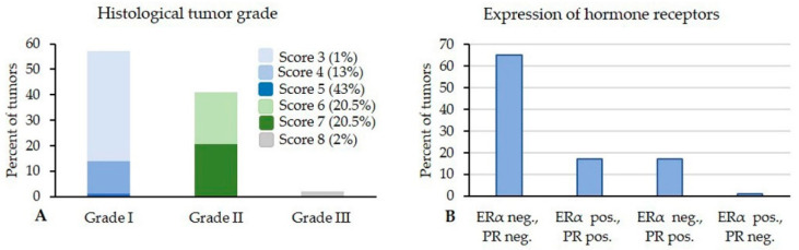
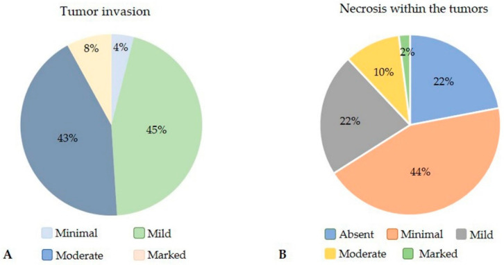
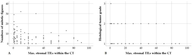
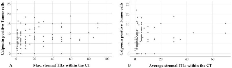
Tumor infiltrating lymphocytes (TILs) serve as prognostic biomarker in human breast cancer. Rabbits have the potential to act as animal model for human breast cancer, and close similarities exist between the rabbit and human immune system. The aim of this study is to characterize TILs in pet rabbit mammary carcinomas and to statistically correlate results with histological and immunohistochemical tumor characteristics. Microscopic evaluation of TILs was performed in hematoxylin and eosin stained sections of 107 rabbit mammary carcinomas according to international guidelines for human breast cancer. Data on histological features of malignancy, estrogen and progesterone receptor status and calponin expression were obtained from the data base. This study revealed a statistical association between stromal TILs in the central tumor (CT) and infiltrative margin. Higher maximal percentages of stromal TILs at the CT were statistically correlated with decreased mitotic count and lower tumor grade. An increased number of calponin positive tumor cells was statistically associated with a lower mitotic count and a higher percentage of stromal TILs. Results suggest that higher percentages of stromal TILs are useful biomarkers that may point toward a favorable prognosis in rabbit mammary carcinomas and support the concept of the use of rabbits for translational research.

Keywords: Oryctolagus cuniculus; animal model; biomarker; breast cancer; comparative pathology; light microscopy; mammary carcinomas; pet rabbit; translational medicine; tumor infiltrating lymphocytes.


Conflict of interest statement

The authors declare no conflict of interest.

Figures

Figure 1
Figure 1
Stromal tumor infiltrating lymphocytes (TILs) within the central tumor (CT) of pet rabbit mammary carcinomas. Illustrated are two representative tumor areas with very few stromal TILs (A,A’) and multifocal aggregates of stromal TILs (B,B’), respectively. (A,A’) Grade I carcinoma with a tubular growth pattern and moderate secretory activity. Occasional stromal TILs (less than 1%) are present in the 20× objective field of view (FOV) shown in A. The rectangular area delineated in A is illustrated in A’ in higher magnification. Individual rare stromal TILs are labelled by arrows. (B,B’) Stromal TILs (asterisks) form multifocal aggregates that dissect between tubular structures lined by tumor cells (arrowheads). The 20× objective FOV present in B contains an estimate of 50% stromal TILs. The area delineated by a rectangle is shown in B’ in higher magnification.
Figure 2
Figure 2
Stromal tumor infiltrating lymphocytes (TILs) within the infiltrative margin (IM) of pet rabbit mammary carcinomas. Shown are two representative tumors with nearly complete absence of stromal TILs (A,A’) and multifocal clusters of stromal TILs (B,B’) at the IM, respectively. (A,A’) Grade II carcinoma with tubular and cystic growth patterns and mild secretory activity. The IM contains no unequivocal stromal TILs. In A, the 10× objective field of view (FOV) is depicted. The rectangle in A delineates the area that is shown in A’ in higher magnification. (B,B’) Grade II carcinoma with a predominantly tubular growth. TILs (green asterisks) are mostly located between and adjacent to the infiltrative tumor cell nests (arrowheads). In B, the 20× objective FOV is shown and contains approximately 25% stromal TILs. The area that is contained within the rectangle is depicted in B’ in close up.
Figure 3
Figure 3
Intra-tumoral infiltrating lymphocytes (IT TILs) of pet rabbit mammary carcinomas. Depicted are two representative tumors with almost lack of IT TILs (A) and some IT TILs (B), respectively. In comparison to stromal TILs (asterisk), IT TILs (arrowheads) are immediately associated with tumor cells (arrowheads). Intra-tumoral cell fragments (arrows) are present (A,B).
Figure 4
Figure 4
(A) Of the 107 rabbit mammary carcinomas, 57% were grade I, 41% grade II, and 2% grade III. (B) The majority (65%) was hormone receptor negative, whereas 17% of the tumors expressed both receptors, 17% showed immunostaining for solely progesterone receptor, and 1% was immunoreactive for only estrogen receptor. ERα = estrogen receptor; PR = progesterone receptor.
Figure 5
Figure 5
(A) Rabbit mammary tumors showed mostly mild (45%) and moderate (43%) invasive behavior; only small percentages of tumors displayed minimal (4%) and marked (8%) tissue invasion. (B) Necrosis was absent in 22% of the tumors, whereas minimal and mild necrosis was detected in 44% and 22% of rabbit mammary carcinomas, respectively. Moderate necrosis was present in 10% of the tumors and 2% of tumors had marked necrosis.
Figure 6
Figure 6
Higher maximal (max.) stromal tumor infiltrating lymphocytes (TILs) per 20× objective field of view (FOV) within the central tumor (CT) are statistically correlated with decreased numbers of mitotic figures per ten 40× HPFs (A) and a lower histological tumor grade (B).
Figure 7
Figure 7
An increased percentage of calponin positive tumor cells is statistically correlated with higher maximal (max.) stromal tumor infiltrating lymphocytes (TILs) per 20× objective FOV within the central tumor (CT), (A) and higher average stromal TILs within the central tumor (B).

References

    1. Mage R.G. Immunology of lagomorphs, Chapter IV. In: Pastoret P.-P., Griebel P., Bazin H., Govaerts A., editors. Handbook of Vertebrate Immunology. Academic Press; San Diego, CA, USA: 1998. pp. 223–260.
    1. Shiomi M. Rabbits as model for the study of human diseases, Chapter 7. In: Houdebine L.-M., Fan J., editors. Rabbit Biotechnology: Rabbit Genomics, Transgenesis, Cloning and Models. Springer Science and Business Media, B.V.; Dordrecht, The Netherlands: Berlin/Heidelberg, Germany: New York, NY, USA: London, UK: 2009. pp. 49–64.
    1. Peng X., Knouse J.A., Hernon K.M. Rabbit models for studying human infectious diseases. Comp. Med. 2015;65:499–507. - PMC - PubMed
    1. Esteves P.J., Abrantes J., Baldauf H.-M., BenMohamed L., Chen Y., Christensen N., González-Gallego J., Giacani L., Hu J., Kaplan G., et al. The wide utility of rabbits as models of human diseases. Exp. Mol. Med. 2018;50:1–10. doi: 10.1038/s12276-018-0094-1. - DOI - PMC - PubMed
    1. Tinkey P.T., Uthamanthil R.K., Weisbroth S.H. Rabbit neoplasia. In: Suckow M.A., Stevens K.A., Wilson R.P., editors. The Laboratory Rabbit, Guinea Pig, Hamster and Other Rodents. Academic Press; London, UK: San Diego, CA, USA: 2012. pp. 448–503.