Review
. 2020 Aug 17;13(8):199.
doi: 10.3390/ph13080199.

A Systematic Review of Molecular Imaging Agents Targeting Bradykinin B1 and B2 Receptors

Affiliations
Review

A Systematic Review of Molecular Imaging Agents Targeting Bradykinin B1 and B2 Receptors

Joseph Lau et al. Pharmaceuticals (Basel). .

Abstract

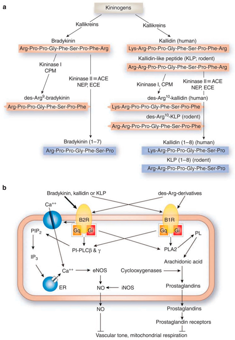
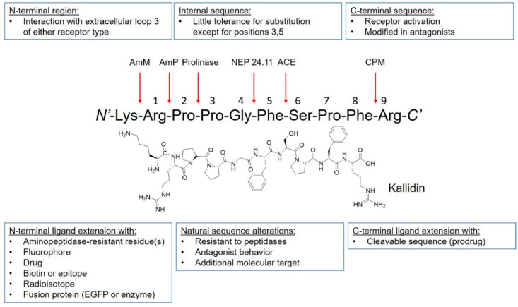
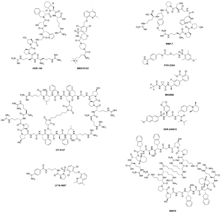
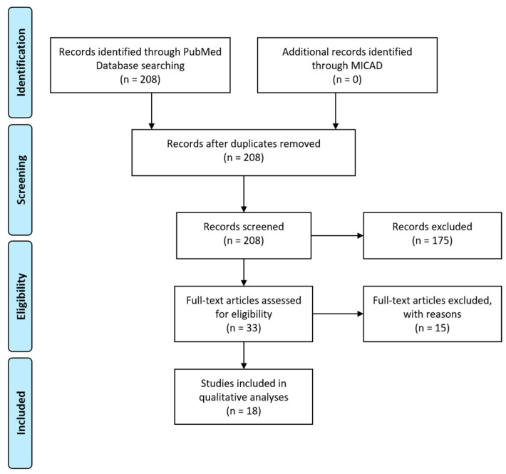
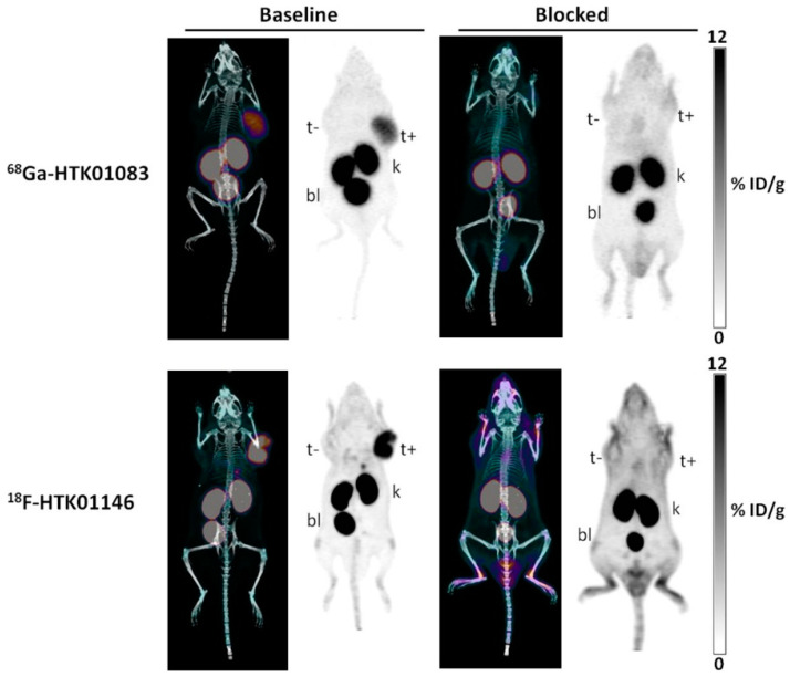
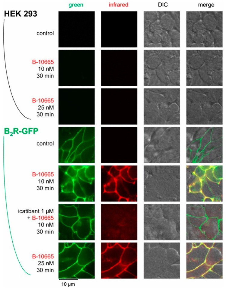
Kinins, bradykinin and kallidin are vasoactive peptides that signal through the bradykinin B1 and B2 receptors (B1R and B2R). B2R is constitutively expressed in healthy tissues and mediates responses such as vasodilation, fluid balance and retention, smooth muscle contraction, and algesia, while B1R is absent in normal tissues and is induced by tissue trauma or inflammation. B2R is activated by kinins, while B1R is activated by kinins that lack the C-terminal arginine residue. Perturbations of the kinin system have been implicated in inflammation, chronic pain, vasculopathy, neuropathy, obesity, diabetes, and cancer. In general, excess activation and signaling of the kinin system lead to a pro-inflammatory state. Depending on the disease context, agonism or antagonism of the bradykinin receptors have been considered as therapeutic options. In this review, we summarize molecular imaging agents targeting these G protein-coupled receptors, including optical and radioactive probes that have been used to interrogate B1R/B2R expression at the cellular and anatomical levels, respectively. Several of these preclinical agents, described herein, have the potential to guide therapeutic interventions for these receptors.

Keywords: bradykinin receptors; kinins; nuclear imaging; optical imaging; personalized medicine.


Conflict of interest statement

François Bénard and Kuo-Shyan Lin are coinventors of a patent (WO 2014/040192 A1) covering the development of radiopharmaceuticals targeting bradykinin B1 receptor.

Figures

Figure 1
Figure 1
Components and signaling of the kallikrein–kinin system. (a) Biosynthesis and metabolism of kinins. Kininogens are processed by kallikreins to produce bradykinin and kallidin. Kininase I removes the carboxyl-Arg from bradykinin and kallidin to yield des-Arg9-bradykinin and des-Arg10-kallidin, respectively. Enzymatic cleavage by kininase II (angiotensin I-converting enzyme), neprilysin, and endothelin-converting enzyme can inactivate kinins. CPM, carboxypeptidase-M; ACE, angiotensin I-converting enzyme; NEP, neprilysin (endopeptidase 24.11); ECE, endothelin-converting enzyme; red, active peptides; blue, inactive peptides. (b) Binding of kinins to bradykinin receptors and intracellular signaling. The thickness of arrows arising from the kinins indicates the relative potency of each peptide to elevate intracellular calcium concentrations. PIP2, phosphatidylinositol-4,5-bisphosphate; PI-PLC, phosphatidylinositol-specific phospholipase C; IP3, 1,4,5-inositol triphosphate; ER, endoplasmic reticulum; PL, phospholipids; PLA2, phospholipase A2; NO, nitric oxide; eNOS, endothelial NO synthase; iNOS, inducible NO synthase. Figure reproduced from Kakoki, M., et al. [18].
Figure 2
Figure 2
Peptide plasticity of kinins. Kinin peptides can be modified to improve bradykinin B1R or bradykinin B2R selectivity, increase resistance to enzymatic cleavage, confer agonistic or antagonistic properties, and prolong pharmacologic effect. Moreover, peptide ligands can be conjugated to fluorophores, drugs, radioisotope complex, and be fused with other proteins. Arrows indicate positions of potential cleavage. AmM: aminopeptidase M; AmP: aminopeptidase P; NEP 24.11: neutral endopeptidase 24.11; ACE: angiotensin-converting enzyme; CPM: carboxypeptidase M. Figure adapted from Marceau, F., et al. [25].
Figure 3
Figure 3
Chemical structures of B1R/B2R agonists and antagonists listed in Table 1. Both peptides and small molecules have been evaluated in clinical trials. The chemical structure of BI-113823, B1R antagonist, has not been disclosed.
Figure 4
Figure 4
Literature search for B1R and B2R imaging agents. Flow diagram to illustrate the process of article selection of this systematic review. Template redrawn from Moher, D.; et al. [69].
Figure 5
Figure 5
Confocal fluorescence imaging in HEK293a cells transiently expressing rabbit B1R and cavelolin-1-CherryFP. Cells were treated with 20 nM B-10376 (antagonist) or B-10378 (agonist). Both peptides labeled plasma membrane, but cells treated with B-10378 displayed enhance punctate fluorescence condensation that colocalized with cavelolin-1 indicated by hashed arrows. Figure reproduced from Bawolak, M.T.; et al. [72].
Figure 6
Figure 6
B1R inflammation imaging. After 1-day starvation, A549 cells were treated with 100 µg P. aeruginosa lysate for 4 h to induce inflammation. Cells were stained with 100 nM of FITC-conjugated Lys-[D-Phe8]des-Arg9-BK for 1 h at 37°C and analyzed with optical imaging. Differential interference contrast (DIC) microscope image of (A) normal and (C) inflamed cells. Fluorescence and DIC merged images of (B) normal and (D) inflamed cells. Figure reproduced from Yeo, K.B.; et al. [75].
Figure 7
Figure 7
B1R positron emission tomography imaging. Maximum intensity projection PET images obtained with 68Ga-P03083 without (left), with enalaprilat (middle), and with co-injection of phosphoramidon (right). The B1R+ tumor (red arrow) was located on the right shoulder (the animal is viewed on a coronal projection, ventral viewpoint). The B1R− tumor (blue arrow) had no significant uptake. The gray scale bar to the right of each image is set in units of %ID/g. Figure reproduced from Lin, K.S.; et al. [77].
Figure 8
Figure 8
Optimization of B1R radiopharmaceuticals. (A) Chemical structure of B1R radioligands. (B) Representative 1 h post-injection MIP (maximum intensity projection) and axial PET/CT images of radiolabeled Z02176, Z02137, and Z04139 in mice bearing both B1R+ (indicated by yellow arrows) and B1R– (indicated by red arrows) tumors without and (C) with co-injection of 100 μg cold standard. The displacement in tumor uptake demonstrates target specificity of the imaging agent. The scale bar is in units of %ID/g. Figure reproduced from Zhang, Z.; et al. [82].
Figure 9
Figure 9
Maximal intensity projection images of PET/CT and PET with 68Ga-HTK01083 and 18F-HTK01146 in mice bearing HEK293T and HEK293T::hB1R tumors at 1 h post-injection. Blocking studies were performed with co-injection of 100 μg of R954. t+, B1R+ tumor; t−, B1R- tumor; k, kidney; bl, bladder. Figure adapted from Kuo, H.T.; et al. [83].
Figure 10
Figure 10
Near-infrared imaging of B2R. Imaging of HEK 293a cells expressing B2R-GFP using Cy7-conjugated B-10665 (infrared fluorescence rendered as red false color). Labelling was performed for 30 min at 37 °C, followed by rinse. Original magnification 95×. Figure reproduced with permission from Infrared-emitting, peptidase-resistant fluorescent ligands of the bradykinin B2 receptor: application to cytofluorometry and imaging

References

    1. Leeb-Lundberg L.M.F., Marceau F., Müller-Esterl W., Pettibone D.J., Zuraw B.L. International union of pharmacology. XLV. Classification of the kinin receptor family: From molecular mechanisms to pathophysiological consequences. Pharmacol. Rev. 2005;57:27–77. doi: 10.1124/pr.57.1.2. - DOI - PubMed
    1. Marceau F., Bachelard H., Bouthillier J., Fortin J.P., Morissette G., Bawolak M.T., Charest-Morin X., Gera L. Bradykinin receptors: Agonists, antagonists, expression, signaling, and adaptation to sustained stimulation. Int. Immunopharmacol. 2020;82 doi: 10.1016/j.intimp.2020.106305. - DOI - PubMed
    1. Regoli D., Barabe J., Park W.K. Receptors for bradykinin in rabbit aortae. Can. J. Physiol. Pharmacol. 1977;55:855–867. doi: 10.1139/y77-115. - DOI - PubMed
    1. Regoli D., Marceau F., Barabe J. De novo formation of vascular receptors for bradykinin. Can. J. Physiol. Pharmacol. 1978;56:674–677. doi: 10.1139/y78-109. - DOI - PubMed
    1. Drouin J.N., St-Pierre S.A., Regoli D. Receptors for bradykinin and kallidin. Can. J. Physiol. Pharmacol. 1979;57:375–379. doi: 10.1139/y79-056. - DOI - PubMed

LinkOut - more resources