. 2020 Aug 17;12(8):900.
doi: 10.3390/v12080900.

Characterization of Signal Sequences Determining the Nuclear/Nucleolar Import and Nuclear Export of the Caprine Arthritis-Encephalitis Virus Rev Protein

Affiliations

Characterization of Signal Sequences Determining the Nuclear/Nucleolar Import and Nuclear Export of the Caprine Arthritis-Encephalitis Virus Rev Protein

Marlène Labrecque et al. Viruses. .

Abstract

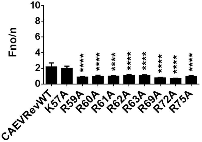
Caprine arthritis-encephalitis virus (CAEV), a lentivirus, relies on the action of the Rev protein for its replication. The CAEV Rev fulfills its function by allowing the nuclear exportation of partially spliced or unspliced viral mRNAs. In this study, we characterized the nuclear and nucleolar localization signals (NLS and NoLS, respectively) and the nuclear export signal (NES) of the CAEV Rev protein. These signals are key actors in the nucleocytoplasmic shuttling of a lentiviral Rev protein. Several deletion and alanine substitution mutants were generated from a plasmid encoding the CAEV Rev wild-type protein that was fused to the enhanced green fluorescent protein (EGFP). Following cell transfection, images were captured by confocal microscopy and the fluorescence was quantified in the different cell compartments. The results showed that the NLS region is localized between amino acids (aa) 59 to 75, has a monopartite-like structure and is exclusively composed of arginine residues. The NoLS was found to be partially associated with the NLS. Finally, the CAEV Rev protein's NES mapped between aa 89 to 101, with an aa spacing between the hydrophobic residues that was found to be unconventional as compared to that of other retroviral Rev/Rev-like proteins.

Keywords: Rev protein; caprine arthritis-encephalitis virus; nuclear export signal (NES); nuclear localization signal (NLS); nucleolar localization signal (NoLS); small ruminant lentivirus (SRLV).


Conflict of interest statement

The authors declare no conflict of interest. The funders had no role in the design of the study; in the collection, analyses, or interpretation of data; in the writing of the manuscript, or in the decision to publish the results.

Figures

Figure 1
Figure 1
The EGFP-CAEV Rev WT fusion protein localizes to the cytoplasm, nucleus and nucleoli of transfected BoMac and HeLa cells. (A) Microscopic analyses of EGFP-CAEV Rev WT fusion protein in BoMac and HeLa cells. Cells were transfected with pEGFP-CAEV Rev WT for 24 h and were treated, where mentioned, with 5 nM of leptomycin B (LMB) 5 h prior to cell fixation and permeabilization. Cells were then immunostained for nucleolin detection, as seen in red, and the nucleus was counterstained with DAPI, as seen in blue. Expression of EGFP-Rev, as seen in green, was visualized by CLSM at 60× magnification. The images are representative of the expression pattern observed in cells from three independent experiments. The merge images represent the superposition of EGFP-Rev, DAPI and nucleolin, and the white bars represent a length of 10 μM. The calculated Fn/c (B) and Fno/n (C) ratios were expressed as means ± SD from three independent experiments, in which 10 cells were analyzed for each of them (n = 30). Using Student’s T-test corrected with the Holm–Sidak method for multiple comparison of the means, each cell line was analyzed for significant differences, which are indicated by * (p < 0.05) and **** (p < 0.0001). Using ANOVA and Tukey’s multiple comparison test, the Fn/c and Fno/n ratios measured for the BoMac cells were compared with those of the HeLa cells, regardless of the LMB treatment. No significant differences were observed between these cells.
Figure 2
Figure 2
The amino acids between positions 56 to 115 are involved in the nuclear localization and nuclear export of the CAEV Rev protein. (A) CAEV Rev deletion mutants (M1 to M6) were generated from pEGFP-CAEV Rev WT using Gibson assembly mastermix. (B) Microscopic analyses of the CAEV Rev mutants M1 to M6 compared to the CAEV Rev WT. BoMac cells were transfected with pEGFP-CAEV Rev WT or each of the pEGFP-CAEV Rev mutants for 24 h and fixed. The nucleus was counterstained with DAPI, as seen in blue. Expression of EGFP-Rev, as seen in green, was visualized by CLSM at 60× magnification. The images are representative of the expression pattern observed in cells from three independent experiments. The merge images represent the superposition of EGFP-Rev and DAPI, and the white bars represent a length of 10 μM. (C) The calculated Fn/c ratios were expressed as means ± SD from three independent experiments, in which 10 cells were analyzed for each of them (n = 30). (D) The nuclear export activities of EGFP-CAEV Rev WT and mutants M1 to M6 were determined using a CAT reporter assay. HeLa cells were cotransfected with pDM148 and pEGFP-C1 or pEGFP-CAEV Rev WT or pEGFP-CAEV Rev mutants or were untransfected (Unt). Following 48 h of transfection, 50 μg of total cell lysate was used for the assay and the CAT expression levels were normalized via immunoblotting using EGFP-specific antibody (bottom of the panel). The Rev activity was expressed as the ratio of EGFP-CAEV Rev WT or mutant protein CAT expression to the basal expression of EGFP alone. The results represent the mean values ± SD of three separate experiments (triplicate samples per experiment). Antibody against GADPH was used as a loading control. According to one-way ANOVA followed by Dunnett’s test, the values significantly different from those of the CAEV Rev WT protein are indicated by *** (p < 0.0005).
Figure 3
Figure 3
The region between amino acids 57 to 82 of the CAEV Rev protein promotes nuclear localization. (A) The sequence aa 57 to 82 of the CAEV Rev protein (CAEV 57–82Rev) was cloned into pEGFP-C1 or pEGFP-βGal. (B) BoMac cells were transfected with the plasmid constructs encoding for EGFP or EGFP-βGal alone or fused to CAEV 57-82Rev. Following 24 h of incubation, cells were fixed and nucleus was counterstained with DAPI, as seen in blue. Expression of EGFP, as seen in green, was visualized by CLSM at 60× magnification. The images are representative of the expression pattern observed in cells from three independent experiments. The merge images represent the superposition of EGFP and DAPI, and the white bars represent a length of 10 μM. (C) The calculated Fn/c ratios were expressed as means ± SD from three independent experiments, in which 10 cells were analyzed for each of them (n = 30). Significant differences, determined using Student’s T-test, between EGFP or EGFP-βGal alone or fused to the CAEV 57–82Rev sequence, are indicated by **** (p < 0.0001). Expression of EGFP (D) or EGFP-βGal (E) alone or fused to CAEV 57–82Rev were visualized via immunoblotting using EGFP-specific antibody. Antibody against GADPH was used as a loading control.
Figure 4
Figure 4
The region encompassing amino acids 57 to 75 is associated with nuclear localization of the CAEV Rev protein. (A) CAEV Rev deletion mutants (RevΔ1 to RevΔ4) were generated from pEGFP-CAEV Rev WT using Gibson assembly mastermix. (B) Microscopic analyses of the CAEV Rev mutants RevΔ1 to RevΔ4 compared to the CAEV Rev WT. BoMac cells were transfected with pEGFP-CAEV Rev WT or each of the pEGFP-CAEV Rev mutants for 24 h and were treated, where mentioned, with 5 nM of leptomycin B (LMB) 5 h prior to cell fixation and permeabilization. Cells were then immunostained for nucleolin detection, as seen in red, and nucleus was counterstained with DAPI, as seen in blue. Expression of EGFP-Rev, as seen in green, was visualized by CLSM at 60× magnification. The images are representative of the expression pattern observed in cells from three independent experiments. The merge images represent the superposition of EGFP-Rev and DAPI, and the white bars represent a length of 10 μM. (C) The calculated Fn/c ratios were expressed as means ± SD from three independent experiments, in which 10 cells were analyzed for each of them (n = 30). (D) The nuclear export activities of EGFP-CAEV Rev WT and mutants RevΔ1 to RevΔ4 were determined using a CAT reporter assay. HeLa cells were cotransfected with pDM148 and pEGFP-C1 or pEGFP-CAEV Rev WT or pEGFP-CAEV Rev mutants or were untransfected (Unt). Following 48 h of transfection, 50 μg of total cell lysate was used for the assay and the CAT expression levels were normalized via immunoblotting using EGFP-specific antibody (bottom of the panel). The Rev activity was expressed as the ratio of EGFP-CAEV Rev WT or mutant protein CAT expression to the basal expression of EGFP alone. The results represent the mean values ± SD of three separate experiments (triplicate samples per experiment). Antibody against GADPH was used as a loading control. According to one-way ANOVA followed by Dunnett’s test, the values significantly different from those of the CAEV Rev WT protein are indicated by **** (p < 0.0001).
Figure 5
Figure 5
The NLS of the CAEV Rev protein has a monopartite-like structure. (A) CAEV Rev lysine (K) or arginine (R) to alanine (A) substitution mutant proteins were generated from pEGFP-CAEV Rev WT by site-directed mutagenesis. (B) Microscopic analyses of the CAEV Rev mutant proteins compared to the CAEV Rev WT. BoMac cells were transfected with pEGFP-CAEV Rev WT or pEGFP-CAEV Rev mutants for 24 h and were treated with 5 nM of leptomycin B (LMB) 5 h prior to cell fixation and permeabilization. Cells were then immunostained for nucleolin detection, as seen in red, and nucleus was counterstained with DAPI, as seen in blue. Expression of EGFP-Rev, as seen in green, was visualized by CLSM at 60× magnification. The images are representative of the expression pattern observed in cells from three independent experiments. The merge images represent the superposition of EGFP-Rev, nucleolin and DAPI, and the white bars represent a length of 10 μM. (C) The calculated Fn/c ratios were expressed as means ± SD from three independent experiments, in which 10 cells were analyzed for each of them (n = 30). (D) The nuclear export activities of EGFP-CAEV Rev WT and mutant proteins were determined using a CAT reporter assay. HeLa cells were cotransfected with pDM148 and pEGFP-C1 or pEGFP-CAEV Rev WT or pEGFP-CAEV Rev mutants or were untransfected (Unt). Following 48 h of transfection, 50 μg of total cell lysate was used for the assay and the CAT expression levels were normalized via immunoblotting using EGFP-specific antibody (bottom of the panel). The Rev activity was expressed as the ratio of EGFP-CAEV Rev WT or mutant protein CAT expression to the basal expression of EGFP alone. The results represent the mean values ± SD of three separate experiments (triplicate samples per experiment). Antibody against GADPH was used as a loading control. According to one-way ANOVA followed by Dunnett’s test, the values significantly different from those of the CAEV Rev WT protein are indicated by ** (p < 0.005), *** (p < 0.0005) and **** (p < 0.0001).
Figure 6
Figure 6
Impact of single alanine substitutions on the nucleolar localization of the CAEV Rev protein. Single alanine substitutions targeting arginine (R) and lysine (K) were generated by site-directed mutagenesis. BoMac cells were transfected with non mutated or mutated pEGFP-CAEV Rev constructs and were treated with 5 nM of leptomycin B (LMB) 5 h prior to cell fixation and immunostaining. Cells were then analyzed by CLSM, as shown in Fig. 5B. The calculated Fno/n ratios were expressed as means ± SD from three independent experiments, in which 10 cells were analyzed for each of them (n = 30). Significant differences, using a one-way ANOVA followed by a Dunnett’s test, are indicated by: **** (p < 0.0001).
Figure 7
Figure 7
The NES of the CAEV Rev protein lies between amino acids 89 to 101. (A) Plasmids encoding HIV-1 Rev(1.4)-EGFP (negative control; Rev1.4), HIV-1 Rev(1.4)-NES3-EGFP (positive control; Rev1.4NES3) or plasmids encoding HIV-1 Rev(1.4) containing either the predicted NES sequence (amino acids 89 to 101) of the CAEV Rev WT protein (CAEVRevNESWT) or (B) each of the CAEV NES mutated sequences were used. (C) HeLa cells were transfected for 24 h and were incubated with or without CHX (10 μg/mL) and ActD (5 μg/mL) 3 h prior to cell fixation. Cells were then fixed and counterstained with DAPI, as seen in blue. Expression of EGFP-Rev, as seen in green, was visualized by CLSM at 60× magnification. The images are representative of the expression pattern observed in cells from three independent experiments. The merge images represent the superposition of EGFP-Rev and DAPI, and the white bars represent a length of 10 μM. (D) The calculated Fn/c ratios were expressed as means ± SD from three independent experiments, in which 10 cells were analyzed for each of them (n = 30). (E) The nuclear export activity of the EGFP-fused HIV-1 Rev protein was determined using a CAT reporter assay. HeLa cells were cotransfected with pDM128 and pRev(1.4)-EGFP or pRev(1.4)-NES3-EGFP or plasmids containing the NES sequence of CAEV Rev WT or each of the CAEV Rev NES mutated sequences or were left untransfected (Unt). Following 48 h of transfection, 50 μg of total cell lysate was used for the assay and the CAT expression levels were normalized via immunoblotting using EGFP-specific antibody (bottom of the panel). Rev activity was then determined as the ratio of HIV-1 Rev protein CAT expression harboring the HIV-1 Rev NES WT (NES3), the CAEV Rev NES WT or the CAEV Rev NES mutant to the basal expression from pDM128 construct co-transfected with empty pEGFP-C1 alone. The results represent the mean values ± SD of three separate experiments (triplicate samples per experiment). Antibody against GADPH was used as a loading control. According to one-way ANOVA followed by Dunnett’s test, the values significantly different from those of CAEV Rev WT protein are indicated by * (p < 0.05), ** (p < 0.005), *** (p < 0.0005) and **** (p < 0.0001).

Similar articles

References

    1. Larruskain A., Jugo B.M. Retroviral infections in sheep and goats: Small ruminant lentiviruses and host interaction. Viruses. 2013;5:2043–2061. doi: 10.3390/v5082043. - DOI - PMC - PubMed
    1. Santry L.A., de Jong J., Gold A.C., Walsh S.R., Menzies P.I., Wootton S.K. Genetic characterization of small ruminant lentiviruses circulating in naturally infected sheep and goats in Ontario, Canada. Virus Res. 2013;175:30–44. doi: 10.1016/j.virusres.2013.03.019. - DOI - PubMed
    1. Peterhans E., Greenland T., Badiola J., Harkiss G., Bertoni G., Amorena B., Eliaszewicz M., Juste R.A., Krassnig R., Lafont J.P., et al. Routes of transmission and consequences of small ruminant lentiviruses (SRLVs) infection and eradication schemes. Vet. Res. 2004;35:257–274. doi: 10.1051/vetres:2004014. - DOI - PubMed
    1. Ramirez H., Reina R., Amorena B., de Andres D., Martinez H.A. Small ruminant lentiviruses: Genetic variability, tropism and diagnosis. Viruses. 2013;5:1175–1207. doi: 10.3390/v5041175. - DOI - PMC - PubMed
    1. Da Cruz J.C.M., Singh D.K., Lamara A., Chebloune Y. Small ruminant lentiviruses (SRLVs) break the species barrier to acquire new host range. Viruses. 2013;5:1867–1884. doi: 10.3390/v5071867. - DOI - PMC - PubMed

Publication types

Substances

LinkOut - more resources