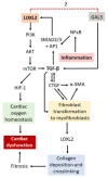
Linking LOXL2 to Cardiac Interstitial Fibrosis
- PMID: 32824630
- PMCID: PMC7460598
- DOI: 10.3390/ijms21165913
Linking LOXL2 to Cardiac Interstitial Fibrosis
Abstract
Cardiovascular diseases (CVDs) are the leading causes of death worldwide. CVD pathophysiology is often characterized by increased stiffening of the heart muscle due to fibrosis, thus resulting in diminished cardiac function. Fibrosis can be caused by increased oxidative stress and inflammation, which is strongly linked to lifestyle and environmental factors such as diet, smoking, hyperglycemia, and hypertension. These factors can affect gene expression through epigenetic modifications. Lysyl oxidase like 2 (LOXL2) is responsible for collagen and elastin cross-linking in the heart, and its dysregulation has been pathologically associated with increased fibrosis. Additionally, studies have shown that, LOXL2 expression can be regulated by DNA methylation and histone modification. However, there is a paucity of data on LOXL2 regulation and its role in CVD. As such, this review aims to gain insight into the mechanisms by which LOXL2 is regulated in physiological conditions, as well as determine the downstream effectors responsible for CVD development.
Keywords: DNA methylation; Lysyl Oxidase-Like 2 (LOXL2); cardiovascular disease (CVD); epigenetics; fibrosis.
Conflict of interest statement
The authors declare no conflict of interest. The funders had no role in the design of the study; in the collection, analyses, or interpretation of data; in the writing of the manuscript, or in the decision to publish the results.
Figures


References
-
- World Health Statistics (WHO) Monitoring Health for the SDGs. WHO; Geneva, Switzerland: 2019.
-
- World Health Statistics (WHO) New Initiative Launched to Tackle Cardiovascular Disease: The World’s Number One Killer. WHO; Geneva, Switzerland: 2016.
-
- World Health Statistics (WHO) Global Hearts Initiative: Working Together to Promote Cardiovascular Health. WHO; Geneva, Switzerland: 2018.
-
- World Health Statistics (WHO) Obesity and Overweight Fact Sheet. [(accessed on 12 November 2018)]; Available online: https://www.who.int/news-room/fact-sheets/detail/obesity-and-overweight.
-
- International Diabetes Federation (IDF) IDF Diabetes Atlas. 9th ed. IDF; Brussels, Belgium: 2019.
Publication types
MeSH terms
Substances
Grants and funding
LinkOut - more resources
Full Text Sources
Other Literature Sources
Medical
Research Materials

