

Manganese Ions Individually Alter the Reverse Transcription Signature of Modified Ribonucleosides
- PMID: 32824672
- PMCID: PMC7466121
- DOI: 10.3390/genes11080950
Manganese Ions Individually Alter the Reverse Transcription Signature of Modified Ribonucleosides
Abstract
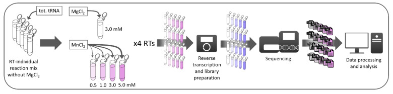
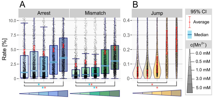
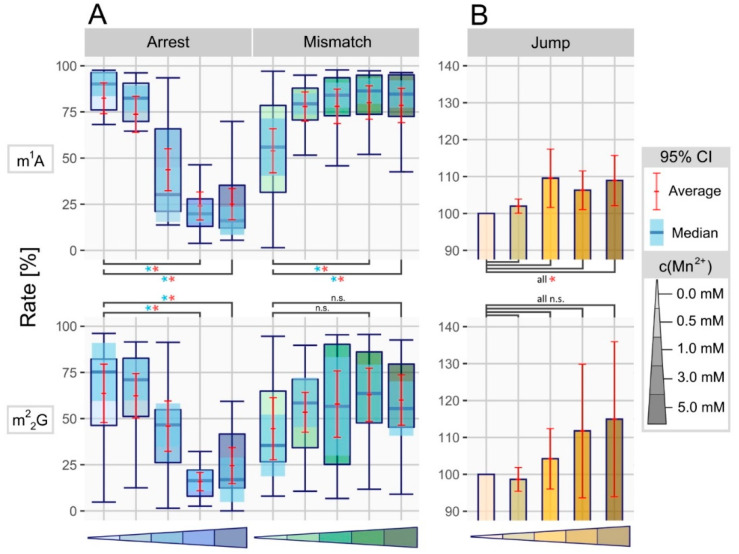
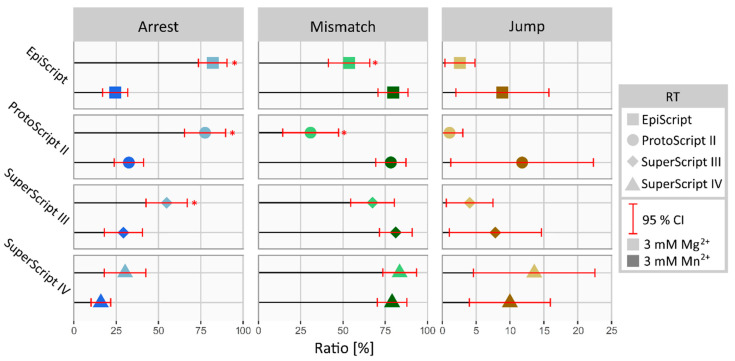
Reverse transcription of RNA templates containing modified ribonucleosides transfers modification-related information as misincorporations, arrest or nucleotide skipping events to the newly synthesized cDNA strand. The frequency and proportion of these events, merged from all sequenced cDNAs, yield a so-called RT signature, characteristic for the respective RNA modification and reverse transcriptase (RT). While known for DNA polymerases in so-called error-prone PCR, testing of four different RTs by replacing Mg2+ with Mn2+ in reaction buffer revealed the immense influence of manganese chloride on derived RT signatures, with arrest rates on m1A positions dropping from 82% down to 24%. Additionally, we observed a vast increase in nucleotide skipping events, with single positions rising from 4% to 49%, thus implying an enhanced read-through capability as an effect of Mn2+ on the reverse transcriptase, by promoting nucleotide skipping over synthesis abortion. While modifications such as m1A, m22G, m1G and m3C showed a clear influence of manganese ions on their RT signature, this effect was individual to the polymerase used. In summary, the results imply a supporting effect of Mn2+ on reverse transcription, thus overcoming blockades in the Watson-Crick face of modified ribonucleosides and improving both read-through rate and signal intensity in RT signature analysis.
Keywords: RNA modifications; RT signature; m1A; manganese chloride; reverse transcription.
Conflict of interest statement
The authors declare no conflict of interest.
Figures
Similar articles
-
Machine learning of reverse transcription signatures of variegated polymerases allows mapping and discrimination of methylated purines in limited transcriptomes.Nucleic Acids Res. 2020 Apr 17;48(7):3734-3746. doi: 10.1093/nar/gkaa113. Nucleic Acids Res. 2020. PMID: 32095818 Free PMC article.
-
Reverse Transcription in the Saccharomyces cerevisiae Long-Terminal Repeat Retrotransposon Ty3.Viruses. 2017 Mar 15;9(3):44. doi: 10.3390/v9030044. Viruses. 2017. PMID: 28294975 Free PMC article. Review.
-
Base modifications affecting RNA polymerase and reverse transcriptase fidelity.Nucleic Acids Res. 2018 Jun 20;46(11):5753-5763. doi: 10.1093/nar/gky341. Nucleic Acids Res. 2018. PMID: 29750267 Free PMC article.
-
Frequency and effect of the binding of Mg2+, Mn2+, and Co2+ ions on the guanine base in Watson-Crick and reverse Watson-Crick base pairs.J Phys Chem B. 2009 Nov 26;113(47):15670-8. doi: 10.1021/jp906847p. J Phys Chem B. 2009. PMID: 19921955
-
Strand transfer events during HIV-1 reverse transcription.Virus Res. 2008 Jun;134(1-2):19-38. doi: 10.1016/j.virusres.2007.12.017. Epub 2008 Feb 14. Virus Res. 2008. PMID: 18279992 Review.
Cited by
-
Characterization and Engineering of Two Novel Strand-Displacing B Family DNA Polymerases from Bacillus Phage SRT01hs and BeachBum.Biomolecules. 2025 Aug 5;15(8):1126. doi: 10.3390/biom15081126. Biomolecules. 2025. PMID: 40867571 Free PMC article.
-
Advances in methods for tRNA sequencing and quantification.Trends Genet. 2024 Mar;40(3):276-290. doi: 10.1016/j.tig.2023.11.001. Epub 2023 Dec 19. Trends Genet. 2024. PMID: 38123442 Free PMC article. Review.
-
CLIP-Seq to identify targets and interactions of RNA binding proteins and RNA modifying enzymes.Methods Enzymol. 2021;658:419-434. doi: 10.1016/bs.mie.2021.08.001. Epub 2021 Aug 23. Methods Enzymol. 2021. PMID: 34517957 Free PMC article.
-
Analysis of RNA Modifications by Second- and Third-Generation Deep Sequencing: 2020 Update.Genes (Basel). 2021 Feb 16;12(2):278. doi: 10.3390/genes12020278. Genes (Basel). 2021. PMID: 33669207 Free PMC article. Review.
-
N1-methylation of adenosine (m1A) in ND5 mRNA leads to complex I dysfunction in Alzheimer's disease.Mol Psychiatry. 2024 May;29(5):1427-1439. doi: 10.1038/s41380-024-02421-y. Epub 2024 Jan 29. Mol Psychiatry. 2024. PMID: 38287100 Free PMC article.
References
-
- Cohn W.E., Volkin E. Nucleoside-5′-Phosphates from Ribonucleic Acid. Nature. 1951;167:483–484. doi: 10.1038/167483a0. - DOI
Publication types
MeSH terms
Substances
LinkOut - more resources
Full Text Sources
Other Literature Sources
