Review
. 2020 Aug 18;21(16):5933.
doi: 10.3390/ijms21165933.

How Do Molecular Dynamics Data Complement Static Structural Data of GPCRs

Affiliations
Review

How Do Molecular Dynamics Data Complement Static Structural Data of GPCRs

Mariona Torrens-Fontanals et al. Int J Mol Sci. .

Abstract

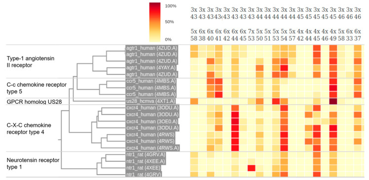
G protein-coupled receptors (GPCRs) are implicated in nearly every physiological process in the human body and therefore represent an important drug targeting class. Advances in X-ray crystallography and cryo-electron microscopy (cryo-EM) have provided multiple static structures of GPCRs in complex with various signaling partners. However, GPCR functionality is largely determined by their flexibility and ability to transition between distinct structural conformations. Due to this dynamic nature, a static snapshot does not fully explain the complexity of GPCR signal transduction. Molecular dynamics (MD) simulations offer the opportunity to simulate the structural motions of biological processes at atomic resolution. Thus, this technique can incorporate the missing information on protein flexibility into experimentally solved structures. Here, we review the contribution of MD simulations to complement static structural data and to improve our understanding of GPCR physiology and pharmacology, as well as the challenges that still need to be overcome to reach the full potential of this technique.

Keywords: GPCRs; drug discovery; ligand binding; molecular dynamics; receptor (in)activation; receptor signaling.


Conflict of interest statement

The authors declare no conflict of interest.

Figures

Figure 1
Figure 1
(a) Number of G protein-coupled receptors (GPCRs) structures available in GPCRdb [4,5] over time. (b) Number of publications per year indexed at Thomson Reuters’ Web of Science that contain the topics “molecular dynamics” and (“GPCR” or “GPCRs”). The exponential growth of successful GPCR research based on molecular dynamics (MD) simulations is evidenced by the rapid upsurge in the number of publications per year related to this subject.
Figure 2
Figure 2
Schematic view of the ligand-protein interaction results that can be obtained with the GPCRmd server [53]. Specifically, the GPCRmd Workbench module of the server enables interactive visualization (GPCRmd Viewer) and analysis (GPCRmd Toolkit) for individual simulations, including ligand-protein interactions among others. Figure obtained from the GPCRmd server [53].
Figure 3
Figure 3
(a) Flowchart summarizing the stages of a MD simulation. (b) Example of a GPCR molecular system, including the β-2 adrenergic receptor (β2AR, blue) with a full agonist in the binding site (orange) in a 1-palmitoyl-2-oleoyl-sn-glycero-3-phosphocholine (POPC) membrane (tails in light brown, heads colored by heteroatom). The system is solvated with water (red) and ionized with sodium (green) and chloride (purple) ions.
Figure 4
Figure 4
Example of different parameters analyzed in a 500 ns-long MD simulation of the A2A receptor (A2AR). (a) Root mean square deviation (RMSD) profile taking as reference the first frame of the simulation, which is superimposed to the rest of the frames. RMSD values (i.e., structural differences with respect to the reference frame) increase over the simulation time until the system reaches a stable conformation after 100 ns. (b) Root mean square fluctuation (RMSF) profile displaying the values of all the alpha carbons in the protein. Higher RMSF values correspond to flexible loops, while lower ones belong to transmembrane helices, where residues are stabilized by the secondary structure. (c) Radius of gyration (RG) profile where the RG fluctuates around the same value during the simulation, indicating that the system does not suffer any big change in compactness. (d) Superimposition of 25 representative frames of the simulated receptor. The relative mobility of loop regions contrasts with the rigidness of the transmembrane helices.
Figure 5
Figure 5
Pattern of total interaction frequency of several MD simulations of GPCRs, extracted from the GPCRmd Receptor Meta-analysis tool (https://submission.gpcrmd.org/contmaps/) of the GPCRmd server [53]. Columns represent interacting residue pairs according to Ballesteros-Weinstein residue numbering [142], whereas rows represent different simulations. The color of each cell shows the frequency in which any type of non-covalent interaction occurs during the simulation. Results are clustered based on the interaction frequencies of the simulations. This clustering is able to separate simulations according to the receptor subtype, showing that different receptor subtypes present differentiated interaction patterns.

References

    1. Hauser A.S., Attwood M.M., Rask-Andersen M., Schiöth H.B., Gloriam D.E. Trends in GPCR drug discovery: New agents, targets and indications. Nat. Rev. Drug Discov. 2017;16:829–842. doi: 10.1038/nrd.2017.178. - DOI - PMC - PubMed
    1. Congreve M., de Graaf C., Swain N.A., Tate C.G. Impact of GPCR Structures on Drug Discovery. Cell. 2020;181:81–91. doi: 10.1016/j.cell.2020.03.003. - DOI - PubMed
    1. Jazayeri A., Andrews S.P., Marshall F.H. Structurally enabled discovery of adenosine a2a receptor antagonists. Chem. Rev. 2017;117:21–37. doi: 10.1021/acs.chemrev.6b00119. - DOI - PubMed
    1. Pándy-Szekeres G., Munk C., Tsonkov T.M., Mordalski S., Harpsøe K., Hauser A.S., Bojarski A.J., Gloriam D.E. GPCRdb in 2018: Adding GPCR structure models and ligands. Nucleic Acids Res. 2018;46:D440–D446. doi: 10.1093/nar/gkx1109. - DOI - PMC - PubMed
    1. Berman H.M., Westbrook J., Feng Z., Gilliland G., Bhat T.N., Weissig H., Shindyalov I.N., Bourne P.E. The Protein Data Bank. Nucleic Acids Res. 2000;28:235–242. doi: 10.1093/nar/28.1.235. - DOI - PMC - PubMed

LinkOut - more resources