. 2020 Aug 19;21(17):5944.
doi: 10.3390/ijms21175944.

The Molecular Basis and Biologic Significance of the β-Dystroglycan-Emerin Interaction

Affiliations

The Molecular Basis and Biologic Significance of the β-Dystroglycan-Emerin Interaction

Wendy Lilián Gómez-Monsivais et al. Int J Mol Sci. .

Abstract

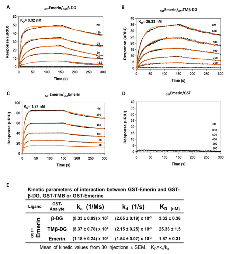
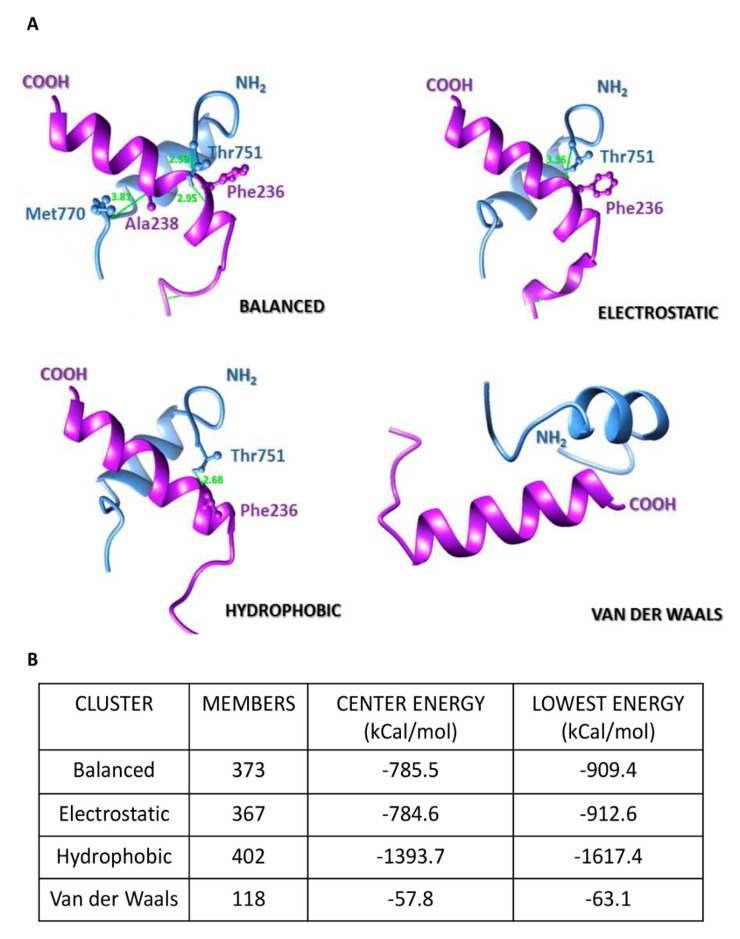
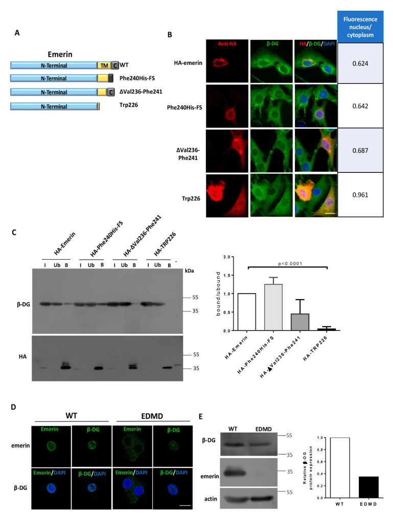
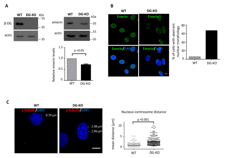
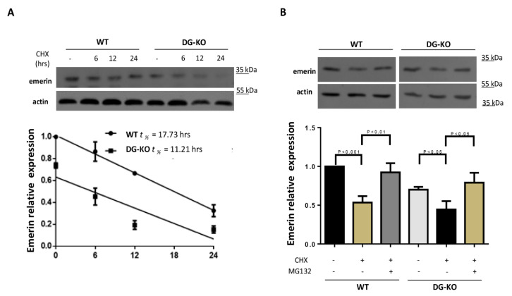
β-dystroglycan (β-DG) assembles with lamins A/C and B1 and emerin at the nuclear envelope (NE) to maintain proper nuclear architecture and function. To provide insight into the nuclear function of β-DG, we characterized the interaction between β-DG and emerin at the molecular level. Emerin is a major NE protein that regulates multiple nuclear processes and whose deficiency results in Emery-Dreifuss muscular dystrophy (EDMD). Using truncated variants of β-DG and emerin, via a series of in vitro and in vivo binding experiments and a tailored computational analysis, we determined that the β-DG-emerin interaction is mediated at least in part by their respective transmembrane domains (TM). Using surface plasmon resonance assays we showed that emerin binds to β-DG with high affinity (KD in the nanomolar range). Remarkably, the analysis of cells in which DG was knocked out demonstrated that loss of β-DG resulted in a decreased emerin stability and impairment of emerin-mediated processes. β-DG and emerin are reciprocally required for their optimal targeting within the NE, as shown by immunofluorescence, western blotting and immunoprecipitation assays using emerin variants with mutations in the TM domain and B-lymphocytes of a patient with EDMD. In summary, we demonstrated that β-DG plays a role as an emerin interacting partner modulating its stability and function.

Keywords: Emery-Dreifuss muscular dystrophy; emerin; nuclear envelope; proteasome; surface plasmon resonance assay; β-dystroglycan.


Conflict of interest statement

The authors declare no conflict of interest. The funders had no role in the design of the study; in the collection, analyses, or interpretation of data; in the writing of the manuscript, or in the decision to publish the results.

Figures

Figure 1
Figure 1
Mapping of protein domains involved in the interaction between β-DG and emerin. (A) Top panel. Schematic representation of GST and GST-fusion proteins containing full-length β-DG or its separate domains, N-terminal domain (NT); C-terminal domain (CT), transmembrane domain (TM) and NT+CT domains. The numbers on the right indicate the amino acid residues of β-DG contained in each construct. Bottom panel. GST-tagged β-DG proteins were expressed in E. coli, purified using glutathione-Sepharose beads and visualized by SDS-PAGE followed by Coomassie blue staining. Mw, Protein molecular weight markers; (B) Top panel. Scheme showing GST and GST-fusion proteins containing full-length emerin or its separate domains, N-terminal (NT); C-terminal domain (CT) and transmembrane (TM) domains. The amino acid residues of emerin present in each construct are indicated on the right. Bottom panel. Representative Coomassie blue-stained gel showing the expression of GST and GST-tagged emerin proteins, expressed in bacteria and further purified as per (A). Mw, protein molecular weight markers; (C) GST and GST-tagged β-DG proteins immobilized on glutathione-Sepharose beads were incubated with in vitro labeled 35S-emerin to perform pull-down assays. Phosphorimaging results documenting interaction of 35S-emerin with GST–β-DG, GST–NT-TM and GST–TM, but not with GST–NT, GST-CT or GST alone are shown; (D) GST and GST-tagged emerin proteins previously immobilized on glutathione-Sepharose beads were incubated with in vitro translated 35S-β-DG to carry out in vitro interaction assays. Interaction of 35S-β-DG with GST–emerin and GST–TM, but not with GST–NT, GST-CT or GST alone was revealed by phosphorimaging analysis. (C,D) The input lanes correspond to 10% of the reticulocyte reaction used in the binding assays.
Figure 2
Figure 2
Transmembrane domain of β-DG interacts with emerin in intact cells. (A) Schematic representation of GFP-fusion proteins containing full-length β-DG (GFP–β-DG) or its separate transmembrane domain (GFP–TMβ-DG), as well as full-length emerin (GFP–emerin) or its separate transmembrane domain (GFP–TM–Eme). (B,C) C2C12 cells were transiently transfected to express GFP alone or the above mentioned GFP-tagged proteins. At 24 h post-transfection, the cells were immunostained for β-DG or emerin as indicated and counterstained with DAPI to decorate nuclei, prior to be analyzed by confocal microscopy. Representative single optical Z-sections are shown Scale bar, 10 µm. The Manders overlap coefficient was calculated on double labeling immunofluorescence (vs, versus); (B) M1 denotes the signal of GFP–β-DG or GFP–TMβ-DG coincident with endogenous emerin signal over its total intensity, while M2 denotes the signal of endogenous emerin coincident with GFP–β-DG and GFP–TMβ-DG signal over their total intensity (right chart); (C) M1 denotes the signal of GFP–emerin or GFP–TM–Eme coincident with endogenous β-DG signal over its total intensity, while M2 denotes the signal of endogenous β-DG coincident with GFP–emerin and GFP–TM–Eme signal over their total intensity (right chart). (D,E). Lysates from transfected cells were subjected to immunoprecipitation using the GFP-trap system and the precipitated proteins were visualized by SDS-PAGE/WB analysis using anti-emerin, anti-β-DG or anti-GFP antibodies. Input (I) correspond to 5% of lysates prior to immunoprecipitation. B, bound fraction; Ub, unbound fraction.
Figure 3
Figure 3
Affinity of emerin for β-DG and the TM domain of β-DG. Surface plasmon (A) resonance (SPR) assays were performed to determine kinetic parameters of the interaction of emerin with full-length β-DG or the TM domain of β-DG alone. GST–β-DG (A), GST–TMβ-DG (B), GST–emerin (C) of GST alone (D) were layered onto an SPR sensor, previously coated with GST–emerin. The responses (µRIU) observed for each protein concentration at different incubation times are shown. Analysis of emerin self-assembly served as positive control, while its interaction with GST alone served as negative control; (E) The kinetic parameters of the interaction of emerin with β-DG, TM of β-DG or emerin are shown.
Figure 4
Figure 4
In silico analyses of emerin–TM-β-DGTM interaction mode. (A) Docking of the TM domains of emerin (purple) and β-DG (blue) and their modes of interaction. The C-terminal region of emerin and the N-terminal region of β-DG at the outer nuclear membrane are indicated. Residues involved in clashes/contacts are displayed in ball-and-stick conformation and colored accordingly to its backbone structure, with clashes/contacts displayed as green lines (distance ≤ 4.0 Å); (B) Conformer members of each interactive cluster and their corresponding values of energy (in kcal/mol) at center of complexes and for the most stable state (lowest energy) between the TM domains are shown.
Figure 5
Figure 5
Effect of emerin mutants on the subcellular localization of β-DG. (A) Schematic representation of emerin and the emerin mutants Phe240His–FS, ΔVal236–Phe241 and Trp226, (B) C2C12 cells grown on coverslips were transiently transfected to express the indicated HA-tagged emerin constructs. At 24-post-transfection, the cells were fixed, double immunostained with anti-HA and anti-β-DG antibodies and counterstained with DAPI to visualize nuclei. Typical single optical Z-sections obtained by CLSM are shown, with arrows indicating subcellular localization of β-DG in transfected cells (scale bar 10 µm). The nuclear to cytoplasmic fluorescence ratio (Fn/c) of β-DG was quantified in the cells transfected with HA-tagged emerin proteins (right chart); (C) Interaction of β-DG with HA-tagged emerin proteins was analyzed by immunoprecipitation using anti-HA antibodies. Bound (B) and unbound (Ub) fractions were analyzed by western blotting using specific antibodies against β-DG and HA. Input corresponds to 5% of total lysates prior to immunoprecipitation. Right. Densitometric analysis of immunoblot autoradiograms was carried out to estimate the relative binding ability of HA-tagged emerin proteins to endogenous β-DG (the band intensity of β-DG in the bound fraction was divided by the band intensity of β-DG in the unbound fraction, and bound/unbound ratio of WT emerin was set at 1. Data correspond to the mean ± SD from three separate experiment, with significant differences calculated by unpaired t-test; (D) Distribution of β-DG in B-lymphocyte cultures derived from a patient with EDMD or from healthy individual. B-lymphocytes grown on coverslips were immunostained for emerin or β-DG and counterstained with DAPI to decorate nuclei. Representative single optical Z-sections obtained by CLSM from two independent experiments are shown (scale bar 10 µm). Right; (E) Lysates from EDMD and control B-lymphocytes were analyzed using antibodies against emerin, β-DG and actin (loading control). Quantification of β-DG relative expression from two independent experiments is shown (right graph).
Figure 6
Figure 6
DG–KO cells showed decreased emerin levels, aberrant nuclear morphology and increased nucleus-centrosome distance. (A) Emerin levels were assessed in WT and DG–KO C2C12 cells by western blotting, using antibodies against β-DG, emerin and actin (loading control). Results correspond to mean +SEM of three separate experiments, with a p-value showing significant differences (unpaired t-test); (B) WT and DG–KO C2C12 cells were immunolabeled for emerin and counterstained with DAPI to decorate nuclei. The percentage of cells with aberrant nuclear morphology was calculated from three independent experiments (n = 150 nuclei), with a p-value showing significant differences (unpaired t-test). Scale bar, 10 µm; (C) WT and DG–KO C2C12 cells were double stained with anti-γ-tubulin antibodies and DAPI to decorate centrosomes and nuclei, respectively. Typical nuclei showing centrosome positioning are shown; scale bar, 10 µm. Nucleus-centrosome distance were measured in overlaid images using Leica Application Suite, Advanced Fluorescence Lite imaging processing software. Data in the graph correspond to the mean ± SD of triplicate experiments (n = 300 nuclei), with p-value denoting significant difference (student’s t-test).
Figure 7
Figure 7
Loss of β-DG accelerates emerin degradation by the proteasome. (A) WT and DG–KO C2C12 myoblasts were treated with cycloheximide (CHX) for the indicated time intervals. Cell lysates were then subjected to western blot analysis using specific antibodies for emerin and actin (loading control). Bottom panel. Emerin half-life (t1/2) was calculated by densitometry analysis of western blots, as described in Methods. Data correspond to mean +SEM of three separate experiments, and the linear regression plot was obtained using Graphpad Prism 6 software; (B) WT and DG–KO C2C12 myoblasts treated with CHX as in (A) were further incubated for 24 h with MG132 (proteasome inhibitor) or vehicle alone (-). Data correspond to mean +SEM of three separate experiments, with p-values indicating significance differences (unpaired t-test).

Similar articles

Cited by

References

    1. Tatli M., Medalia O. Insight into the functional organization of nuclear lamins in health and disease. Curr. Opin. Cell Biol. 2018;54:72–79. doi: 10.1016/j.ceb.2018.05.001. - DOI - PubMed
    1. Dechat T., Gesson K., Foisner R. Lamina-independent lamins in the nuclear interior serve important functions. Cold Spring Harb. Symp. Quant. Biol. 2010;75:533–543. doi: 10.1101/sqb.2010.75.018. - DOI - PubMed
    1. Ungricht R., Kutay U. Mechanisms and functions of nuclear envelope remodelling. Nat. Rev. Mol. Cell Biol. 2017;18:229–245. doi: 10.1038/nrm.2016.153. - DOI - PubMed
    1. Martínez-Vieyra I.A., Vásquez-Limeta A., González-Ramírez R., Morales-Lázaro S.L., Mondragón M., Mondragón R., Ortega A., Winder S.J., Cisneros B. A role for β-dystroglycan in the organization and structure of the nucleus in myoblasts. Biochim. Et Biophys. Acta. 2013;1833:698–711. doi: 10.1016/j.bbamcr.2012.11.019. - DOI - PubMed
    1. Ervasti J.M., Campbell K.P. A role for the dystrophin-glycoprotein complex as a transmembrane linker between laminin and actin. J. Cell Biol. 1993;122:809–823. doi: 10.1083/jcb.122.4.809. - DOI - PMC - PubMed

MeSH terms